ABSTRACT
Objectives: The aim of this systematic review was to critically analyze previously published
studies of the effects of dentin surface pretreatment with deproteinizing agents on the bonding of self-etch (SE) adhesives to dentin. Additionally, a meta-analysis was conducted to quantify the effects of the above-mentioned surface pretreatment methods on the bonding of SE adhesives to dentin.
Materials and Methods: An electronic search was performed using the following databases:
Scopus, PubMed and ScienceDirect. The online search was performed using the following keywords: ‘dentin’ or ‘hypochlorous acid’ or ‘sodium hypochlorite’ and ‘self-etch adhesive.’
The following categories were excluded during the assessment process: non-English articles, randomized clinical trials, case reports, animal studies, and review articles. The reviewed studies were subjected to meta-analysis to quantify the effect of the application time and concentration of sodium hypochlorite (NaOCl) and hypochlorous acid (HOCl) deproteinizing agents on bonding to dentin.
Results: Only 9 laboratory studies fit the inclusion criteria of this systematic review. The
results of the meta-analysis revealed that the pooled average microtensile bond strength values to dentin pre-treated with deproteinizing agents (15.71 MPa) was significantly lower than those of the non-treated control group (20.94 MPa).
Conclusions: In light of the currently available scientific evidence, dentin surface
pretreatment with deproteinizing agents does not enhance the bonding of SE adhesives to dentin. The HOCl deproteinizing agent exhibited minimal adverse effects on bonding to dentin in comparison with NaOCl solutions.
Keywords: Deproteinizing agents; Hypochlorous acid; Self-etch adhesives; Smear layer;
Sodium hypochlorite
INTRODUCTION
One of the ultimate goals of modern dentistry is to link basic research findings with their clinical significance. This can be achieved by synthesizing state-of-the-art scientific knowledge from conflicting results, which may limit the translation of research findings into daily clinical practice. The philosophy of evidence-based dentistry was developed to support both clinicians and academicians in making ‘well-justified’ decisions and judgments. This
Research Article
Received: Aug 25, 2017 Accepted: Jan 20, 2018
Alshaikh KH, Hamama HHH, Mahmoud SH
*Correspondence to
Hamdi H. H. Hamama, BDS, MDS, PhD Clinical Assistant Professor, Operative Dentistry Discipline, Faculty of Dentistry, The University of Hong Kong, Room 3B–53B, Operative Dentistry Discipline, Prince Philip Dental Hospital, Faculty of Dentistry, The University of Hong Kong, 34 Hospital Road, Sai Ying Pun, Hong Kong.
E-mail: [email protected] Copyright © 2018. The Korean Academy of Conservative Dentistry
This is an Open Access article distributed under the terms of the Creative Commons Attribution Non-Commercial License (https://
creativecommons.org/licenses/by-nc/4.0/) which permits unrestricted non-commercial use, distribution, and reproduction in any medium, provided the original work is properly cited.
Conflict of Interest
No potential conflict of interest was reported by the authors.
Author Contributions
Conceptualization: Hamama HHH, Mahmoud SH; Data curation: Alshaikh KH, Hamama HHH, Mahmoud SH; Funding acquisition:
Alshaikh KH, Hamama HHH, Mahmoud SH; Investigation: Alshaikh KH, Hamama HHH, Mahmoud SH; Methodology: Alshaikh KH, Hamama HHH, Mahmoud SH; Project administration: Hamama HHH, Mahmoud SH; Resources: Alshaikh KH, Hamama HHH, Mahmoud SH; Supervision: Alshaikh KH,
Khaldoan H. Alshaikh,
1Hamdi H. H. Hamama ,
1,2*Salah H. Mahmoud
11Department of Operative Dentistry, Faculty of Dentistry, Mansoura University, Mansoura, Egypt
2Operative Dentistry Discipline, Faculty of Dentistry, The University of Hong Kong, Hong Kong S.A.R., China
Effect of smear layer deproteinization on bonding of self-etch adhesives to dentin: a systematic review and
meta-analysis
Hamama HHH, Mahmoud SH; Validation:
Hamama HHH, Mahmoud SH; Visualization:
Alshaikh KH; Writing - original draft: Alshaikh KH; Writing - review & editing: Hamama HHH, Mahmoud SH.
ORCID iDs
Hamdi H. H. Hamama
https://orcid.org/0000-0003-3205-345X
medical philosophy incorporates standardized scientific skills and tools (e.g., systematic reviews and meta-analyses) to strengthen the current scientific evidence on controversial research topics [1]. Systematic reviews and meta-analyses depend mainly on analyzing the current available scientific knowledge to reach the highest level of evidence [2].
In the past 3 decades, the field of adhesive dentistry has been comprehensively investigated.
This has led to significant developments in the chemistry of dental adhesives, allowing greater preservation of the tooth substrate. The dental substrate is a complex structure that consists of enamel, dentin, and cementum. Enamel is a homogenous hard tissue consisting of hydroxyapatite (HAp) (96 Wt%) crystals [3]. Conversely, dentin is a heterogeneous tissue consisting of 20 Wt% inorganic crystals (HAp) that envelop the dentinal collagen fibers (mainly type I fibers) [4]. Previous laboratory studies [5-10] reported that enamel exhibited higher bond strength values than dentin. The most difficult challenge in bonding to dentin is its relatively high-water content, which may interfere with the bonding of hydrophobic dental adhesives to the collagen scaffolds of dentin [5]. This problem seems to be more obvious in bonding to caries-affected dentin, which has a porous nature and contains high water percentages [11].
A thin layer, referred to as the ‘smear layer,’ is generated when cutting into dentin [12-14].
This layer covers the superficial dentin surface and may extend into dentinal tubules, forming smear ‘plugs.’ This layer consists of depleted hydroxyapatite crystals, denatured collagen fibrils, saliva, and blood and food debris. The smear layer plays a significant role in the bonding of resin-based adhesives to dentin. Current dental adhesives can be classified into 4 categories according to how they deal with the smear layer: etch-and-rinse (E&R), self-etch (SE), multi-mode ‘universal,’ and resin-modified glass ionomer adhesives. In E&R adhesive systems, dentin surface treatment is performed using phosphoric acid etching gels to totally remove the smear layer and open the dentinal tubules. Theoretically, this technique can enhance resin infiltration into the partially demineralized collagen network.
However, the surface treatment of dentin with phosphoric acid solutions faces 2 major challenges. The first challenge is the excessive dehydration of the dentin collagen caused by over-air-drying, which is referred to as dentin desiccation. Prolonged air drying of dentin collapses the micro-spaces (created after the demineralization of dentin) of its collagenous fibril network and subsequently reduces the infiltration of resin adhesives. The second challenge associated with dentin etching is deep demineralization beyond the resin- infiltration level, which leads to poor hybridization with dentin.
SE adhesives were introduced to overcome the problems of E&R adhesives. SE adhesives depend on a smear layer-modifying (dissolving) bonding strategy. Nevertheless, the demineralization depth of SE adhesives is less than that of E&R adhesives, and many studies have shown that the quality of the hybrid layer produced by SE adhesives is much better than that generated using E&R adhesives [15-17]. The presence of water is essential for the ionization of the acidic moieties of SE adhesives to form oxonium ions (H
3O
+) [18], which demineralize the dentin surface [18]. Currently, the SE approach is widely accepted by practicing dentists, and most manufacturers claim that these categories of adhesives are more user-friendly, have fewer application steps and a shorter application time, and do not require complicated technique-sensitive procedures [19,20]. Due to the incomplete removal of the smear layer, SE adhesives exhibit a marked reduction in postoperative sensitivity [21,22]. SE adhesives can be classified as either one-step (1-S) or two-step (2-S) adhesives.
The acidulated primer can be either provided in a separate bottle (2-S) or combined with
the hydrophobic resin adhesive in the same bottle as ‘all-in-one’ (1-S) SE adhesives [23].
Furthermore, SE adhesives can be classified according to their acidity: ultra-mild (pH > 2.5), mild (pH ≈ 2), intermediately strong (pH 1 to 2), and strong (pH ≤ 1) [24].
An important technique aiming to enhance resin/dentin hybridization involves pretreatment of the dentin surface with a deproteinizing agent, such as sodium hypochlorite (NaOCl) or hypochlorous acid (HOCl) solution [25]. This dentin surface pretreatment method is referred to as the smear layer deproteinizing process. Deproteinizing agents can dissolve the organic content of the smear layer, and they exhibit marked antibacterial activity [26]. Several studies have reported that the pretreatment of dentin with either NaOCl or HOCl deproteinizing agents could dissolve the organic components of the smear layer, leaving the inorganic crystals to act as filler with the hybrid layer [27-29]. Nevertheless, NaOCl exhibited a strong non-specific proteolytic response, and it has been reported that it may adversely affect the intact ‘sound’ collagen [30,31].
The aim of this systematic review was to critically analyze previously published studies of the effects of dentin surface pretreatment with deproteinizing agents on the bonding of SE adhesives to dentin. Additionally, a meta-analysis was performed to quantify the effects of the above-mentioned surface pretreatment methods on the bonding of SE adhesives to dentin. The key questions of this systematic review were “Do deproteinizing agents promote bonding of SE adhesives to dentin?” and “What are the ideal smear layer deproteinizing protocols (concentration and application time) to obtain adequate bond strength?”
MATERIALS AND METHODS
Protocol development and eligibility criteria
The protocol of this systematic review was designed following the Preferred Reporting Items Systematic Review and Meta-Analysis (PRISMA) guidelines [32]. The methodologies of the previous laboratory studies were comprehensively assessed. The reviewed studies were subjected to a meta-analysis to quantify the effects of the application time and concentration of NaOCl and HOCl deproteinizing agents on bonding to dentin. The meta-analysis was conducted using Comprehensive Meta-Analysis software (version 5, Biostat, Englewood, NJ, USA) with 95% confidence intervals.
Search strategies/inclusion and exclusion criteria
The initial online search was performed by 1 of the authors (K.A.) using the following databases: Scopus, PubMed, and ScienceDirect. The online search was performed using the following keywords: ‘dentin’ or ‘hypochlorous acid’ or ‘sodium hypochlorite’ and ‘self-etch adhesive.’ An additional hand search was performed to check for non-online resources. The initial screening of the articles depended on the title, abstract, and full text (when needed).
All articles found by both electronic and hand searching were collected onto a single sheet, of which 3 copies were printed and distributed among the 3 authors. Each author individually checked the eligibility criteria for each study, and the agreement of at least 2 authors
was essential for exclusion/inclusion of the study for the systematic review. The selected manuscripts were discussed and the selections were made in face-to-face meetings.
This review included studies that stated clear objectives and detailed testing methodologies.
The selected studies had at least a 2-arm design; in the test group, the dentin surface
pretreatment was performed using a deproteinizing agent, while in the control group, no dentin surface pretreatment was used. Studies that utilized carious or bovine teeth were excluded. The bond strength testing of included studies was performed by a standard microtensile bond strength (μTBS) method. Accordingly, studies that utilized other bond strength testing methodologies (e.g., macro-tensile or shear bond strength) were excluded.
The following categories were excluded during the assessment process: non-English articles, randomized controlled trials (RCTs), case reports, animal studies, and review articles. The studies that were included investigated the bonding of SE adhesive to dentin; therefore, studies that evaluated the bonding of E&R adhesives to dentin were excluded. Any study that failed to present an appropriate and logical statistical analysis was excluded. The goal was to include studies that evaluated the bonding of resin composite to dentin; therefore, studies that were conducted to evaluate the bonding of glass ionomer cements, resin-modified glass ionomer cements, or compomer to dentin were excluded.
RESULTS
Search results
The initial search of the ScienceDirect, PubMed, and Scopus databases resulted in 124 articles being identified. Three review articles were excluded. Another 8 studies were also excluded because they were conducted to evaluate the bonding of resin luting cement to dentin. Of the remaining 113 studies, 1 was an animal study, 6 utilized bovine teeth, and 3 were RCTs; these 10 studies were excluded. In addition, 22 studies were excluded because they utilized laser-treated (2 studies), bleached (5 studies), carious (12 studies), or deciduous (3 studies) teeth. Moreover, 1 oral bioscience study that evaluated stem cells and 2 studies that used ethylenediaminetetraacetic acid (EDTA)-treated teeth were excluded. All studies that evaluated the bonding of E&R adhesives to deproteinized dentin surfaces were excluded (12 studies).
From the remaining 63 articles, 11 were excluded because they used NaOCl as a storage medium, not for dentin surface treatment. Another 23 endodontic studies that utilized root canal-treated teeth were also excluded. Of the remaining 29 studies, 3 that did not use deproteinizing agents to optimize the hybrid layer and 1 that evaluated hardness properties were excluded. In addition, 16 studies were excluded for the following reasons: 8 studies evaluated nanoleakage patterns, while the remaining 8 used a shear bond strength testing method. Finally, 9 studies fit the inclusion criteria of this systematic review (Table 1). The detailed study selection procedure is illustrated in Figure 1.
Assessment of the deproteinizing agent concentrations/application time periods Only laboratory studies were included in this systematic review, regardless of the
concentration of the deproteinizing agent and exposure time. Two studies used HOCl
solution with different concentrations, while the remaining 7 studies used NaOCl solution
with different concentrations. All included studies used SE adhesive; 8 of them used 2-S
SE adhesives and 4 used 1-S SE adhesives. In the reviewed studies, NaOCl solution was
used at the following concentrations: 6% (4 studies), 1% (2 studies), 5% (1 study), and
0.5% (1 study). Only 1 study used a molar concentration formula (806.02 mM) to describe
the concentration of the NaOCl solution. The application time varied among the reviewed
studies. Four studies applied 6% NaOCl for 30 seconds, while 2 studies applied the same
concentration for 15 seconds. Only 2 studies applied NaOCl and HOCl for 5 seconds [33,34].
Moreover, the following application times were used for smear layer deproteinization by NaOCl solution: 60 seconds [35], 30 minutes [36], 40 minutes [37], and 1 hour [38].
Assessment of μTBS: testing setup
Six of the included studies used a crosshead speed of 1 mm/min [28,33-35,39,40], and 3 used a crosshead speed of 0.5 mm/min [36-38]. Seven of the included studies used hourglass- shape specimens for microtensile testing [28,33,34,37-40], while the remaining 2 used rectangular beams [35,36]. Seven of the selected studies used bonded specimens with a surface area of 1 mm
2, while the remaining 2 utilized bonded specimens with a surface area of 0.7 mm
2.
Table 1. Summary of methodologies and results of the included studies Study Sample size
(molars) Method, test machine,
and speed Adhesive
system Number, diameter,
and shape of beam Storage
time NaOCl concentration
and time Result
Taniguchi
et al. [28] 40 - µTBS
- Testing machine: EZ-test, Shimadzu Co., Kyoto, Japan - Cross-head speed: 1.0 mm/min
1-S SE and
2-S SE Three hourglass- shaped specimens with a cross-sectional area of approximately 1 mm2
24 hr water
storage a. 6% NaOCl for 30 and 15 sec
b. Control group: rinse with water
Pretreatment of dentin with NaOCl for 30 sec adversely affected the bonding of SE adhesives to dentin Kunawarote
et al. [33] 39 - µTBS
- Testing machine: EZ-test, Shimadzu Co., Kyoto, Japan - Cross-head speed: 1 mm/min
2-S SE Five hourglass-shaped specimens with a cross-sectional area of approximately 1 mm2
24 hr water
storage a. 6% NaOCl b. 50 ppm HOCl for 30,
15, and 5 sec c. Control group: rinse
with water
The longer the dentin pretreatment time with NaOCl, the lower µTBS values were obtained
Cecchin
et al. [38] 30 - µTBS
- Universal testing machine (Emic DL 2000) at a cross-head speed of 0.5 mm/min
1-S SE Four hourglass-shaped specimens with a cross-sectional area of approximately 1 mm2
24 hr water
storage a. 1% NaOCl applied to the dentin for 1 hr b. Control group: DI
water
The deproteinizing did not deteriorate the bonding of SE adhesive (XENO III, DENTSPLY, Tulsa, OK, USA) to dentin Farina
et al. [37] 60 - µTBS
- Universal testing machine (Emic DL 2000) at a cross-head speed of 0.5 mm/min
2-S SE Four hourglass-shaped specimens with a cross-sectional area of approximately 1 mm2
24 hr water
storage a. 1% NaOCl was applied to the dentin surface for 40 min b. Control group: DI
water
Dentin surface pretreatment with 1 % NaOCl reduced the bonding of SE to dentin
Ozturk
and Ozer [35] 40 - µTBS
- Testing apparatus (Bencor- Multi T, Danville Engineering Co., Danville, CA, USA) at a cross-head speed of 1 mm/min
2-S SE Three rectangular
sticks (1.0 ± 0.03 mm2) 24 hr water
storage a. 5% NaOCl for 1 min b. Control group: DI
water
Dentin surface pretreatment with NaOCl reduced the bonding of SE to dentin
Prasansuttiporn
et al. [39] 24 - µTBS
- Universal testing machine (EZ-test, Shimadzu Crop., Kyoto, Japan) at a cross-head speed of 1 mm/min
2-S SE Four hourglass-shaped specimens with a cross-sectional area of approximately 1 mm2
24 hr water
storage a. 6% NaOCl for 30 sec b. Control group: DI
water
The NaOCl-treated group exhibited lower bond strength than the control group
Kunawarote
et al. [34] 40 - µTBS
- Testing machine (EZ-test, Shimadzu, Kyoto, Japan) at a cross-head speed of 1 mm/min
2-S SE Five hourglass-shaped specimens with a cross-sectional area of approximately 1 mm2
24 hr water
storage a. 806 mM NaOCl, b. 0.95 or 1.91 mM HOCl
for 5 sec c. Control group: DI
water
None of the pretreatments demonstrated a negative influence on the bonding of SE adhesives to normal dentin
Prasansuttiporn
et al. [40] 36 - µTBS
- Universal testing machine (EZ-test, Shimadzu Crop., Kyoto, Japan) at a cross-head speed of 1 mm/min
1-S SE and
2-S SE Five hourglass-shaped specimens with a cross-sectional area of approximately 1 mm2
24 hr water
storage a. 6% NaOCl for 30 sec b. Control group: DI
water
The recorded bond strength values of the deproteinized dentin group were significantly lower than those of the control group Sacramento
et al. [36] 90 - µTBS
- Universal testing machine (Instron model 4411, Canton, MA, USA) at a cross-head speed of 0.5 mm/min.
1-S SE and
2-S SE Fourteen sticks with a surface area of about 1.0 mm2
24 hr water
storage a. 0.5% NaOCl for 30 min b. Control group: DI
water
The NaOCl-treated group exhibited lower bond strength than the control group
NaOCl, sodium hypochlorite; μTBS, microtensile bond strength; 1-S, one-step; 2-S, two-step; SE, self-etch; HOCl, hypochlorous acid; DI, distilled water.
In all the included studies, bonded specimens were tested after 24-hour water storage;
then, the fractured dentin surfaces were gold sputter-coated and observed under a scanning electron microscope to assess the fracture modes. The failure modes were classified into;
1) adhesive if 100% of the bonded interface failed between the dentin and bonding agent,
121Studies identified through: Scopus, PubMed, and ScienceDirect (n = 124) Search words (terms): “dentin” OR “hypochlorous acid”
OR “sodium hypochlorite” OR “self-etch adhesive”
Review articles (n = 3)
Studies conducted to evaluate bounding of GIC to dentin (n = 8) 113
Studies that utilized non-human teeth (bovine teeth) (n = 7) 106
Clinical studies (n = 3) 103
Studies that utilized laser-treated teeth (n = 5) 98
Studies that used bleached teeth (n = 5) 93
Studies that used EDTA as a deproteinizing agent (n = 2) 91
Studies that evaluated bonding to endodontically treated (n = 23) 68
Basic oral bioscience studies mainly focusing on stem cells (n = 1) 67
Studies that evaluated bonding to caries-affected dentin (n = 12) 55
Studies that evaluated bonding of E&R adhesives to deproteinized dentin (n = 12) 43
Studies evaluating bond durability, in which NaOCl was used as a storage medium (n = 11) 32
Studies conducted on deciduous teeth (n = 3) 29
Studies that did not evaluate deproteinizing methods (n = 3) 26
Studies that evaluated hardness (n = 1) 25
Studies that used other bonding strength testing methods than µTBS (n = 8) 17
Nanoleakage studies (n = 8) 9
Figure 1. Flow chart of the study selection procedure.
GIC, glass ionomer cement; EDTA, ethylenediaminetetraacetic acid; NaOCl, sodium hypochlorite; µTBS, microtensile bond strength.
2) cohesive in dentin if 100% of the failure occurred within the dentin, 3) cohesive in resin composite if 100% of the failure occurred within a resin composite restoration, or 4) mixed failure if a combination of adhesive and cohesive failures in the dentin and/or resin composite was observed. A significant increase in the number of mixed failures was observed after dentin surface treatment with 6% NaOCl for prolonged times [28,33,39,40]. Two studies [33,34] reported that the surface treatment of dentin with 50 ppm HOCl showed more mixed failures than were observed in the NaOCl groups. The use of 1% NaOCl for 40 minutes showed more adhesive failures, and similar findings were reported when using 5% NaOCl for 60 seconds [35,37].
The results of this review revealed that μTBS values significantly decreased following dentin surface treatment with high concentrations of HOCl. Additionally, the adhesive failure mode was the predominant fracture pattern in this group. Moreover, the concentration of deproteinizing solution had a significant effect on the failure mode. Studies that utilized NaOCl showed a significant increase in the mixed failure percentage associated with increased NaOCl concentration. It was also shown that 2-S SE adhesives exhibited significantly higher μTBS values than 1-S SE adhesives [28,33-40]. The μTBS results of the reviewed studies are shown in Table 2.
Meta-analysis results
The results of this meta-analysis revealed that the pooled average μTBS values of dentin pre-treated with deproteinizing agents (15.71 MPa) were significantly lower than those of the non-treated control group (20.94 MPa) (Figures 2 and 3). However, since the majority of the reviewed studies were performed using NaOCl solution, the overall average seems to be an inappropriate basis for making judgments. Therefore, a specific meta-analysis for each deproteinizing solution was conducted. This analysis revealed that the mean μTBS values of the HOCl group (40.17 MPa) were significantly higher than those of the NaOCl group
Table 2. Overall analysis of µTBS and fracture modes reported in the reviewed studiesStudy SE adhesive system Deproteinizing agent Time Mean µTBS
(MPa) Mode of failure (%)
Cohesive in resin Cohesive in dentin Mixed Adhesive
Taniguchi et al. [28] Bond Force (1-S) 6% NaOCl 30 sec 30.4 4 4 83 4
15 sec 43.7
Clearfil SE Protect (2-S) 6% NaOCl 30 sec 34.4 0 4 91.5 4
15 sec 42.0
Kunawarote et al. [33] Clearfil SE Bond (2-S) 6% NaOCl 30 sec 27.19 0 0 90 10
15 sec 38.43 0 20 65 15
5 sec 40.34 0 35 58 7
50 ppm HOCl 30 sec 36.87 0 17 55 28
15 sec 37.64 0 60 23 17
5 sec 41.97 38 10 25 27
Cecchin et al. [38] XENO III (1-S) 1% NaOCl 1 hr 19.41 NA NA NA NA
Farina et al. [37] Clearfil SE Bond (2-S) 1% NaOCl 40 min 19.08 0 0 27 73
Ozturk and Ozer [35] Clearfil SE Bond (2-S) 5% NaOCl 60 sec 15.58 13.5 6.5 80
Prasansuttiporn et al. [39] Clearfil Protect Bond (2-S) 6% NaOCl 30 sec 43.6 7.5 7.5 85 0
Kunawarote et al. [34] Clearfil SE Bond (2-S) 806.02 mM NaOCl 5 sec 40.87 0 40 50 10
0.95 mM HOCl 5 sec 41.93 35 15 35 15
1.91 mM HOCl 5 sec 41.24 27 7 38 28
Prasansuttiporn et al. [40] Clearfil s3 bond (1-S) 6% NaOCl 30 sec 33.6 7 14.5 78.5 0
Bond force (1-S) 6% NaOCl 30 sec 26.9 22 0 64 14
Clearfil protect bond (2-S) 6% NaOCl 30 sec 43.6 7.5 7.5 85 0
Sacramento et al. [36] Clearfil protect bond (2-S) 0.5% NaOCl 30 min 30.60 70 0 30 0
Adper Prompt L-Pop (1-S) 0.5% NaOCl 30 min 20.67 25 0 75 0
μTBS, microtensile bond strength; SE, self-etch; 1-S, one-step; 2-S, two-step; NaOCl, sodium hypochlorite; HOCl, hypochlorous acid; NA, not available.
0 30 60
Study name Statistics for each study Mean and 95% CI
Group SE Mean Standard
error Variance Lower limit Upper
limit z-value p value Taniguchi et al. [28] A-30 sec 1-step 30.40 2.22 4.94 26.04 34.75 13.68 0.00
A-15 sec 43.70 2.86 8.17 38.10 49.30 15.29 0.00
A-30 sec 2-step 34.40 1.01 1.02 32.42 36.38 34.05 0.00
A-15 sec 42.00 1.04 1.08 39.96 44.00 40.41 0.00
Kunawarote et al. [33] A-30 sec 2-step 27.19 1.78 3.16 23.71 30.67 15.30 0.00
A-15 sec 38.43 1.59 2.52 35.32 41.54 24.23 0.00
A-5 sec 40.34 1.67 2.77 37.08 43.61 24.22 0.00
B-30 sec 36.87 1.82 3.31 33.31 40.43 20.28 0.00
B-15 sec 37.64 1.83 3.36 34.05 41.23 20.53 0.00
B-5 sec 41.97 1.49 2.22 39.05 44.89 28.18 0.00
Cecchin et al. [38] C-1 hr 1-step 19.41 1.68 2.82 16.12 22.70 11.56 0.00 Farina et al. [37] C-40 min 2-step 19.08 1.23 1.51 16.67 21.49 15.51 0.00 Ozturk and Ozer [35] D-60 sec 2-step 15.58 2.58 6.67 15.53 15.63 603.41 0.00 Prasansuttiporn et al. [39] A-30 sec 2-step 43.60 1.34 1.79 40.98 46.22 32.63 0.00 Kunawarote et al. [34] E-5 sec 2-step 40.87 1.69 2.84 37.57 44.17 24.25 0.00
F-5 sec 41.93 1.56 2.43 38.87 44.99 26.90 0.00
G-5 sec 41.24 2.25 5.04 36.84 45.64 18.37 0.00
Prasansuttiporn et al. [40] A-30 sec 1-step 33.60 0.78 0.60 32.08 35.12 43.35 0.00
A-30 sec 26.90 1.26 1.58 24.44 29.36 21.42 0.00
A-30 sec 2-step 43.60 1.34 1.79 40.98 46.22 32.63 0.00 Sacramento et al. [36] H-30 min 2-step 30.60 1.29 1.67 28.07 33.13 23.68 0.00 H-30 min 1-step 20.67 1.68 2.81 17.38 23.96 12.33 0.00
15.71 2.57 6.62 15.66 15.76 610.80 0.00
Figure 2. Overall meta-analysis results of the mean µTBS of SE adhesives bonded to NaOCl/HOCl-treated dentin.
µTBS, microtensile bond strength; SE, self-etch; NaOCl, sodium hypochlorite; HOCl, hypochlorous acid; CI, confidence interval; A, 6% NaOCl; B, 50 ppm HOCl;
C, 1% NaOCl; D, 5% NaOCl; E, 806.02 mM NaOCl; F, 0.95 mM HOCl; G, 1.91 mM HOCl; H, 0.5% NaOCl.
−60 −30 0 30 60
Study name Statistics for each study Mean and 95% CI
Group SE Mean Standard
error Variance Lower limit Upper
limit z-value p value Taniguchi et al. [28] C 2-step 44.00 1.03 1.07 41.98 46.02 42.60 0.00
C 1-step 40.90 1.55 2.40 37.86 43.94 26.40 0.00
Kunawarote et al. [33] C 2-step 41.26 1.75 3.07 37.87 44.70 23.53 0.00
Cecchin et al. [38] C 1-step 11.89 1.33 1.78 9.27 14.51 8.91 0.00
Farina et al. [37] C 2-step 26.88 1.21 1.45 24.52 29.24 22.31 0.00
Ozturk and Ozer [35] C 2-step 20.87 0.03 0.00 20.82 20.92 808.29 0.00 Prasansuttiporn et al. [39] C 2-step 50.50 1.28 1.65 47.99 53.01 39.37 0.00 Kunawarote et al. [34] C 2-step 41.56 1.70 2.91 38.22 44.90 24.38 0.00 Prasansuttiporn et al. [40] C 1-step 39.30 0.99 0.98 37.36 41.24 39.74 0.00
C 1-step 34.20 1.09 1.20 32.05 36.35 31.21 0.00
C 2-step 50.50 1.28 1.65 47.99 53.01 39.37 0.00
Sacramento et al. [36] C 2-step 27.91 1.92 3.70 24.14 31.68 14.51 0.00
C 1-step 23.21 2.08 4.33 19.13 27.29 11.16 0.00
20.94 0.03 0.001 20.89 20.99 813.00 0.00
Figure 3. Meta-analysis results of µTBS for control groups.
µTBS, microtensile bond strength; SE, self-etch; C, no dentin surface treatment was performed; CI, confidence interval.
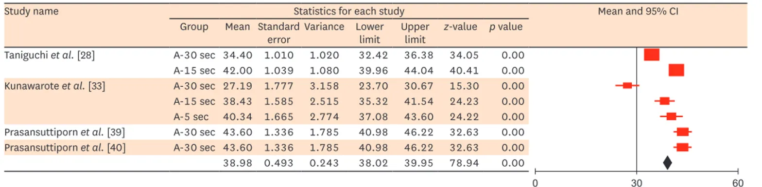
(15.87 MPa) (Table 3, Figures 4 and 5). Additionally, long exposure to the deproteinizing agent adversely affected bonding to dentin (Table 3, Figures 6 and 7). For the deproteinizing groups, the results of the meta-analysis showed that the 2-S SE adhesives exhibited higher mean bond strength values than the 1-S SE adhesives (Table 3, Figures 8 and 9).
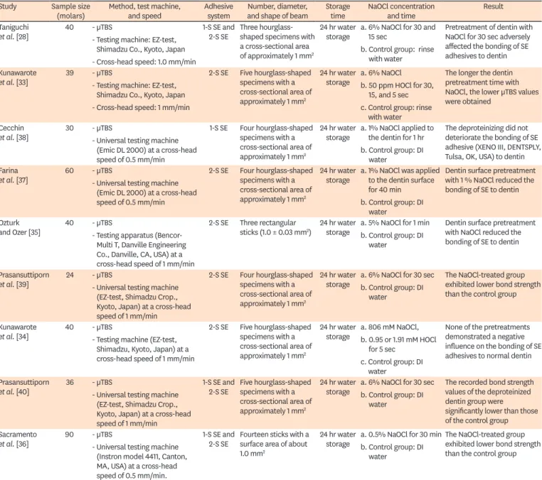
Table 3. Results of applying the medical statistical model of Borenstein et al. [70] to the meta-analysis outcomes
Factor No. of study µTBS (MPa)
Deproteinizing agent NaOCl 8 16.21 ± 0.02b
HOCl 2 40.17 ± 0.76a
Application time (sec) 5 2 41.33 ± 0.70a
15 2 40.56 ± 0.70a
30 4 34.75 ± 0.40b
SE adhesive 1-S 4 38.98 ± 0.49a
2-S 2 32.21 ± 0.62b
Data are shown as means ± standard deviations. Groups identified by different superscript letters within the rows for each factor were significantly different at p < 0.05.
µTBS, microtensile bond strength; NaOCl, sodium hypochlorite; HOCl, hypochlorous acid; SE, self-etch; 1-S, one- step; 2-S, two-step.
0 30 60
Study name Statistics for each study Mean and 95% CI
Group SE Mean Standard
error Variance Lower limit Upper
limit z-value p value Taniguchi et al. [28] A-30 sec 1-step 30.40 2.22 4.94 26.04 34.76 13.68 0.00
A-15 sec 43.70 2.86 8.167 38.10 49.30 15.29 0.00
A-30 sec 2-step 34.40 1.01 1.02 32.42 36.38 34.05 0.00
A-15 sec 42.00 1.04 1.08 39.96 44.04 40.42 0.00
Kunawarote et al. [33] A-30 sec 2-step 27.19 1.78 3.16 23.71 30.67 15.23 0.00
A-15 sec 38.43 1.59 2.52 35.32 41.54 24.23 0.00
A-5 sec 40.34 1.67 2.77 37.08 43.61 24.22 0.00
Cecchin et al. [38] B-1 hr 1-step 19.41 1.68 2.82 16.12 22.70 11.56 0.00 Farina et al. [37] B-40 min 2-step 19.08 1.23 1.51 16.67 21.49 15.51 0.00 Ozturk and Ozer [35] C-60 sec 2-step 15.58 0.06 0.001 15.53 15.63 603.41 0.00 Prasansuttiporn et al. [39] A-30 sec 2-step 43.60 1.34 1.79 40.98 46.22 32.63 0.00 Kunawarote et al. [34] D-5 sec 2-step 40.87 1.69 2.84 37.57 44.17 24.25 0.00 Prasansuttiporn et al. [40] A-30 sec 1-step 33.60 0.78 0.60 32.08 35.12 43.35 0.00
A-30 sec 26.90 1.26 1.58 24.44 29.36 21.42 0.00
A-30 sec 2-step 43.60 1.34 1.79 40.98 46.22 32.63 0.00 Sacramento et al. [36] E-30 min 2-step 30.60 1.29 1.67 28.07 33.13 23.68 0.00 E-30 min 1-step 20.67 1.68 2.81 17.38 23.96 12.24 0.00 15.69 0.03 0.001 15.64 15.74 609.42 0.00
Figure 4. Meta-analysis results of the mean µTBS for SE adhesive bonded to NaOCl-treated dentin.
µTBS, microtensile bond strength; SE, self-etch; NaOCl, sodium hypochlorite; CI, confidence interval; A, 6% NaOCl; B, 1% NaOCl; C, 1% NaOCl; D, 806.02 mM NaOCl; E, 0.5% NaOCl.
0 30 60
Study name Statistics for each study Mean and 95% CI
Group Mean Standard
error Variance Lower limit Upper
limit z-value p value Kunawarote et al. [33] A-30 sec 36.87 1.82 3.31 33.31 40.43 20.28 0.00 A-15 sec 37.64 1.83 3.36 34.05 41.23 20.53 0.00
A-5 sec 41.97 1.49 2.22 39.05 44.89 28.18 0.00
Kunawarote et al. [34] B-5 sec 41.93 1.56 2.43 38.87 44.99 26.90 0.00
C-5 sec 41.24 2.25 5.04 36.84 45.64 18.37 0.00
40.17 0.78 0.60 38.65 41.69 51.76 0.00
Figure 5. Meta-analysis results of the mean µTBS for SE adhesive bonded to HOCl-treated dentin.
µTBS, microtensile bond strength; SE, self-etch; HOCl, hypochlorous acid; CI, confidence interval; A, 50 ppm HOCl; B, 0.95 mM HOCl; C, 1.91 mM HOCl.
0 30 60
Study name Statistics for each study Mean and 95% CI
Group Mean Standard
error Variance Lower limit Upper
limit z-value p value
Kunawarote et al. [33] A 40.34 1.67 2.77 37.08 43.60 24.22 0.00
B 41.97 1.49 2.22 39.05 44.89 28.18 0.00
Kunawarote et al. [34] C 40.87 1.69 2.84 37.57 44.17 24.25 0.00
D 41.93 1.56 2.43 38.87 44.99 26.90 0.00
E 41.24 2.25 5.04 36.84 45.64 18.37 0.00
41.33 0.75 0.56 39.86 42.80 55.03 0.00
Figure 6. Meta-analysis results of the mean µTBS for 5 second dentin surface treatment with a deproteinizing agent.
µTBS, microtensile bond strength; CI, confidence interval; A, 6% NaOCl; B, 50 ppm HOCl; C, 806.02 mM NaOCl; D, 0.95 mM HOCl; E, 1.91mM HOCl.
0 30 60
Study name Statistics for each study Mean and 95% CI
Group SE Mean Standard
error Variance Lower limit Upper
limit z-value p value Taniguchi et al. [28] A 1-step 30.40 2.22 4.94 26.04 34.76 13.68 0.00
A 2-step 34.40 1.01 1.021 32.42 36.38 34.05 0.00
Kunawarote et al. [33] A 2-step 27.19 1.778 3.16 23.71 30.67 15.30 0.00
B 36.87 1.82 3.31 33.31 40.43 20.28 0.00
Prasansuttiporn et al. [39] A 2-step 43.60 1.34 1.79 40.98 46.22 32.63 0.00 Prasansuttiporn et al. [40] A 1-step 33.60 0.78 0.60 32.08 35.12 43.35 0.00
A 26.90 1.26 1.58 24.44 29.36 21.42 0.00
A 2-step 43.60 1.34 1.79 40.98 46.22 32.63 0.00
34.75 0.44 0.19 33.90 35.62 79.41 0.00
Figure 7. Meta-analysis results of the mean µTBS for 30 second dentin surface treatment with a deproteinizing agent.
µTBS, microtensile bond strength; SE, self-etch; CI, confidence interval; A, 6% NaOCl; B, 50 ppm HOCl.
0 30 60
Study name Statistics for each study Mean and 95% CI
Group SE Mean Standard
error Variance Lower limit Upper
limit z-value p value Taniguchi et al. [28] A-30 sec 1-step 30.40 2.22 4.94 26.04 34.76 13.67 0.00
A-15 sec 43.70 2.86 8.17 38.09 49.30 15.29 0.00
Prasansuttiporn et al. [40] A-30 sec 1-step 33.60 0.78 0.60 32.08 35.12 43.35 0.00
A-30 sec 26.90 1.26 1.58 24.43 29.36 21.41 0.00
32.21 0.62 0.38 30.99 33.41 52.16 0.00
Figure 8. Meta-analysis results of the mean µTBS for one-step SE adhesive bonded to deproteinized dentin.
µTBS, microtensile bond strength; SE, self-etch; CI, confidence interval; A, 6% NaOCl.
0 30 60
Study name Statistics for each study Mean and 95% CI
Group Mean Standard
error Variance Lower limit Upper
limit z-value p value Taniguchi et al. [28] A-30 sec 34.40 1.010 1.020 32.42 36.38 34.05 0.00 A-15 sec 42.00 1.039 1.080 39.96 44.04 40.41 0.00 Kunawarote et al. [33] A-30 sec 27.19 1.777 3.158 23.70 30.67 15.30 0.00 A-15 sec 38.43 1.585 2.515 35.32 41.54 24.23 0.00 A-5 sec 40.34 1.665 2.774 37.08 43.60 24.22 0.00 Prasansuttiporn et al. [39] A-30 sec 43.60 1.336 1.785 40.98 46.22 32.63 0.00 Prasansuttiporn et al. [40] A-30 sec 43.60 1.336 1.785 40.98 46.22 32.63 0.00 38.98 0.493 0.243 38.02 39.95 78.94 0.00
Figure 9. Meta-analysis results of the mean µTBS for two-step SE adhesive bonded to deproteinized dentin.
µTBS, microtensile bond strength; SE, self-etch; CI, confidence interval; A, 6% NaOCl.
Discussion
Currently, evidence-based dentistry is an essential approach for detecting research gaps and synthesizing conclusions from the current literature despite conflicting opinions.
The ultimate goal of this scientific approach is to summarize, disseminate, and critique the currently available scientific knowledge, while aiming to translate this knowledge into clinical recommendations. A systematic review is a powerful tool in this scientific approach that helps achieve its objectives [1]. The majority of selected studies in this review did not follow methodologically ideal testing techniques, and consequently, considerable variation in the results was observed among the studies. Thus, the rationale behind conducting this review was to obtain well-justified conclusions, which may help both researchers and clinicians to judge the efficacy of using deproteinizing agents as a dentin surface pretreatment method for modifying the smear layer.
Dentin is a natural composite structure and is considered a challenging substrate for dental adhesion. Dentin has a heterogeneous nature and consists of a complex inorganic/
organic structure [4]. The presence of the smear layer represents another major challenge for successful bonding to dentin [41,42]. It is well known that a micromechanical adhesion mechanism plays an essential role in the adhesion of resin-based bonding agents to dentin, in which adhesive primers infiltrate into the superficial demineralized collagen fibers of
‘hybridized’ dentin [43]. However, previous studies showed that resin primers could not totally infiltrate the demineralized dentin, leaving behind some gaps and denuded collagen.
These negative spaces can act as pathways for microorganisms and may influence bond stability, particularly when water seeps in [44-48].
The results of this systematic review showed that the surface pretreatment of dentin with either NaOCl or HOCl solutions led to low μTBS values compared with non-treated surfaces.
Additionally, it showed that the μTBS values of dentin treated with HOCl solution were significantly higher than those of NaOCl-pretreated dentin. This may be attributed to the chemistry of the NaOCl solution, which has a low surface tension and a high potential to disrupt both sound and denatured collagen. It has been reported that applying NaOCl to the smear layer removed only the superficial ‘loosely attached organic component, without opening the dentinal tubules’ [28,49-51]. However, it may deteriorate the mechanical properties of dentin via the degradation of the sound collagen fibers [31]. NaOCl solutions may degrade the collagen scaffolds of dentin, consequently reducing the number of bonding sites for adhesive primers. This impairs resin hybridization with dentin, leading to a marked reduction in the μTBS [43,52-54].
Furthermore, the low bond strength of NaOCl-treated dentin may be attributed to the strong oxidizing action of NaOCl, which leads to the formation of chloramine-derived radicals. These reactive radicals could interfere with the free radical polymerization of resin- based adhesives [26,55-58]. Additionally, bonding to dentin might be influenced by the residual NaOCl entrapped in the porous structure of mineralized dentin [59]. The residual chemical substances in the fluid may interact with the adhesive system and affect the light polymerization of the monomer in the demineralized dentin, causing a marked reduction in bond strength [37,60].
Moreover, Taniguchi et al. [28] investigated the surface pH of NaOCl-treated dentin and
reported that these surfaces exhibited significantly higher pH values than non-treated
dentin surfaces, even after copious rinsing with water for sufficient time periods. The high alkalinity of NaOCl-treated surfaces could be explained by the high concentration of hydroxyl (OH) groups on the dentin surface [51,61,62]. The alkalinity of NaOCl might buffer the acidity of SE adhesives and thus reduce their hybridization with the underlying dentin [33].
These results are in agreement with many previous studies [28,43,55] that reported that the application of NaOCl to dentin had an adverse effect on the bonding of SE to dentin.
Nonetheless, a few studies have reported that NaOCl treatment increased the bond strength of some adhesive systems, and they attributed their results to the effects of NaOCl on the removal of the collagen layer, which may be beneficial for some resins to create proper dentinal bonding [63-65]. However, most of those studies neglected the adverse effects of NaOCl on bonding to dentin and did not provide logical explanations for the high bond strength results that they obtained.
It is well known that the hydration reaction of NaOCl leads to the formation of HOCl, which is a potent deproteinizing agent as well as an effective biological oxidizing agent [49]. In aqueous solution, HOCl partially dissociates into the anion hypochlorite (OCl
−) and cation hydrogen (H
+). The pH of HOCl is slightly acidic, which could partially demineralize the dentin and allow it to achieve a better resin hybridization than NaOCl solutions [66,67].
Furthermore, it was stated that the higher reactivity of NaOCl to amino acids makes it resistant to washing (even after copious rinsing with water), leaving high concentrations of chlorine on the surface [68,69]. Unlike NaOCl, HOCl solutions can be easily rinsed off, and this might provide a logical explanation of the relatively high μTBS values of HOCl-pretreated dentin surfaces in comparison with NaOCl-pretreated dentin.
The results of this study showed that long surface treatment with deproteinizing agents adversely affected the bonding of SE to dentin. Application of deproteinizing agents for an extended period may lead to the destruction of more collagen scaffolds, resulting in a marked reduction in binding sites for adhesive primers. Additionally, 2-S SE adhesives showed higher μTBS values than 1-S SE adhesives. This may be due to the contamination of 1-S SE adhesives by NaOCl byproducts that affect the free-radical polymerization reaction.
Moreover, the alkalinity of NaOCl may neutralize the acidity of ultra-mild 1-S SE, whereas this buffering action has a minimal effect on the intermediate pH 2-S SE adhesives. These results are in agreement with those of the study of Hamama et al. [11], in which nanoleakage results revealed that the silver nitrate intake was higher in sound dentin treated with Carisolv (a NaOCl-based chemomechanical caries removal agent) and bonded with a 1-S SE adhesive than in the corresponding groups bonded with a 2-S SE adhesive. They attributed the higher silver uptake to the contamination of the hybrid layer by NaOCl residues, which affected the free-radical polymerization reaction and consequently led to a reduction in μTBS.
An unavoidable limitation of the current systematic review was that one of its exclusion criteria was non-English manuscripts; however, some of those excluded studies may have contained useful information for this review.
Conclusions
In light of the currently available scientific evidence, pretreatment of dentin surfaces with
deproteinizing agents does not enhance the bonding of SE adhesives to dentin. HOCl as a
deproteinizing agent exhibits minimal adverse effects on bonding to dentin in comparison with
NaOCl solutions. Accordingly, when needed, it is preferable to use HOCl as a deproteinizing agent for dentin surface pretreatment prior to the application of SE adhesives. The 2-S SE adhesives show more reliable bonding to deproteinized dentin than 1-S SE adhesives. Long exposure to deproteinizing agents significantly impairs the bonding of SE agents to dentin.
REFERENCES
1. Jourdan M, Gagne S, Dubois-Laurent C, Maghraoui M, Huet S, Suel A, Hamama L, Briard M, Peltier D, Geoffriau E. Carotenoid content and root color of cultivated carrot: a candidate-gene association study using an original broad unstructured population. PLoS One 2015;10:e0116674.
PUBMED | CROSSREF
2. Miller SA, Forrest JL. Translating evidence-based decision making into practice: appraising and applying the evidence. J Evid Based Dent Pract 2009;9:164-182.
PUBMED | CROSSREF
3. Fuentes V, Toledano M, Osorio R, Carvalho RM. Microhardness of superficial and deep sound human dentin. J Biomed Mater Res A 2003;66:850-853.
PUBMED | CROSSREF
4. Ritter AV, Eidson RS, Donovan TE. Dental caries: etiology, clinical characteristics, risk assessment, and management. In: Heymann HO, Swift EJ, Ritter AV. editors. Sturdevant's art & science of operative dentistry. 6th ed. St. Louis (MO): Elsevier Mosby; 2013. p41-88.
5. Swift EJ, Perdigao J, Heymann HO. Bonding to enamel and dentin: a brief history and state of the art, 1995. Quintessence Int 1995;26:95-110.
PUBMED
6. Van Meerbeek B, De Munck J, Yoshida Y, Inoue S, Vargas M, Vijay P, Van Landuyt K, Lambrechts P, Vanherle G. Buonocore memorial lecture. Adhesion to enamel and dentin: current status and future challenges. Oper Dent 2003;28:215-235.
PUBMED
7. Hikita K, Van Meerbeek B, De Munck J, Ikeda T, Van Landuyt K, Maida T, Lambrechts P, Peumans M.
Bonding effectiveness of adhesive luting agents to enamel and dentin. Dent Mater 2007;23:71-80.
PUBMED | CROSSREF
8. Bowen RL. Adhesive bonding of various materials to hard tooth tissues. II. Bonding to dentin promoted by a surface-active comonomer. J Dent Res 1965;44:895-902.
PUBMED | CROSSREF
9. Xie J, Powers JM, McGuckin RS. In vitro bond strength of two adhesives to enamel and dentin under normal and contaminated conditions. Dent Mater 1993;9:295-299.
PUBMED | CROSSREF
10. Reis AF, Giannini M, Kavaguchi A, Soares CJ, Line SR. Comparison of microtensile bond strength to enamel and dentin of human, bovine, and porcine teeth. J Adhes Dent 2004;6:117-121.
PUBMED
11. Hamama HH, Yiu CK, Burrow MF. Effect of chemomechanical caries removal on bonding of self-etching adhesives to caries-affected dentin. J Adhes Dent 2014;16:507-516.
PUBMED | CROSSREF
12. Pashley DH. Smear layer: overview of structure and function. Proc Finn Dent Soc 1992;88:215-224.
PUBMED
13. Spencer P, Ye Q, Park J, Topp EM, Misra A, Marangos O, Wang Y, Bohaty BS, Singh V, Sene F, Eslick J, Camarda K, Katz JL. Adhesive/dentin interface: the weak link in the composite restoration. Ann Biomed Eng 2010;38:1989-2003.
PUBMED | CROSSREF
14. Ishioka S, Caputo AA. Interaction between the dentinal smear layer and composite bond strength. J Prosthet Dent 1989;61:180-185.
PUBMED | CROSSREF
15. Giachetti L, Bambi C, Scaminaci Russo D. SEM qualitative evaluation of four self-etching adhesive systems. Minerva Stomatol 2005;54:415-428.
PUBMED
16. Waidyasekera K, Nikaido T, Weerasinghe DS, Ichinose S, Tagami J. Reinforcement of dentin in self-etch adhesive technology: a new concept. J Dent 2009;37:604-609.
PUBMED | CROSSREF
17. Ozer F, Blatz MB. Self-etch and etch-and-rinse adhesive systems in clinical dentistry. Compend Contin Educ Dent 2013;34:12-14.
PUBMED
18. Perdigão J, Reis A, Loguercio AD. Dentin adhesion and MMPs: a comprehensive review. J Esthet Restor Dent 2013;25:219-241.
PUBMED | CROSSREF
19. Peumans M, Kanumilli P, De Munck J, Van Landuyt K, Lambrechts P, Van Meerbeek B. Clinical effectiveness of contemporary adhesives: a systematic review of current clinical trials. Dent Mater 2005;21:864-881.
PUBMED | CROSSREF
20. van Dijken JW, Sunnegårdh-Grönberg K, Lindberg A. Clinical long-term retention of etch-and-rinse and self- etch adhesive systems in non-carious cervical lesions. A 13 years evaluation. Dent Mater 2007;23:1101-1107.
PUBMED | CROSSREF
21. Perdigão J, Geraldeli S, Hodges JS. Total-etch versus self-etch adhesive: effect on postoperative sensitivity.
J Am Dent Assoc 2003;134:1621-1629.
PUBMED | CROSSREF
22. Tay FR, King NM, Chan KM, Pashley DH. How can nanoleakage occur in self-etching adhesive systems that demineralize and infiltrate simultaneously? J Adhes Dent 2002;4:255-269.
PUBMED
23. Van Landuyt KL, Mine A, De Munck J, Countinho E, Peumans M, Jaecques S, Lambrechts P, Van Meerbeek B. Technique sensitivity of water-free one-step adhesives. Dent Mater 2008;24:1258-1267.
PUBMED | CROSSREF
24. Tay FR, Pashley DH. Aggressiveness of contemporary self-etching systems. I: depth of penetration beyond dentin smear layers. Dent Mater 2001;17:296-308.
PUBMED | CROSSREF
25. Yamauti M, Hashimoto M, Sano H, Ohno H, Carvalho RM, Kaga M, Tagami J, Oguchi H, Kubota M.
Degradation of resin-dentin bonds using NaOCl storage. Dent Mater 2003;19:399-405.
PUBMED | CROSSREF
26. Nikaido T, Takano Y, Sasafuchi Y, Burrow MF, Tagami J. Bond strengths to endodontically-treated teeth.
Am J Dent 1999;12:177-180.
PUBMED
27. Zehnder M, Grawehr M, Hasselgren G, Waltimo T. Tissue-dissolution capacity and dentin-disinfecting potential of calcium hydroxide mixed with irrigating solutions. Oral Surg Oral Med Oral Pathol Oral Radiol Endod 2003;96:608-613.
PUBMED | CROSSREF
28. Taniguchi G, Nakajima M, Hosaka K, Iwamoto N, Ikeda M, Foxton RM, Tagami J. Improving the effect of NaOCl pretreatment on bonding to caries-affected dentin using self-etch adhesives. J Dent 2009;37:769-775.
PUBMED | CROSSREF
29. Montes MA, de Goes MF, Sinhoreti MA. The in vitro morphological effects of some current pre-treatments on dentin surface: a SEM evaluation. Oper Dent 2005;30:201-212.
PUBMED
30. Boyde A, Jones SJ. Backscattered electron imaging of dental tissues. Anat Embryol (Berl) 1983;168:211-226.
PUBMED | CROSSREF
31. Carvalho RM, Chersoni S, Frankenberger R, Pashley DH, Prati C, Tay FR. A challenge to the conventional wisdom that simultaneous etching and resin infiltration always occurs in self-etch adhesives.
Biomaterials 2005;26:1035-1042.
PUBMED | CROSSREF
32. Moher D, Liberati A, Tetzlaff J, Altman DG, Altman D, Antes G, Atkins D, Barbour V, Barrowman N, Berlin JA, Clark J, Clarke M, Cook D, D'Amico R, Deeks JJ, Devereaux PJ, Dickersin K, Egger M, Ernst E, Gøtzsche PC, Grimshaw J, Guyatt G, Higgins J, Ioannidis JP, Kleijnen J, Lang T, Liberati A, Magrini N, McNamee D, Moja L, Moher D, Mulrow C, Napoli M, Oxman A, Pham B, Rennie D, Sampson M, Schulz KF, Shekelle PG, Tetzlaff J, Tovey D, Tugwell P. Preferred reporting items for systematic reviews and meta- analyses: the PRISMA statement. Int J Surg 2010;8:336-341.
PUBMED | CROSSREF
33. Kunawarote S, Nakajima M, Shida K, Kitasako Y, Foxton RM, Tagami J. Effect of dentin pretreatment with mild acidic HOCl solution on microtensile bond strength and surface pH. J Dent 2010;38:261-268.
PUBMED | CROSSREF
34. Kunawarote S, Nakajima M, Foxton RM, Tagami J. Effect of pretreatment with mildly acidic hypochlorous acid on adhesion to caries-affected dentin using a self-etch adhesive. Eur J Oral Sci 2011;119:86-92.
PUBMED | CROSSREF
35. Ozturk B, Ozer F. Effect of NaOCl on bond strengths of bonding agents to pulp chamber lateral walls. J Endod 2004;30:362-365.
PUBMED | CROSSREF
36. Sacramento PA, Sampaio CS, de Carvalho FG, Pascon FM, Borges AF, Alves MC, Hosoya Y, Puppin- Rontani RM. Influence of NaOCl irrigation and water-storage on degradation and microstructure of resin- dentin interface. Int J Adhes Adhes 2013;47:117-124.
PUBMED | CROSSREF
37. Farina AP, Cecchin D, Barbizam JV, Carlini-Júnior B. Influence of endodontic irrigants on bond strength of a self-etching adhesive. Aust Endod J 2011;37:26-30.
PUBMED | CROSSREF
38. Cecchin D, Farina AP, Galafassi D, Barbizam JV, Corona SA, Carlini-Júnior B. Influence of sodium hypochlorite and edta on the microtensile bond strength of a self-etching adhesive system. J Appl Oral Sci 2010;18:385-389.
PUBMED | CROSSREF
39. Prasansuttiporn T, Nakajima M, Kunawarote S, Foxton RM, Tagami J. Effect of reducing agents on bond strength to NaOCl-treated dentin. Dent Mater 2011;27:229-234.
PUBMED | CROSSREF
40. Prasansuttiporn T, Nakajima M, Foxton RM, Tagami J. Scrubbing effect of self-etching adhesives on bond strength to NaOCl-treated dentin. J Adhes Dent 2012;14:121-127.
PUBMED | CROSSREF
41. Boyde A, Switsur VR, Stewart AD. An assessment of two new physical methods applied to the study of dental tissues. Arch Oral Biol 1962;7 Supplement:185-193.
42. Eick JD, Wilko RA, Anderson CH, Sorensen SE. Scanning electron microscopy of cut tooth surfaces and identification of debris by use of the electron microprobe. J Dent Res 1970;49:1359-1368.
PUBMED | CROSSREF
43. Perdigão J, Lopes M, Geraldeli S, Lopes GC, García-Godoy F. Effect of a sodium hypochlorite gel on dentin bonding. Dent Mater 2000;16:311-323.
PUBMED | CROSSREF
44. Hashimoto M, Ohno H, Kaga M, Sano H, Endo K, Oguchi H. The extent to which resin can infiltrate dentin by acetone-based adhesives. J Dent Res 2002;81:74-78.
PUBMED | CROSSREF
45. Chan KM, Tay FR, King NM, Imazato S, Pashley DH. Bonding of mild self-etching primers/adhesives to dentin with thick smear layers. Am J Dent 2003;16:340-346.
PUBMED
46. Spencer P, Wang Y. Adhesive phase separation at the dentin interface under wet bonding conditions. J Biomed Mater Res 2002;62:447-456.
PUBMED | CROSSREF
47. Wang Y, Spencer P. Hybridization efficiency of the adhesive/dentin interface with wet bonding. J Dent Res 2003;82:141-145.
PUBMED | CROSSREF
48. De Munck J, Vargas M, Iracki J, Van Landuyt K, Poitevin A, Lambrechts P, Van Meerbeek B. One-day bonding effectiveness of new self-etch adhesives to bur-cut enamel and dentin. Oper Dent 2005;30:39-49.
PUBMED
49. Boyde A. Methodology of calcified tissue specimen preparation for scanning electron microscopy. In:
Dickson GR, editor. Methods of calcified tissue preparation. Amsterdam: Elsevier; 1984. p251-307.
50. Fawzy AS, Amer MA, El-Askary FS. Sodium hypochlorite as dentin pretreatment for etch-and-rinse single-bottle and two-step self-etching adhesives: atomic force microscope and tensile bond strength evaluation. J Adhes Dent 2008;10:135-144.
PUBMED
51. Wang L, Bassiri M, Najafi R, Najafi K, Yang J, Khosrovi B, Hwong W, Barati E, Belisle B, Celeri C, Robson MC. Hypochlorous acid as a potential wound care agent: part I. Stabilized hypochlorous acid: a component of the inorganic armamentarium of innate immunity. J Burns Wounds 2007;6:e5.
PUBMED
52. Vargas MA, Cobb DS, Armstrong SR. Resin-dentin shear bond strength and interfacial ultrastructure with and without a hybrid layer. Oper Dent 1997;22:159-166.
PUBMED
53. Prati C, Chersoni S, Pashley DH. Effect of removal of surface collagen fibrils on resin-dentin bonding.
Dent Mater 1999;15:323-331.
PUBMED | CROSSREF
54. Saboia VP, Rodrigues AL, Pimenta LA. Effect of collagen removal on shear bond strength of two single- bottle adhesive systems. Oper Dent 2000;25:395-400.
PUBMED
55. Lai SC, Mak YF, Cheung GS, Osorio R, Toledano M, Carvalho RM, Tay FR, Pashley DH. Reversal of compromised bonding to oxidized etched dentin. J Dent Res 2001;80:1919-1924.
PUBMED | CROSSREF
56. Morris MD, Lee KW, Agee KA, Bouillaguet S, Pashley DH. Effects of sodium hypochlorite and RC-prep on bond strengths of resin cement to endodontic surfaces. J Endod 2001;27:753-757.
PUBMED | CROSSREF
57. Hawkins CL, Davies MJ. Hypochlorite-induced oxidation of proteins in plasma: formation of chloramines and nitrogen-centred radicals and their role in protein fragmentation. Biochem J 1999;340:539-548.
PUBMED | CROSSREF
58. Rueggeberg FA, Margeson DH. The effect of oxygen inhibition on an unfilled/filled composite system. J Dent Res 1990;69:1652-1658.
PUBMED | CROSSREF
59. Mountouris G, Silikas N, Eliades G. Effect of sodium hypochlorite treatment on the molecular composition and morphology of human coronal dentin. J Adhes Dent 2004;6:175-182.
PUBMED
60. Vongphan N, Senawongse P, Somsiri W, Harnirattisai C. Effects of sodium ascorbate on microtensile bond strength of total-etching adhesive system to NaOCl treated dentine. J Dent 2005;33:689-695.
PUBMED | CROSSREF
61. Hiraishi N, Kitasako Y, Nikaido T, Nomura S, Burrow MF, Tagami J. Effect of artificial saliva contamination on pH value change and dentin bond strength. Dent Mater 2003;19:429-434.
PUBMED | CROSSREF
62. Haapasalo M, Qian W. Irrigants and intracanal medicaments. In: Ingle JI, Bakland L, Baumgartner J, editors. Ingle's endodontics. 6th ed. Hamilton: BC Decker; 2008. p992-1018.
63. Inoue S, Murata Y, Sano H, Kashiwada T. Effect of NaOCl treatment on bond strength between indirect resin core-buildup and dentin. Dent Mater J 2002;21:343-354.
PUBMED
64. Pioch T, Kobaslija S, Schagen B, Götz H. Interfacial micromorphology and tensile bond strength of dentin bonding systems after NaOCl treatment. J Adhes Dent 1999;1:135-142.
PUBMED
65. de Castro AK, Hara AT, Pimenta LA. Influence of collagen removal on shear bond strength of one-bottle adhesive systems in dentin. J Adhes Dent 2000;2:271-277.
PUBMED
66. Mainnemare A, Mégarbane B, Soueidan A, Daniel A, Chapple IL. Hypochlorous acid and taurine-N- monochloramine in periodontal diseases. J Dent Res 2004;83:823-831.
PUBMED | CROSSREF
67. Christensen CE, McNeal SF, Eleazer P. Effect of lowering the pH of sodium hypochlorite on dissolving tissue in vitro. J Endod 2008;34:449-452.
PUBMED | CROSSREF
68. Guentzel JL, Liang Lam K, Callan MA, Emmons SA, Dunham VL. Reduction of bacteria on spinach, lettuce, and surfaces in food service areas using neutral electrolyzed oxidizing water. Food Microbiol 2008;25:36-41.
PUBMED | CROSSREF
69. Mishra P, Palamara JE, Tyas MJ, Burrow MF. Effect of static loading of dentin beams at various pH levels.
Calcif Tissue Int 2006;79:416-421.
PUBMED | CROSSREF
70. Borenstein M, Hedges LV, Higgins JP, Rothstein HR. Introduction to meta-analysis. Chichester: John Wiley & Sons; 2009.


![Table 3. Results of applying the medical statistical model of Borenstein et al. [70] to the meta-analysis outcomes](https://thumb-ap.123doks.com/thumbv2/123dokinfo/5283906.146734/9.892.286.829.265.383/table-results-applying-medical-statistical-borenstein-analysis-outcomes.webp)