서 론
(systemic lupus erythematosus, SLE)
1,2)
. SLE
, ,
, , ,
, , , , ,
1-3)
. 20-40
3,4)
,
5)
.
1,6,7)
,
T, B
2007 1 17
2008 3 4
,
(Tel:+82-42-229-6880, Fax:+82-42-254-3403, E-mail:[email protected])
滋腎活血湯 全身性紅斑性狼瘡 影響
최훈섭, 조충식, 김철중 Original Article
Effect of Jasinwhalhyul-tang on MRL/MpJ-Ipr/Ipr Mouse Model with Systemic Lupus Erythematosus
Hoon-Seob Choi, Chung-Sik Cho, Cheol-Jung Kim
Department of Internal Medicine, College of Oriental Medicine, Daejeon University, Daejeon, Korea.
Objective : The main purpose of this study was to evaluate the effect of Jasinwhalhyul-tang (Zishenhuoxue-tang, JWT) on MRL/MpJ-Ipr/Ipr mouse model with systemic lupus erythematosus.
Methods: The effect of JWT on MRL/MpJ-Ipr/Ipr mice that have autoimmune disease similar to SLE in humans was evaluated after JWT per oral in the present study. Mice were administered with Jasinwhalhyul-tang (Zishenh- uoxue-tang, JWT) (80 or 400mg/kg) or distilled water for control group from experimental week 10 for 22 weeks.
Results : The amount of erythematosus skin lesion and proteinuria were significantly decreased. The size and weight of cervical lymph nodes and spleen were significantly reduced. The ratio between activated CD3
+CD69
+T-cells and undifferentiated CD3
+CD4
-CD8
-T-cells in lymph nodes, spleen and kidney was effectively reduced. The gene expression of TGF-β in spleen and kidney was increased. The amount of anti-dsDNA IgG in blood was decreased.
The gene expression of TGF-β in normal mouse spleen cells was increased depending on concentration by treatment of with T cell stimulating agent. In the histological examination of skin and kidney, the amount of infiltration of immune cells involved in the inflammatory response was decreased.
Conclusions : According to the above results, JWT should be considered as an applicable therapeutic agent to SLE in clinical practice. Further research is required to investigate other efficacies of JWT on SLE.
Key Words : SLE, Jasinwhalhyul-tang ( Zishenhuoxue-tang , JWT), MRL/MpJ-Ipr/Ipr mouse model
1)
, T
8,9)
. SLE
, , ,
, , , ,
10,11)
. ,
, , , ,
, , ,
,
10,11).
12)
SLE 1 ,
13-15)
SLE , ,
.
16)
SLE
, ,
, , ,
, , , , , ,
SLE .
, SLE
MRL/MpJ-lpr/lpr
17-19)
,
, , , ,
,
, cytokine,
, .
실 험
1.
재료 1)
MRL/MpJ-lpr/lpr 30g 7 8
SLC Inc.(Hamamatsu, Japan)
, ( )
22±2 1
.
20g 6 8 Balb/c (
)
. kg
(Table 1).
2)
diethyl pyrocarbonate(DEPC), 3-4,5- dimethyl-thiazol-2,5-carboxymethoxyphenyl-2,4-s ulfophenyl-2H-tetrazolim(MTS), Freund’s complete adjuvant, chloroform, collagenase, RPMI -1640
IMDM medium, isopropanol,
(RBC lysis solution), ethidium bromide(EtBr), Dulbecco’s phosphate buffered saline(D-PBS), calf thymus DNA, formaldehyde, lamide, mag- nesium chloride(MgCl
2) Sigma (U.S.A),
(fetal bovine serum, FBS) Hyclone (U.S.A), anti-CD3-PE(phycoerythrin), anti- CD4- FITC(fluorescein isothiocyanate), anti-Gr1-PE, anti- CD8-FITC, anti-CD25-PE, anti-CD28-PE, anti-
조단백질 22.1%
조지방 8.0%
조섬유 5.0%
조회분 8.0%
칼슘 0.6%
인 0.4%
Table 1.
CD11b-FITC, anti-IgE-FITC anti-B220-PE, anti- CD69-FITC, goat anti-mouse IgG HRP conjugated, goat anti-TGFβ, propidium iodide(PI) RNase
Pharmingen (U.S.A) .
3)
( , Korea), rotary vaccum evaporator(Büchi B-480, Switzerland), freeze dryer(EYELA FDU-540, Japan), CO
2incubator (Forma scientific Co., U.S.A), clean bench(Vision scientific Co., Korea), autoclave(Sanyo, Japan), micro-pipet(Gilson, France), water bath(Vision scientific Co., Korea), vortex mixer(Vision scientific Co., Korea), spectrophotometer(Shimazue, Japan), centrifuge (Sigma, U.S.A), deep-freezer(Sanyo, Japan), thermocycler system (MWG Biotech., Germany), ice-maker(Vision scientific Co., Korea), homogenizer(OMNI, U.S.A), plate shaker(Lab-Line, U.S.A) ELISA reader(Molecular Devices, U.S.A)
.
4)
, 1
(Table 2).
2.
방법 1)
1 5,000
2
rotary vacuum evaporator .
freeze dryer 1
25g .
.
MRL/MpJ-lpr/lpr 10
( )
80mg/kg (JWT- ), 400mg/kg
(JWT- ) 3 . 10
22 13
0.2ml
韓 藥 名 生 藥 名 用量(g)
生地黃 Rehmanniae Radix 7.50
牧丹皮 Moutan Cortex 7.50
赤芍藥 Paeonia Radix Rubra 7.50
桃 仁 Persicae Semen 15.00
白花蛇舌草 Oldenlndiae Diffusae Herba 15.00
半枝蓮 Scutellariae Barbatae Herba 15.00
白 朮 Atractylodis Macrocephalae Rhizoma 6.00
茯 Poria cocos 6.00
山 藥 Dioscorea Rhizoma 7.50
Smilax Chinae Rhizoma 15.00
薏苡仁 Coicic Semen 15.00
丹 參 Salvia Miltriorrhizae Radix 7.50
黨 參 Codonopsis Pilosulae Radix 7.50
Total amount 132.00
Table 2.
, . 2)
MRL/MpJ-lpr/lpr JWT
10 22
. 3)
MRL/MpJ-lpr/lpr SLE
. ,
20)
1 .
0 :
1 : 1 0.5cm
2 : 2 0.5cm
3 : 2 0.5cm
4)
enzyme- linked immuno- sorbent assay(ELISA)
.
96well-ELISA plate 0.1M carbonate buffer goat anti- murine albumin(Bethyl, U.S.A.)
24 , 0.1%
Bovine serum albumin(BSA)
(PBS) 2 blocking
plate . plate well
2000 20,000 100
2 3
, 50,000 anti-murine
albumin/HRP conjugated(Bethyl, U.S.A.) 100 2
. well 5 , tetram-
ethylbenzidine(TMB) 100
15 30 1N
, ELISA reader
450nm .
0 500ng/ml murine
albumin .
4
20,21)
. grade 0 : 0 0.3mg/ml grade 1 : 0.3 1mg/ml grade 2 : 1 3mg/ml grade 3 : 3 20mg/ml
grade 4 : 20mg/ml , death
5) , ,
10 22 MRL
/MpJ-lpr/lpr ethyl ether , ,
.
6) , ,
MRL/MpJ-lpr/lpr Normal
Balb/c spleen, lymph node, kidney
100mesh (kidney
collagenase 30 2 )
D-PBS 5 (1700rpm) 2
cell strainer (FALCON)
. ACK (8.3g NH4Cl, 1g
KHCO3, in 1L of demineralized water+0.1mM
EDTA) 5
D-PBS 2 0.04%
trypan blue .
2×10⁵ 4 (immuno-fluorescence staining) . anti-CD3-PE(phycoerythrin), anti-CD4-FITC(fluorescein isothiocyanate), anti-Gr1 -PE, anti-CD8-FITC, anti-CD25-PE, anti-CD28- PE, anti-CD11b-FITC, anti-IgE-FITC anti-B220- PE, anti-CD69-FITC, fluorescein isothio- cyanate(FITC)-anti-mouse CD19 30
. 3
(flow cytometer, Becton Dickinson, U.S.A) MRL
/MpJ-lpr/lpr ,
CD3e, CD4, CD8, CD25, CD69,
CD19 .
CellQuest CD3
+, CD19
+, CD4
+, CD8
+, CD4
+CD25
+, CD3
+CD69
+, CD3
+CD4
-CD8
-(gated, %) .
7) (1) RNA MRL/MpJ-lpr/lpr
RNAzolB .
chloroform(CHCl3) 15
. 15
13,000rpm 200
2-propanol 200
15 .
13,000rpm 80% EtOH
3 vaccum pump
RNA . RNA DEPC
20 heating block 75
first strand cDNA .
(2) -
(reverse transcription) total
RNA 3 75 5 ,
2.5 10mM dNTPs mix, 1 random sequence hexanucleotides(25pmole/25 ), RNA inhibitor
1 RNase inhibitor(20U/ ), 1 100mM DTT, 4.5 5×RT buffer (250mM Tris-HCl, pH 8.3, 375mM KCl, 15mM MgCl
2) , 1
M-MLV RT(200U/ ) DEPC
20
. 20 2,000
rpm 5 37
60 first-strand cDNA
, 95 5 M-MLV
RT cDNA
polymerase chain reaction(PCR) . PCR iCycler(Biorad, U.S.A)
. 3 cDNA
, primer L32,
Foxp3, TGF-β, IL-10, IL-2, γ-IFN, IL-12, GITR,
CTLA-4, CD25
sense primer(20pmole/ ) antisense primer
(20pmole/ ) 1 , 3
2.5mM dNTPs, 3 10×PCR buffer(100mM Tris-HCl, pH 8.3, 500 mM KCl, 15mM MgCl
2), 1X SyberGreen, 0.18 Taq polymerase
(5U/ ) 30
pre-denaturation; 95 , 5 , denaturation; 95 , 30 , annealing; 58 , 30 , elongation; 72 , 30 40cycles post-
elongation 72 3 PCR
. Real-time PCR iCycler
.
8) IgG Anti-dsDNA
cytokine ELISA
.
96well-ELISA plate 0.1M carbonate buffer goat anti- mouse IgG (total IgG ) calf thymus DNA (anti- dsDNA )
24 , 0.1%
BSA (Bovine serum albumin) PBS
2 blocking plate
. plate well 100 100,000 100
2 3 ,
1,000 100,000 anti-mouse IgG/HRP conjugated 100
2 . well 5
, TMB (tetramethyl-benzidine)
100 15 30
1N , ELISA
reader 450nm
. Total IgG 0
1,000ng/ml murine IgG
. Anti-dsDNA
. 9)
T-
, 2×10⁶/ml
Balb/c PMA
(phorbol myristate acetate; 10ng/ml) IoM(iono-
mycin; 1μM) T-
JWT . 1
13,000rpm Realtime RT-
PCR .
10)
MRL/MpJ-lpr/lpr
10% formalin-
. 4-μm
, H&E . 11)
± , Student’s T-test
22)p<0.05 .
성 적
1.
체중에 미치는 영향
10 22
(Table 3) JWT
, JWT .
Group Mean Body Weight (g)
10 week 22 week Δ(22week∼ 10week)
Control 31.4 ± 2.1 41.2 ± 2.4 9.6 ± 2.7
JWT- Ⅰ 31.5 ± 1.6 40.9 ± 2.7 9.6 ± 2.9
JWT- Ⅱ 30.8 ± 2.6 40.0 ± 2.7 9.2 ± 3.6
The MRL/MpJ-lpr/lpr mice were administered with JWT (JWT- Ⅰ, 80mg/kg, p.o and JWT-Ⅱ, 400mg/kg, p.o) for 13weeks daily. Control group mice were administered with D.W. instead of JWT. Statistically significant differences in mean body weight of mice were not observed between the groups (n=10).
Table 3.
2.
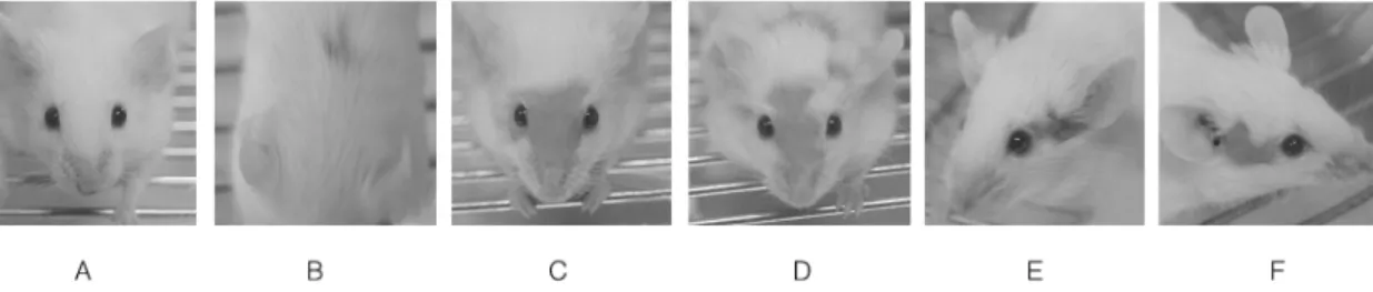
홍반에 미치는 영향
2 2.1±0.9
, JWT
, 0.5cm
1 .
JWT- 0.8±0.5 (p<0.05), JWT- 0.2±0.4(p<0.05)
(Fig. 1, 2).
3.
요단백에 미치는 영향
(100mg/ , grade 2
Fig. 1.
4
3
2
1
0
Clinical score
Control JWT-I JWT-II
*
*
100%
90%
80%
70%
60%
50%
40%
30%
20%
10%
0%
Control JWT-I JWT-II
0 1 2 3 4
Fig. 2. Fig. 3.
) 60%
, JWT- 40%
. JWT- 20% (p<0.05) (Fig. 3).
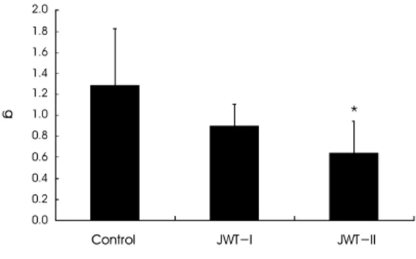
4.
비장
,경부림프절
,신장의 중량 및 크기에 미 치는 영향
JWT- , JWT-
(Fig. 4, 6).
, JWT- 0.64±0.31g(p<0.05) (Fig. 5, 7).
JWT- , JWT- (Fig. 8).
1.4
1.2 1.0
0.8
0.6
0.4
0.2
0.0
Control JWT-I JWT-II
g
2.0 1.8 1.6 1.4 1.2 1.0 0.8 0.6 0.4 0.2 0.0
Control JWT-I JWT-II
g *
Fig. 6. Fig. 7.
Fig. 4. Fig. 5.
5.
비장
,경부림프절
,신장에서의 세포 조성비에 미치는 영향
, JWT-
CD4
+CD25
+(p<0.05) , JWT- CD3
+, CD3
+CD69
+, CD3
+CD4
-CD8
-(p<0.05) ,
CD4
+CD25
+(p<0.05)
(Table 4).
, JWT-
CD3
+CD69
+, CD4
+, CD8
+, JWT- CD3
+CD69
+, CD4
+, CD8
+, CD4
+CD25
+, CD3
+CD4
-CD8
-(p<0.05)
(Table 5).
, JWT-
0.8
0.7
0.6 0.5
0.4 0.3 0.2
0.1
0.0
g
Control JWT-I JWT-II
14
12
10
8
6
4
2
0
Control JWT-I JWT-II
(mg/ml)
Fig. 8. Fig. 9.
Leucocytes
type Organ Normal
Balb/c(%)
MRL-lpr lupus model mice(%)
Control JWT- Ⅰ JWT- Ⅱ
CD3
+(T) LN# 73.0±0.85 94.6±1.20 93.7±.0.58 91.8±1.30
*CD19
+(B) LN 25.6±0.90 1.7±0.12 1.7±0.86 4.0±0.48
CD3
+CD69
+LN 4.1±0.29 35.2±1.35 28.1±4.15 14.9±2.06
*CD4
+LN 66.2±2.00 9.6±2.15 8.7±2.05 7.6±0.65
CD8
+LN 17.7±0.30 2.0±0.20 2.0±0.30 2.0±0.45
CD4
+CD25
+LN 5.0±0.44 2.1±0.15 2.6±0.06
*2.6±0.07
*CD3e
+CD4
-CD8
-LN 3.6±0.45 76.1±2.25 73.2±0.20 61.0±2.45
*#LN : Lymphnode
The MRL/MpJ-lpr/lpr mice were administered with JWT (JWT- Ⅰ, 80mg/kg, p.o and JWT-Ⅱ, 400mg/kg, p.o) for 13weeks daily.
Results are mean ± SD (n=5 mice per group). *p<0.05 versus control group.
Table 4.
CD3
+, CD4
+, CD4
+CD25
+, CD3
+CD4
-CD8
-, JWT- CD3
+, CD3
+CD69
+, CD4
+, CD8
+, CD4
+CD25
+, CD3
+CD4
-CD8
-(p<0.05) (Table 6).
6.
비장 및 신장에서의 유전자 발현에 미치는 영향
L32 , TNF-α,
TGF-β, IL-10, IL-2, IL-12, IL-13, γ-IFN
cytokines Foxp3, CD25, CTLA-4, GITR regulatory T-cell
, JWT
, TGF-β JWT
(p<0.05) (Table 7).
Leucocytes
type Organ Normal
Balb/c(%)
MRL-lpr lupus model mice(%)
Control JWT- Ⅰ JWT- Ⅱ
CD3
+(T) SP
#49.6±5.10 86.4±2.45 84.9±1.25 84.4±3.10
CD19
+(B) SP 40.9±5.75 9.6±1.20 12.2±0.15 12.4±2.35
CD3
+CD69
+SP 2.9±0.60 18.4±0.40 12.6±2.50
*13.5±0.60
*CD4
+SP 25.5±3.75 12.2±0.25 9.2±0.50
*11.2±0.05
*CD8
+SP 13.6±2.35 7.4±0.10 4.6±0.35
*4.9±0.65
*CD4
+CD25
+SP 2.6±0.26 2.3±0.17 1.9±0.37 1.4±0.20
*CD3e
+CD4
-CD8
-SP 3.7±2.06 69.1±0.70 65.5±1.85 61.8±1.00
*#SP : Spleen B/T Rate 0.824 0.111 0.144 0.147
The MRL/MpJ-lpr/lpr mice were administered with JWT (JWT- Ⅰ, 80mg/kg, p.o and JWT-Ⅱ, 400mg/kg, p.o) for 13weeks daily.
Results are mean ± SD (n=5 mice per group). *p<0.05 versus control group.
Table 5.
Leucocytes
type Organ Normal
Balb/c(%)
MRL-lpr lupus model mice(%)
Control JWT- Ⅰ JWT- Ⅱ
CD3
+(T) KD
#20.8±2.15 91.3±2.10 83.6±0.80
*82.7±2.55
*CD19
+(B) KD 3.0±1.15 0.4±0.09 1.2±0.45 1.4±0.26
CD3
+CD69
+KD 6.0±0.44 57.7±1.30 56.9±1.55 52.5±2.20
*CD4
+KD 6.7±0.43 67.9±0.60 56.5±2.25
*57.8±1.30
*CD8
+KD 5.1±1.65 20.5±0.20 22.2±0.65 13.6±1.15
*CD4
+CD25
+KD 0.8±0.10 1.5±0.43 0.50±0.04
*0.9±0.14
*CD11b
+Gr-1
+KD 0.42±0.02 1.24±0.14 1.16±0.39 0.62±0.27
CD3e
+CD4
-CD8
-KD 0.1±0.01 3.6±0.64 1.0±0.07
*1.6±0.19
*#KD : Kidney B/T Rate 0.144 0.004 0.014 0.017
The MRL/MpJ-lpr/lpr mice were administered with JWT (JWT- Ⅰ, 80mg/kg, p.o and JWT-Ⅱ, 400mg/kg, p.o) for 13weeks daily.
Results are mean ± SD (n=5 mice per group). *p<0.05 versus control group.
Table 6.
7.
혈액내
IgG및
Anti-dsDNA정량 결과 IgG
(Fig. 9), Anti-dsDNA IgG JWT- (p<0.05) (Fig. 10).
8.
세포의 유전자 발현에 미치는 영향
TGF-β , L32
TNF-α, IL-10, IL-2, IL-12, IL-13, γ-IFN
cytokines Foxp3, CD25, CTLA-4, GITR regulatory T-cell
, TGF-β
, JWT
(p<0.05) (Fig. 11).
9.
피부 및 신장의 조직학적 소견
JWT
Genes Control JWT-I JWT-II
(Spleen)
TNF- α 0.15 ± 0.07 0.20 ± 0.05 0.21 ± 0.06
TGF- β 0.46 ± 0.09 0.76 ± 0.06
*0.66 ± 0.04
*IL-10 0.26 ± 0.15 0.31 ± 0.06 0.36 ± 0.10
IL-2 2.12 ± 0.55 2.53 ± 0.62 1.95 ± 0.33
IL-12 0.02 ± 0.01 0.02 ± 0.02 0.02 ± 0.01
IL-13 1.90 ± 0.49 2.24 ± 0.33 2.60 ± 0.93
γIFN 0.10 ± 0.03 0.15 ± 0.07 0.12 ± 0.03
Foxp3 0.21 ± 0.07 0.28 ± 0.10 0.27 ± 0.14
CD25 0.30 ± 0.23 0.29 ± 0.05 0.38 ± 0.06
CTLA-4 0.61 ± 0.08 0.59 ± 0.05 0.69 ± 0.11
GITR 0.02 ± 0.01 0.03 ± 0.02 0.05 ± 0.02
(Kidney)
TNF- α 0.74 ± 0.02 0.84 ± 0.24 0.68 ± 0.07
TGF- β 2.23 ± 052 6.19 ± 0.64
*3.56 ± 0.37
*IL-10 0.38 ± 0.19 0.37 ± 0.03 0.39 ± 0.08
IL-2 0.49 ± 0.05 0.58 ± 0.15 0.60 ± 0.11
IL-12 0.06 ± 0.02 0.05 ± 0.04 0.03 ± 0.03
IL-13 1.59 ± 0.16 2.46 ± 0.62 1.51 ± 0.12
γIFN 0.32 ± 0.05 0.24 ± 0.07 0.29 ± 0.06
Foxp3 0.21 ± 0.17 0.40 ± 0.17 0.39 ± 0.18
CD25 0.10 ± 0.04 0.12 ± 0.05 0.09 ± 0.05
CTLA-4 0.16 ± 0.07 0.19 ± 0.04 0.14 ± 0.06
GITR 0.05 ± 0.02 0.08 ± 0.03 0.03 ± 0.02
The MRL/MpJ-lpr/lpr mice were administered with JWT (JWT-I, 80mg/kg, p.o and JWT-II, 400mg/kg, p.o) for 13weeks daily. Gene expression levels were analyzed by real-time RT-PCR method as described in Methods section. Results are mean ± SD (n=5 mice per group). *p<0.05 versus control group. Results mean normalized fold increase in specific mRNA levels, that is expression levels of specific genes normalized against those of L32 gene.
Table 7.
,
(Fig. 12).
, JWT glomeruli
(Fig. 13).
고 찰
SLE
1,23)
, , , ,
4,23)
,
, , ,
6)
. SLE
, ,
1,6,15)
,
1,4,6,15)
. SLE
(ANA)
, DNA, , RNA
ANA
25)
. ANA
, ANA
*
Control JWT-I JWT-II
10 9 8 7 6 5 4 3 2 1 0
Ratio to normal level
Normal
*
Control JWT-5 JWT-50
80 90
70 60 50 40 30 20 10 0
norm alized fold increase in TGF-mRNA levels
*
Fig. 10. Fig. 11. β
μ μ
β
μ
μ
(Immune Complexes)
SLE
6).
,
, , , , ,
,
, , ,
ANA
25).
SLE 1982
(American Rheumatism Association, ARA)
11 4
SLE
1,3,24-27).
, DNA , LE
, , γ-globulin ,
, Extractable
, Reumatic Factor ,
,
, FDP , albumin ,
ESR CRP
25,28).
SLE
,
7,29)
.
30)
,
31)
.
,
Fig. 12. Fig. 13.
,
32,33)
.
34)
,
SLE
35).
SLE
35 90%
24,36), ARA
0.5gm 3
+( ,
, , )
3,12,24)
.
SLE 2/3 ,
globulin
3,12,24,36)
.
SLE , , ,
, , , ,
10-12)
, SLE
, ,
11,29).
SLE
, , , ,
, ,
10-12,37). , ,
, , , , ,
11-12)
,
, , ,
37)
.
,
, SLE
, , ,
, , , , ,
12)
.
MRL/MpJ-lpr/lpr SLE
, fas
38), fas apoptosis
38)
. Fas
SLE
38).
T ,
. 3
75 ,
anti-dsDNA
. 22
17-19)
. SLE
MRL/MpJ-lpr/lpr
17-19). , JWT
, JWT
.
JWT- , JWT-
(Fig. 1, 2).
. , (100mg/dL, grade 2 )
60% , JWT-
40%, JWT- 20%
(Fig. 3).
.
(Fig. 4, 5), JWT
, JWT-
.
.
JWT ,
,
JWT- CD3
+, CD3
+CD69
+, CD3
+CD4
-CD8
-, CD4
+CD25
+, JWT- CD4
+CD25
+(Table 4), JWT- CD3
+CD69
+, CD4
+, CD8
+, JWT-
CD3
+CD69
+, CD4
+, CD8
+, CD4
+CD25
+, CD3
+CD4
-CD8
-(Table 5),
JWT- CD3
+, CD4
+, CD4
+CD25
+, CD3
+CD4
-CD8
-, JWT- CD3
+, CD4
+, CD4
+CD25
+, CD3
+CD4
-CD8
-, CD3
+CD69
+, CD8
+(Table 6). CD4
+CD25
+regulatory T-
, CD
+CD69
+, CD3
+CD4
-CD8
-SLE T-
T- , JWT-
. T- ,
. JWT
, L32 , TNF-
α, TGF-β, IL-10, IL-2, IL-12, IL-13, γ-IFN cytokines Foxp3, CD25, CTLA-4, GITR
regulatory T-cell
JWT
, TGF-β
JWT . TGF-
β pleiotropic cytokine
,
39).
TGF-β ,
,
DNA chip analysis
. TGF
-β
40),
. ,
IgG , SLE
anti-dsDNA IgG
41)JWT-
. FACS
CD3
+CD4
-CD8
-T- CD3
+CD69
+T- ,
MRL/MpJ-lpr/lpr
20,21)
. TGF-β
, JWT
. TGF-β
, TGF-β
membrane-bound TGF-β
41)
. JWT MRL/MpJ-lpr/lpr
TGF-β in vitro
, L32
TNF-α, IL-10, IL-2, IL-12, IL-13, γ -IFN cytokines Foxp3, CD25, CTLA-4, GITR regulatory T-cell
. , TGF-β JWT
PMA/ionomycin
50 μg/ml 2 .
T-
.
, JWT
,
.
. ,
, JWT glomeruli
.
.
.
MRL/MpJ-
lpr/lpr SLE ,
, , CD3
+CD4
-CD8
-T- CD3
+CD69
+T- , anti-dsDNA IgG,
, TGF-β , SLE
, .
결 론
MRL/MpJ-lpr/lpr
1. .
2.
.
3. , , CD3
+CD69
+T- CD3
+CD4
-CD8
-T-
.
4. TGF-β
.
5. anti-dsDNA IgG
.
6. TGF-β
. 7.
. SLE
, .
참고문헌