255
Arg72Pro Polymorphism and Exon 7 Codon 249 Mutation of Plasma DNA p53 Gene in Early Hepatocellular Carcinoma
Patients with Hepatitis B Virus Infection in a Korean population
Yongjung Park, Jong-Han Lee, Eun Young Lee, and Hyon-Suk Kim
Department of Laboratory Medicine, Yonsei University College of Medicine, Seoul, Korea
국내 B형 간염에 의한 간암 초기 환자에서의 혈장 DNA p53 유전자 Arg72Pro 다형성 및 exon 7 codon 249 돌연변이
박용정․이종한․이은영․김현숙
연세대학교 의과대학 진단검사의학교실
교신저자:김현숙
우) 120-752 서울시 서대문구 성산로 250번지, 연세대학교 의과대학 진단검사의학교실
전화:02)2228-2443 FAX:02)364-1583 E-mail:[email protected]
Background: This study was performed to investigate on the genotypic frequencies of p53 Arg72Pro polymorphism and the prevalence of p53 codon 249 mutation in hepatocellular carcinoma patients.
Methods: Plasma DNAs were extracted from the samples of 44 early HCC cases, 24 chronic B-viral hepatitis patients and 27 healthy individuals. Serum levels of AFP, PIVKA-II, and HBV DNA-positive rates among the study groups were also compared. PCR-based restriction fragment length polymorphism method was used to determine p53 Arg72Pro genotype and to detect codon 249 mutation.
Results: Serum AFP and PIVKA-II level, Edmondson grade, tumor size and frequency of HBV DNA-positivity among HCC group according to Arg72Pro genotypes showed no statistically significant difference. The frequencies of Arg72Pro genotypes (Arg/Arg, Arg/Pro, Pro/Pro) were respectively as follows:
34.1%, 47.7%, 18.2% in HCC group; 29.2%, 54.2%, 16.7% in hepatitis group;
29.6%, 55.6%, 14.8% in control group. Pro homozygote genotype had a higher risk for developing HCC by adjusted OR (1.529, 95% CI 0.325-7.193), but not statistically significant (P=0.591). No codon 249 mutation was found among 44 HCC cases.
Conclusions: Pro homozygote was around 16% in all study groups, and did not statistically increase risks to developing HCC. We suggest that Arg72Pro polymorphism of p53 gene is not a significant risk factor in early hepatocarcinogenesis.
Key Words:Hepatocellular carcinoma, p53, Hepatitis B virus, Polymorphism
INTRODUCTION
Hepatocellular carcinoma (HCC) is one of the most common malignancies, especially in Southeast Asia and South Africa [1]. Hepatocarcinogenesis is
a complex, slow and multi-step process [2,3], and the etiologic agents of HCC are well known to be hepatitis B virus (HBV), hepatitis C virus and exposure to aflatoxin [1,4,5]. In addition, some genetic alterations were reported as potential risk
J Lab Med Qual Assur 2010 ; 32:255-62
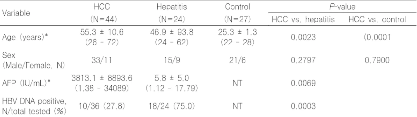
Table 1. Characteristics of study groups
Variable HCC Hepatitis Control P-value
(N=44) (N=24) (N=27) HCC vs. hepatitis HCC vs. control
Age (years)* 55.3 ± 10.6 (26 - 72)
46.9 ± 93.8 (24 - 62)
25.3 ± 1.3
(22 - 28) 0.0023 <0.0001
Sex
(Male/Female, N) 33/11 15/9 21/6 0.2797 0.7900
AFP (IU/mL)* 3813.1 ± 8893.6 (1.38 - 34089)
5.8 ± 5.0
(1.12 - 17.79) NT 0.0069
HBV DNA positive,
N/total tested (%) 10/36 (27.8) 18/24 (75.0) NT 0.0003
* Data are shown as 'mean ± SD (min - max)'.
All patients of HCC and hepatitis groups are HBsAg-positive and all individuals of control groups are HBsAg-negative.
Abbreviations: HCC, hepatocellular carcinoma; NT, not tested.
factors for HCC, and mutations or polymorphisms of p53 gene are one of the frequent genetic alterations accompanied with HCC [3]. p53 gene alterations were also investigated in some other human malignancies, and were associated with invasiveness, recurrence of cancer, or susceptibility to develop cancer in some studies [6-16].
However, the role of p53 polymorphisms in human carcinogenesis is still unclear and even con- troversial in other study [17].
In the case of HCC, some ‘hot spots’ including exon 7 codon 249 mutation (Arg to Ser) and exon 4 codon 72 polymorphism (Arg72Pro) of p53 gene were reported as risk factors for developing HCC [18-29]. Furthermore, p53 codon 249 mutation was reported to be associated with aflatoxin exposure [30-33], and incidences of mutation were reported to be variable according to the racial or geographic differences [18,20,26,27,34-39].
In this study, we investigated on the genotypic frequencies of p53 Arg72Pro polymorphism and the prevalence of p53 codon 249 mutation in early HCC patients to evaluate the role of p53 gene alterations in hepatocarcinogenesis.
MATERIALS AND METHODS 1. Subjects and specimens
Blood samples from 44 early HCC patients with HBV infection who underwent hepatectomy (HCC group), 24 chronic B-viral hepatitis patients (hepatitis group) and 27 young healthy volunteers (control group) were collected. All patients in HCC and hepatitis groups were HBsAg-positive.
Individuals in the control group were tested for complete blood counts, routine chemistries, liver enzymes and hepatitis viral markers, and no abnormal laboratory finding and past medical history were observed. Serum levels of alpha-fetoprotein (AFP) and protein induced by vitamin K absence or antagonist-II (PIVKA-II), and HBV DNA-positive rates in HCC and hepatitis groups were also evaluated. AFP levels were measured by using the UniCel DxI 800 Access Immunoassay system with Access AFP assay kits (Beckman Coulter GmbH, Krefeld, Germany).
PIVKA-II concentrations were determined using Haicatch PIVKA-II enzyme-linked immunosorbent assay kits (Sanko Junyaku Co., Tokyo, Japan).
HBV DNA was detected with Cobas Amplicor HCV monitor 2.0 assay (Roche Molecular Systems, Inc., Pleasanton, CA, USA). Edmondson grades and tumor sizes as the widest diameter in HCC group were analyzed by pathologist with liver tissues collected after hepatectomy.
2. Plasma DNA extraction
Plasma derived from blood samples anti- coagulated with EDTA were processed immediately after collection and stored at -70℃ until used for DNA extraction. Cell-free DNA was extracted from 140 μL of plasma using QIAcube with QIAamp viral RNA Mini Kit (QIAGEN GmbH, Hilden, Germany) which contains carrier RNA for enhancing DNA extraction yield.
3. Genotyping of p53 Arg72Pro polymorphism The genotypes of Arg72Pro were determined by
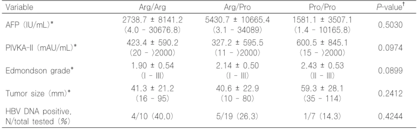
Table 2. Characteristics of genotypic groups of p53 Arg72Pro in the HCC patients
Variable Arg/Arg Arg/Pro Pro/Pro P-value†
AFP (IU/mL)* 2738.7 ± 8141.2
(4.0 - 30676.8)
5430.7 ± 10665.4 (3.1 - 34089)
1581.1 ± 3507.1
(1.4 - 10165.8) 0.5030 PIVKA-II (mAU/mL)* 423.4 ± 590.2
(20 - >2000)
327.2 ± 595.5 (11 - >2000)
600.5 ± 845.1
(15 - >2000) 0.0974
Edmondson grade* 1.90 ± 0.54
(I - III)
2.14 ± 0.50 (I - III)
2.43 ± 0.53
(II - III) 0.0899
Tumor size (mm)* 41.3 ± 21.2
(16 - 95)
40.6 ± 22.9 (10 - 80)
59.3 ± 28.1
(35 - 114) 0.2412
HBV DNA positive,
N/total tested (%) 4/10 (40.0) 5/19 (26.3) 1/7 (14.3) 0.4244
* Data are shown as 'mean ± SD (min - max)'.
† P-values were calculated by ANOVA tests.
Table 3. Genotypic frequencies of p53 Arg72Pro
Genotype HCC (N=44)
Hepatitis (N=24)
Control (N=27)
Crude OR* Adjusted OR*,†
HCC vs.
non-HCC
HCC vs.
Hepatitis
HCC vs.
Control
HCC vs.
non-HCC Arg/Arg,
N (%) 15 (34.1) 7 (29.2) 8 (29.6) 1 1 1 1
Arg/Pro,
N (%) 21 (47.7) 13 (54.2) 15 (55.6) 1.008 (0.299-3.398)
0.892 (0.258-3.083)
0.098 (0.000-32.762)
1.008 (0.299-3.398) Pro/Pro,
N (%) 8 (18.2) 4 (16.7) 4 (14.8) 1.237 (0.570-2.682)
1.065 (0.481-2.360)
0.634 (0.009-45.075)
1.529 (0.325-7.193)
* Data are shown as 'OR (95% confidence interval)'.
† Adjusted for age and sex.
Abbreviations: HCC, hepatocellular carcinoma; OR, odds ratio.
PCR-based restriction fragment length poly- morphism (RFLP) method. Specific primers for PCR amplification of p53 gene exon4 were used (forward: 5’-TTGCCGTCCCAAGCAATGGATGA-3’, re- verse: 5’-TCTGGGAAGGGACAGAAGATGAC-3’). PC R thermo-cycling condition was 5 min at 94℃, followed by 40 cycles of 94℃ for 30 sec, 60℃ for 40 sec, 72℃ for 40 sec, and with a final extension at 72℃ for 7 min. 20 μL of the reaction mixture included 6 μL of extracted DNA, each 1 μL of forward and reverse primers, 1.3 μL of form amide, and distilled water. 6-μL aliquots of PCR products were electrophoresed on 2% agarose gel for 25 min at 110 V to confirm the amplification of the target gene and the product DNA size was 199 bp. Each 14 μL of remaining PCR products was digested with enzyme BstUI (New England BioLabs, Ipswich, MA, USA) at 37℃ for 2 hours and digested products were electrophoresed on 2%
agarose gel for 30 min. Homozygotes for Pro were determined by the single 199 bp non-digested
band, whereas homozygotes for Arg were represented by 113 and 86 bp sized bands.
Heterozygote genotype was identified by representing combined pattern bands (86, 113 and 199 bp) (Fig. 1).
4. p53 codon 249 mutation detection
p53 codon 249 mutation analysis was performed using PCR-based RFLP method. Primers for p53 gene exon 7 were as follows; forward:
5’-CTTGCCACAGGTCTCCCCAA-3’, reverse: 5’-AGGGG TCAGCGGCAAGCAGA-3’. Reaction condition was 5 min at 94℃, followed by 40 cycles of 94℃ for 30 sec, 58℃ for 30 sec, 72℃ for 30 sec, and with a final extension at 72℃ for 10 min. Target DNA amplification was confirmed as follows; 8-μL aliquot of the PCR product from 20-μL reaction mixture was loaded on 2% agarose gel to electrophorese for 25 min at 110 V. Remaining PCR products were digested with HaeIII for overnight at 37℃ and electrophoresed for 25 min
(a) (b)
100 500
200 000
bp
100 500
200 1000
bp
3
1 2
Fig. 1. Agarose gel electrophoresis of P53 Arg72Pro region PCR products. (a) Undigested PCR products with 199 bp in size, (b) PCR products digested with BstUI, 1: Arg/Arg homozygote, 2: Pro/Pro homozygote, 3: Arg/Pro heterozygote.
100 500
200
bp
100 500
200
bp
(a) (b)
Fig. 2. Agarose gel electrophoresis of P53 exon 7 codon 249 region PCR products. (a) Undigested PCR products with 254 bp in size, (b) PCR products digested with HaeIII, all PCR products were cleaved to 92 and 66 bp fragments.
at 110 V. Non mutated codon 249 caused cleavage of 254-bp sized bands to 66 and 92 bp (Fig. 2). If there is AGG to AGT mutation at codon 249, uncleaved 158 bp fragment would be displayed.
5. Statistical analysis
Characteristics of study groups were compared with T-test (for continuous variables) or chi-squired test (for categorical variables). ANOVA test was performed to compare genotypic differences of p53 Arg72Pro polymorphism among HCC group. Odds ratios (OR) for HCC and 95%
confidence intervals (CI) were calculated using logistic regression with univariate and multivariate analysis, and age and sex were included as covariates for multivariate analysis. All statistical analyses were performed using SPSS 12.0 software (SPSS, Chicago, IL, USA), and a P-value less than 0.05 was considered as statistically significant.
RESULTS
There were 44 HCC and 51 non-HCC cases (24
hepatitis and 27 healthy control cases) in this study. Characteristics including age, sex, serum AFP level, HBV DNA positivity of study groups are shown in Table 1. The age (mean ± SD) of HCC, hepatitis and control groups were 55.3 ± 10.6, 46.9 ± 93.8, 25.3 ± 1.3 years respectively, and they were significantly different (HCC vs.
hepatitis, P=0.0023; HCC vs. control, P<0.0001).
Serum AFP level of HCC group was higher than that of hepatitis group (P=0.0069) and the frequency of HBV DNA-positivity in the hepatitis group was higher than that in the HCC group (P=0.0003).
For the HCC group, serum AFP and PIVKA-II level, Edmondson grade, tumor size and frequency of HBV DNA-positivity among groups according to genotypes of Arg72Pro polymorphism are shown in Table 2, and no statistically significant difference between Arg72Pro genotypes was found.
Genotypic frequencies of p53 Arg72Pro, OR and 95% CI for HCC are shown in Table 3. The frequencies of Arg72Pro genotypes were as follows:
Arg/Arg 34.1%, Arg/Pro 47.7%, Pro/Pro 18.2% in HCC group; Arg/Arg 29.2%, Arg/Pro 54.2%, Pro/Pro 16.7% in hepatitis group; Arg/Arg 29.6%, Arg/Pro 55.6%, Pro/Pro 14.8% in control group.
Pro homozygote genotype had a higher risk for HCC by crude OR (1.237, 95% CI 0.570-2.682) and adjusted OR (1.529, 95% CI 0.325-7.193) when HCC group was compared to non-HCC group, but no statistical significance was revealed (P=0.591 for crude and adjusted OR).
PCR-based RFLP method was used to detect p53 exon 7 codon 249 mutation, and among 44 HCC cases, no mutated exon 7 codon 249 was detected (Fig. 2).
DISCUSSION
Hepatocarcinogenesis is a slow and complex process with multi-step stages and various risk factors including HBV, HCV infection, alcohol drinking, aflatoxin exposure, familial history and various genetic background [2,3]. Advances in molecular techniques have been made to reveal the carcinogenetic processes of some human malignancies, but genetic or epigenetic factors for developing HCC are still unclear. p53 gene is one
of well-studied tumor suppressor genes.
HCC is the third most frequent malignancy in Korean male population, and the incidence rate of HCC in Korea is about 50 cases/100,000 person/year. HBV infection is more prevalent in Korea compared to western countries. From these backgrounds, we investigated the frequency or prevalence of p53 gene alterations in Korean population with early HCC or B viral hepatitis, or in good health.
Regarding Arg72Pro polymorphisms of p53 gene in HCC patients, some previous studies suggested that Pro homozygotes had higher relative risk of HCC than Arg homozygotes, and they reported the frequencies of Pro homozygotes as around 18% to 52% in HCC cases and 12% to 44% in control cases [21,23,40]. In our study, the frequencies of Pro homozygote were around 16% in HCC, hepatitis and control groups, which are lower than those in other studies, and there was no statistically significant increase of risk to developing HCC in Pro homozygote cases compared to Arg homozygote cases.
We limited HCC cases as patients with hepatitis B virus infections and as who underwent hepatectomy, which means the patients were operable and in the early stage of cancer, to evaluate the role of Arg72Pro polymorphism of p53 gene in early HCC cases. According to another study on the same polymorphism in HCC cases, no correlation between HCC and the Arg72Pro genotypes was found when comparing the HBsAg- positive HCC subgroup to control [19]. Although not regarding HCC, some studies concluded that there was no evidence of association between the Arg72Pro polymorphism and increased risk to develop cancer [14,17].
The frequencies of Arg72Pro genotypes in our study were different to those of other studies, and this could imply that there are racial or geographical variations in Arg72Pro polymorphism, and our study results support that there may be no or little role of this polymorphism in early hepatocarcinogenesis.
In addition to Arg72Pro polymorphism of p53 gene, we investigated the prevalence of p53 exon 7 codon 249 mutation in early HCC patients. As previously mentioned, the prevalence of codon 249
mutation was reported to be variable and was known to be associated with aflatoxin exposure.
We found no mutated gene among 44 HCC cases and this result is similar with those investigated by Shi et al., and low codon 249 mutation frequency was also reported by another investigators [38,39]. Consequently, our result suggests low aflatoxin exposure in Korean population.
On the other hand, the number of subjects enrolled in this study was restricted, and this could cause a limited statistical power.
Beta-statistical errors, which mean errors causing to interpret actually significant results as non-significant, are generally related to the limited number of study subjects. Therefore, further studies with larger populations would help to define the exact status and nature of polymorphisms in our study.
There is no single main genetic risk factor for HCC proven up to now, and p53 gene alteration may play important roles in hepatocarcinogenesis.
However, the p53 alterations investigated in our study were not significantly associated with early hepatocarcinogenesis. Racial and geographical differences of populations should be also considered in further researches for proving the mechanisms of hepatocarcinogenesis.
요 약
배경: 본 연구는 간암 초기 환자에서의 p53 유전자 Arg72Pro 다형성 및 codon 249 돌연변이의 빈도를 조사하 여 간암 발생과 이들 유전자 변이의 관계를 규명하고자 하 였다.
방법: 44명의 초기 간암, 24명의 만성 B형 간염, 27명의 건강인으로부터 혈액을 채취하여 혈장 유리 DNA를 추출하 였다. 혈청 내 AFP, PIVKA-II, HBV DNA를 측정하여 각 군별로 비교하였다. 제한절편길이다형성분석(RFLP)을 통하 여 p53 Arg72Pro 유전형과 exon 7 codon 249 돌연변이를 검출하였다.
결과: 간암 환자에서 Arg72Pro 유전형에 따른 혈청 내 AFP, PIVKA-II 농도, 간암의 크기와 Edmondson 등급 및 HBV DNA 양성률은 통계적으로 차이가 없었다. Arg72Pro 유전형(Arg/Arg, Arg/Pro, Pro/Pro)의 빈도는 각각 다음과 같다: 간암군 34.1%, 47.7%, 18.2%; 만성 B형 간염군 29.2%, 54.2%, 16.7%; 대조군 29.6%, 55.6%, 14.8%. Pro 동형접합체 유전형은 비간암군과 비교하여 간암 발생 교차비가 높았으
나(crude OR=1.237, 95% CI=0.570-2.682, adjusted OR=
1.529, 95% CI= 0.325-7.193) 통계적으로 유의하지 않았다 (P=0.591). 44명의 간암환자에서 codon 249 돌연변이는 발 견되지 않았다.
결론: 본 연구에서는 Pro 동형접합체 유전형이 모든 실 험 대상군에서 16% 내외로 비슷하게 분포하였으며, Pro 동 형접합체가 간암 발생의 위험도를 유의하게 증가시키지 않 아, 간암 발생의 주요 요인으로 작용하지 않을 것으로 생각 된다.
참 고 문 헌
1. Hagymasi K and Tulassay Z. Epidemiology, risk factors and molecular pathogenesis of primary liver cancer. Orv Hetil 2008;149:541-8.
2. Thorgeirsson SS and Grisham JW. Molecular pathogenesis of human hepatocellular carcinoma. Nat Genet 2002;31:339-46.
3. Feitelson MA, Sun B, Satiroglu Tufan NL, Liu J, Pan J, Lian Z. Genetic mechanisms of hepatocarcinogenesis.
Oncogene 2002;21:2593-604.
4. Xue KX. Molecular genetic and epigenetic mechanisms of hepatocarcinogenesis. Ai Zheng 2005;24:757-68.
5. Keehn DM and Frank-Stromborg M. A worldwide perspective on the epidemiology and primary prevention of liver cancer. Cancer Nurs 1991;14:163-74.
6. Cox DG, Deer D, Guo Q, Tworoger SS, Hankinson SE, Hunter DJ, et al. The p53 Arg72Pro and MDM2 -309 polymorphisms and risk of breast cancer in the nurses' health studies. Cancer Causes Control 2007;18:621-5.
7. Horikawa Y, Nadaoka J, Saito M, Kumazawa T, Inoue T, Yuasa T, et al. Clinical implications of the MDM2 SNP309 and p53 Arg72Pro polymorphisms in transitional cell carcinoma of the bladder. Oncol Rep 2008;20:49-55.
8. Popanda O, Edler L, Waas P, Schattenberg T, Butkiewicz D, Muley T, et al. Elevated risk of squamous-cell carcinoma of the lung in heavy smokers carrying the variant alleles of the TP53 Arg72Pro and p21 Ser31Arg polymorphisms. Lung Cancer 2007;55:25-34.
9. Lind H, Ekstrom PO, Ryberg D, Skaug V, Andreassen T, Stangeland L, et al. Frequency of TP53 mutations in relation to Arg72Pro genotypes in non small cell lung cancer. Cancer Epidemiol Biomarkers Prev 2007;16:2077-81.
10. Zhang X, Miao X, Guo Y, Tan W, Zhou Y, Sun T, et al.
Genetic polymorphisms in cell cycle regulatory genes MDM2 and TP53 are associated with susceptibility to lung cancer. Hum Mutat 2006;27:110-7.
11. Costa S, Pinto D, Pereira D, Rodrigues H,
Cameselle-Teijeiro J, Medeiros R, et al. Importance of TP53 codon 72 and intron 3 duplication 16bp polymorphisms in prediction of susceptibility on breast cancer. BMC Cancer 2008;8:32.
12. Yang M, Guo Y, Zhang X, Miao X, Tan W, Sun T, et al.
Interaction of P53 Arg72Pro and MDM2 T309G polymorphisms and their associations with risk of gastric cardia cancer. Carcinogenesis 2007;28:1996-2001.
13. Kyndi M, Alsner J, Hansen LL, Sorensen FB, Overgaard J.
LOH rather than genotypes of TP53 codon 72 is associated with disease-free survival in primary breast cancer. Acta Oncol 2006;45:602-9.
14. Hamajima N, Matsuo K, Suzuki T, Nakamura T, Matsuura A, Hatooka S, et al. No associations of p73 G4C14-to-A4T14 at exon 2 and p53 Arg72Pro polymorphisms with the risk of digestive tract cancers in Japanese. Cancer Lett 2002;181:81-5.
15. Koushik A, Tranah GJ, Ma J, Stampfer MJ, Sesso HD, Fuchs CS, et al. p53 Arg72Pro polymorphism and risk of colorectal adenoma and cancer. Int J Cancer 2006;119:1863-8.
16. Dakouras A, Nikiteas N, Papadakis E, Perakis M, Valis D, Rallis G, et al. P53Arg72 homozygosity and its increased incidence in left-sided sporadic colorectal adenocarcinomas, in a Greek-Caucasian population. Anticancer Res 2008;28:1039-43.
17. Toruner GA, Ucar A, Tez M, Cetinkaya M, Ozen H, Ozcelik T. P53 codon 72 polymorphism in bladder cancer:
no evidence of association with increased risk or invasiveness. Urol Res 2001;29:393-5.
18. Kimbi GC, Kew MC, Yu MC, Arakawa K, Hodkinson J.
249ser p53 mutation in the serum of black southern African patients with hepatocellular carcinoma. J Gastroenterol Hepatol 2005;20:1185-90.
19. Zhu ZZ, Cong WM, Zhu GS, Liu SF, Xian ZH, Wu WQ, et al. Association of p53 codon 72 polymorphism with genetic susceptibility to hepatocellular carcinoma in Chinese population. Zhonghua Yi Xue Yi Chuan Xue Za Zhi 2005;22:632-5.
20. Huang XH, Sun LH, Lu DD, Sun Y, Ma LJ, Zhang XR, et al. Codon 249 mutation in exon 7 of p53 gene in plasma DNA: maybe a new early diagnostic marker of hepatocellular carcinoma in Qidong risk area, China. World J Gastroenterol 2003;9:692-5.
21. Zhu ZZ, Cong WM, Liu SF, Dong H, Zhu GS, Wu MC.
Homozygosity for Pro of p53 Arg72Pro as a potential risk factor for hepatocellular carcinoma in Chinese population.
World J Gastroenterol 2005;11:289-92.
22. Teramoto T, Satonaka K, Kitazawa S, Fujimori T, Hayashi K, Maeda S. p53 gene abnormalities are closely related to hepatoviral infections and occur at a late stage of hepatocarcinogenesis. Cancer Res 1994;54:231-5.
23. Zhu ZZ, Cong WM, Liu SF, Dong H, Zhu GS, Wu MC.
p53 gene codon 72 polymorphism and genetic susceptibility to hepatocellular carcinoma in Chinese population. Zhonghua Yi Xue Za Zhi 2005;85:76-9.
24. Yu MW, Yang SY, Chiu YH, Chiang YC, Liaw YF, Chen CJ. A p53 genetic polymorphism as a modulator of hepatocellular carcinoma risk in relation to chronic liver disease, familial tendency, and cigarette smoking in hepatitis B carriers. Hepatology 1999;29:697-702.
25. Zhu ZZ, Cong WM, Liu SF, Xian ZH, Wu WQ, Wu MC, et al. A p53 polymorphism modifies the risk of hepatocellular carcinoma among non-carriers but not carriers of chronic hepatitis B virus infection. Cancer Lett 2005;229:77-83.
26. Dong-Dong L and Xi-Ran Z. Plasma 249Ser p53 mutation in patients with hepatocellular carcinoma residing in a high risk area. J Cell Mol Med 2003;7:89-92.
27. Kirk GD, Camus-Randon AM, Mendy M, Goedert JJ, Merle P, Trepo C, et al. Ser-249 p53 mutations in plasma DNA of patients with hepatocellular carcinoma from The Gambia.
J Natl Cancer Inst 2000;92:148-53.
28. Vautier G, Bomford AB, Portmann BC, Metivier E, Williams R, Ryder SD. p53 mutations in british patients with hepatocellular carcinoma: clustering in genetic hemochromatosis. Gastroenterology 1999;117:154-60.
29. El-Kafrawy SA, Abdel-Hamid M, El-Daly M, Nada O, Ismail A, Ezzat S, et al. P53 mutations in hepatocellular carcinoma patients in Egypt. Int J Hyg Environ Health 2005;208:263-70.
30. Kew MC. Synergistic interaction between aflatoxin B1 and hepatitis B virus in hepatocarcinogenesis. Liver Int 2003;23:405-9.
31. Soini Y, Chia SC, Bennett WP, Groopman JD, Wang JS, DeBenedetti VM, et al. An aflatoxin-associated mutational hotspot at codon 249 in the p53 tumor suppressor gene occurs in hepatocellular carcinomas from Mexico.
Carcinogenesis 1996;17:1007-12.
32. El-Shanawani FM, Abdel-Hadi AA, Abu Zikri NB, Ismail A, El-Ansary M, El-Raai A. Clinical significance of aflatoxin, mutant P53 gene and sIL-2 receptor in liver cirrhosis and hepatocellular carcinoma. J Egypt Soc Parasitol 2006;36:221-39.
33. Aguilar F, Hussain SP, Cerutti P. Aflatoxin B1 induces the transversion of G-->T in codon 249 of the p53 tumor suppressor gene in human hepatocytes. Proc Natl Acad Sci
U S A 1993;90:8586-90.
34. Ming L, Thorgeirsson SS, Gail MH, Lu P, Harris CC, Wang N, et al. Dominant role of hepatitis B virus and cofactor role of aflatoxin in hepatocarcinogenesis in Qidong, China. Hepatology 2002;36:1214-20.
35. Kirk GD, Lesi OA, Mendy M, Szymanska K, Whittle H, Goedert JJ, et al. 249(ser) TP53 mutation in plasma DNA, hepatitis B viral infection, and risk of hepatocellular carcinoma. Oncogene 2005;24:5858-67.
36. Murakami Y, Hayashi K, Hirohashi S, Sekiya T.
Aberrations of the tumor suppressor p53 and retinoblastoma genes in human hepatocellular carcinomas. Cancer Res 1991;51:5520-5.
37. Oda T, Tsuda H, Scarpa A, Sakamoto M, Hirohashi S. p53 gene mutation spectrum in hepatocellular carcinoma. Cancer
Res 1992;52:6358-64.
38. Ng IO, Chung LP, Tsang SW, Lam CL, Lai EC, Fan ST, et al. p53 gene mutation spectrum in hepatocellular carcinomas in Hong Kong Chinese. Oncogene 1994;9:985-90.
39. Shi CY, Phang TW, Lin Y, Wee A, Li B, Lee HP, et al.
Codon 249 mutation of the p53 gene is a rare event in hepatocellular carcinomas from ethnic Chinese in Singapore.
Br J Cancer 1995;72:146-9.
40. Yoon YJ, Chang HY, Ahn SH, Kim JK, Park YK, Kang DR, et al. MDM2 and p53 polymorphisms are associated with the development of hepatocellular cargcinoma in patients with chronic hepatitis B virus infection.
Carcinogenesis 2008;29:1192-6.