ISSN 0378-6471 (Print)⋅ISSN 2092-9374 (Online)
http://dx.doi.org/10.3341/jkos.2016.57.11.1801
Case Report
전신홍반성 루프스와 동반된 맥락망막병증 및 이차성 급성 폐쇄각 발작 1예
Choroidoretinopathy and Secondary Angle Closure Attack in Systemic Lupus Erythematosus: A Case Report
안현민1,2⋅최경섭2
Hyun Min Ahn, MD1,2, Kyoung Sub Choi, MD, PhD2
연세대학교 의과대학 안과학교실1, 국민건강보험 일산병원 안과2 Department of Ophthalmology, Yonsei University College of Medicine1, Seoul, Korea Department of Ophthalmology, National Health Insurance Service Ilsan Hospital2, Goyang, Korea
Purpose: Systemic lupus erythematosus (SLE) is a chronic autoimmune disorder with widespread manifestations that rarely in- clude the eye. We present a case of SLE-associated choroidoretinopathy and secondary angle closure attack in both eyes.
Case summary: A 58-year-old male was admitted into the urologic department complaining of right scrotal swelling, and then consulted with the ophthalmology department regarding both ocular pain and eye injection. The patient was diagnosed with acute angle closure attack using a slit lamp test and tonometry secondary to choroidoretinitis with choroidal detachment at fun- dus examination in both eyes. The rheumatologist performed systemic evaluation, including serologic tests, and then diagnosed the patient with SLE. After systemic steroid therapy, intraocular pressure was decreased and choroidal detachment disappeared with improvements of choroidoretinitis in both eyes.
Conclusions: Patients with systemic lupus erythematosus choroidopathy can develop secondary angle closure attack, which can be effectively treated using systemic steroid therapy and antiglaucoma drugs.
J Korean Ophthalmol Soc 2016;57(11):1801-1805
Keywords: Choroidal detachment, Choroiditis, Secondary glaucoma, Systemic lupus erythematosus
■Received: 2016. 7. 21. ■ Revised: 2016. 9. 6.
■Accepted: 2016. 10. 24.
■Address reprint requests to Kyoung Sub Choi, MD, PhD Department of Ophthalmology, National Health Insurance Service Ilsan Hospital, #100 Ilsan-ro, Ilsandong-gu, Goyang 10444, Korea
Tel: 82-31-900-0590, Fax: 82-31-900-0049 E-mail: [email protected]
ⓒ2016 The Korean Ophthalmological Society
This is an Open Access article distributed under the terms of the Creative Commons Attribution Non-Commercial License (http://creativecommons.org/licenses/by-nc/3.0/) which permits unrestricted non-commercial use, distribution, and reproduction in any medium, provided the original work is properly cited.
전신홍반성 루프스(루프스)는 만성 자가면역 질환으로, 다양한 장기를 침범하는 것으로 알려져 있다.1 루프스는 American College of Rheumatology criteria (1982)를 기준 으로 진단하며, 4개 이상의 기준에 해당될 경우 진단할 수 있다.2 루프스는 관절, 피부, 콩팥, 뇌를 포함하여 다양한 장
기를 침범할 수 있는데, 관절이나 피부를 경하게 침범하는 경우부터 콩팥, 심장, 신경계를 심각하게 침범하는 경우까 지 매우 다양하게 나타날 수 있다. 안과적으로도 안검, 각 결막, 공막, 망막, 시신경 등을 포함한 모든 부분을 침범한 다고 알려져 있다.3 그러나 루프스 환자에서 맥락막을 침범 하는 경우는 매우 드문 것으로 알려져 있고, 특히 맥락막병 증에 의한 급성 폐쇄각 발작이 발생하는 경우는 손에 꼽힐 정도로 매우 드물다.4-7 본 증례에서는 맥락망막염에 의한 급성폐쇄각 발작과 고환-부고환염이 합병된 전신홍반성 루 프스를 보고하고자 한다.
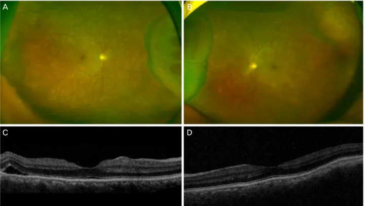
Figure 1. Initial ultra-wide fundus photography of the right eye, left eye and optical coherence tomography (OCT) of the right eye
and left eye. Diffuse choroiditis foci with choroidal detachment in both eyes were found in fundus examination(A, B). Irregular cho- roidal folding with subretinal fluid was found in OCT (C, D).증례보고
전신적 열, 무력감 및 우측 음낭 통증과 부종으로 비감염 성 고환-부고환염이 진단된 58세 남자가 1일 전부터 양안 충혈, 안구 통증이 발생하여 협진 의뢰되었다. 당뇨와 대장 암 과거력이 있었고, 안과적으로는 15년 전 양안 백내장 수 술을 시행 받았었다. 이전에는 양안 교정시력이 0.9였으나, 협진의뢰 시 측정한 최대교정시력은 우안 0.3, 좌안 0.05로 감소한 소견을 보였고, 골드만 압평안압계로 측정한 안압 은 우안 37 mmHg, 좌안 38 mmHg로 상승되어 있었다. 세 극등 검사상에서 van Herick 방법을 통하여 관찰한 주변부 전방은 양안 모두 grade I으로 얕아져 있었고, 우안 전방에 서는 염증반응은 관찰되지 않았으며 좌안 전방에 1+ 정도 의 경한 염증반응이 관찰되었다. 안저소견상 양안 맥락망막 염증이 동반된 맥락막박리 소견이 있었다(Fig. 1A, B). 빛간 섭단층촬영(optical coherence tomography)에서 맥락막주름 과 동반된 망막하삼출이 발견되었다(Fig. 1C, D). 양안 이 차 급성 폐쇄각 발작이 동반된 맥락망막염으로 진단하였다.
전신 감별 검사를 위하여 류마티스 내과에 진료의뢰되었 으며, 피부발진, 경한 용혈성 빈혈, 경한 혈소판 감소증 소견 과 anti-nuclear antibody (ANA) 양성, 홍반성항응고제(Lupus coagulase) 검사 양성이 발견되었다. 본 증례의 경우 고환-
부고환염으로 나타난 장막염(serositis)의 임상소견과 피부 발진용혈성 빈혈, 혈소판 감소증과 ANA 및 홍반성 항응고 제 항체반응검사 양성 소견으로 루프스를 진단할 수 있었다.
흉부 방사선 검사 및 복부 초음파 검사상 폐실질 및 콩팥에 는 이상 소견이 관찰되지는 않았다. 환자 안압 및 염증 조절 을 위하여 안압하강제(도졸라마이드-티몰롤[dorzolamide-to- molol]) 복합 점안제 하루 2회, 브리모니딘(brimonidine) 점 안제 하루 2회, 아세타졸아마이드(경구 1,000 mg/일)와 메 칠프레드니솔론(경구 60 mg/일)을 사용하였다. 비뇨기과에 서는 심한 고환-부고환염에 의한 조직 괴사가 발생하여 우 측 고환-부고환 절제술을 시행하였다.
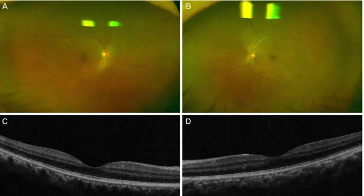
치료 3일째, 최대교정시력 우안 0.7, 좌안 0.2로 상승하 고, 양안 안압 12 mmHg로 하강하는 추세를 보여 안압하강 제를 중단하였다. 치료 7일째, 양안 교정시력 0.9, 양안 안 압 17 mmHg로 측정되었으며, 안저소견 및 빛간섭단층촬 영에서 맥락망막염 감소소견과 맥락막박리가 소실되었다 (Fig. 2). 스테로이드를 40 mg/일로 줄여 사용하였으며 치 료 2주째 20 mg/일로 줄였다. 치료 1개월째, 전신증상의 호 전과 더불어 특이 안과적 이상 소견이 없기에 스테로이드 복용을 중지하였으며, 3개월째 내원 시 재발 소견 없이 경 과관찰 중이다.
A B
C D
Figure 2. Seven days after the initial visit. Choroiditis and choroidal detachment in both eyes were decreased; right eye (A, C), and
left eye (B, D).고 찰
루프스는 눈의 거의 모든 영역을 침범할 수 있는 것으로 알려져 있으나, 맥락막의 침범은 매우 드물며, 신장이나 중 추신경계 침범 등의 상대적으로 심한 루프스 합병증과 동 반되는 것으로 알려져 있다.8-10 68%의 환자에게서 양측성 으로 나타나며, 36%의 환자에게서 중추신경계 침범이 발 생한다고 보고된 바 있다.8-9 본 증례의 경우에도 비록 중추 신경계 검사가 이루어지지 않아 중추신경계의 침범 여부는 명확히 확인할 수 없으나, 심한 고환-부고환염이 발생하여, 조직 괴사로 인한 고환 절제술을 시행하였다. 루프스는 만 성 특발성 자가면역질환으로 자가항체와 보체의 면역 결합 체에 의해 발생한다고 알려져 있으나 명확한 기전은 알지 못한다.11 맥락막의 경우 콩팥과 구조적 병리적 유사성이 많아 면역 결합체의 축적이 발생하며, 이로 인해 맥락막 수 분 저류가 발생한다고 알려져 있다.10,12 하지만 본 증례에서 와 같은 고환-부고환염의 경우는 매우 드물게 보고되어, 맥 락망막염과의 직접적 연관성을 알기 위해서는 추가적인 연 구가 필요할 것으로 보인다.13
루프스 맥락망막병증은 Vogt-Koyanagi-Harada 증후군, 중심맥락망막병증, 고혈압성 망막병증 및 흰점증후군(white dot syndrome)과 같은 기타 맥락망막병증과 감별이 필요하 다. 본 환자의 경우 신경증상 및 피부증상이 동반되지 않은 점 등이 Vogt-Koyanagi-Harada 증후군의 진단기준과 맞지
않았으며, 고혈압의 기왕력이 없고 혈압이 정상범위여서 고 혈압성 망막병증의 경우와 맞지 않았다. 기타 맥락막모세혈 관, 망막색소상피세포, 그리고 망막외층을 침범하는 급성, 아급성 염증성 질환들로 알려진 여러 맥락망막병증과도 그 임상양상이 차이가 보였다. 특히 루프스 환자는 기존에 스 테로이드를 복용하는 경우가 많아 중심맥락망막병증과의 감별이 중요하다.8 본 증례에서는 중심부뿐만 아니라 주변 맥락막과 망막에 다발성 삼출 소견, 맥락막 혹은 망막박리 소견이 나타났으며 이러한 점에서 중심맥락망막병증과 감 별될 수 있었다. 형광안저촬영술 및 인도시아닌그린 조영 술에 의한 진단은 다른 맥락망막병증과의 감별에 도움이 된다 알려져 있다.9 본 증례의 경우 이러한 검사들을 시행 하지는 않았으나 세극등 검사 안저소견과 빛간섭단층촬영 술을 통하여 맥락망막의 삼출 및 염증 양상을 확인하였고, 초기에 전신 질환 감별을 위한 검사를 시행하여 이른 시기 에 루프스 맥락망막병증으로 진단할 수 있었다.
또한 본 증례에서는 루프스 맥락망막염에 의한 섬모체와 맥락막 삼출로 인해 양측성 폐쇄각 발작이 발생하였다. 루 프스에 의한 맥락망막병증으로 폐쇄각 발작이 발생하는 경 우는 매우 드물게 보고된다.5-7 섬모체와 맥락막의 삼출은 맥락막모세혈관층(choriocapillaris)으로부터 주변의 맥락막 위공간(suprachoroidal space)으로의 누출로 발생한다고 생 각되며, 이것은 부종과 맥락막박리를 유발시킨다고 알려져 있다.14 이는 섬모체의 전방회전과 수정체홍채가로막(lens-
A B
C D
iris-diaphragm)의 전방이동(anterior movement)을 유발하며 이로 인해 전방의 깊이가 얕아지게 되어 안압의 상승이 유 발된다.15
급성 폐쇄각 발작이 동반된 루프스 맥락망막염을 치료하 기 위해서는 전신 면역억제 치료가 안압하강제와 함께 반 드시 병행되어야 한다.16 맥락망막병증에 대해 면역억제치 료만으로 충분히 호전되지 않을 경우, 맥락막삼출이 발생 하는 곳에 국소레이저치료를 시행하여 삼출성망막박리 및 시력 호전이 있었다는 연구도 있다.17 안압하강제만으로 안 압하강이 원활하지 않을 경우 레이저홍채절개술이나 맥락 막유출을 위한 수술적 방법이 고려될 수도 있다.7 또한 치 료초기 조절마비제의 경우 섬모체윤근을 마비시킴으로써 섬모체소대를 팽팽하게 하여 앞쪽으로 밀려나있던 수정체 홍채가로막을 본래의 위치인 뒤쪽으로 이동하게 함에 따라 도움이 될 수 있으므로 안압하강제와 병행하여 쓰일 수도 있다.15
본 증례는 전신홍반성 루프스에서 드물지만 맥락망막병 증이 발생한다는 것과 급성 폐쇄각 발작이 일반적인 폐쇄 각 녹내장 외에도 전신질환과 관련하여 맥락망막의 이차적 인 염증에 의해 발생할 수 있음을 보여준다. 또한 전신적 스테로이드 치료와 안압하강제 병행 치료가 루프스와 동반 된 맥락망막병증 및 이차적 폐쇄각 발작에 도움이 됨을 보 여주었다. 추가적으로 질병을 진단 및 치료하는 데 있어 눈 뿐만 아니라 전신적 상태를 감별하는 것 또한 중요하다는 것을 확인할 수 있었다.
REFERENCES
1) Sivaraj RR, Durrani OM, Denniston AK, et al. Ocular manifes- tations of systemic lupus erythematosus. Rheumatology (Oxford) 2007;46:1757-62.
2) Hochberg MC. Updating the American college of rheumatology revised criteria for the classification of systemic lupus erythematosus.
Arthritis Rheum 1997;40:1725.
3) Yoon CK, Park JH, Yu HG. Retinopathy associated with systemic lupus erythematosus. J Korean Ophthalmol Soc 2009;50:1215-20.
4) Edouard S, Douat J, Sailler L, et al. Bilateral choroidopathy in sys- temic lupus erythematosus. Lupus 2011;20:1209-10.
5) Han YS, Yang CM, Lee SH, et al. Secondary angle closure glauco- ma by lupus choroidopathy as an initial presentation of systemic lupus erythematosus: a case report. BMC ophthalmol 2015;15:148.
6) Lavina AM, Agarwal A, Hunyor A, Gass JD. Lupus choroidopathy and choroidal effusions. Retina 2002;22:643-7.
7) Wisotsky BJ, Magat-Gordon CB, Puklin JE. Angle-closure glauco- ma as an initial presentation of systemic lupus erythematosus.
Ophthalmology 1998;105:1170-2.
8) Gäckle HC, Lang GE, Freissler KA, Lang GK. Central serous chorioretinopathy. Clinical, fluorescein angiography and demo- graphic aspects. Ophthalmologe 1998;95:529-33.
9) Chaine G, Haouat M, Menard-Molcard C, et al. Central serous cho- rioretinopathy and systemic steroid therapy. J Fr Ophtalmol 2001;24:139-46.
10) Baglio V, Gharbiya M, Balacco-Gabrieli C, et al. Choroidopathy in patients with systemic lupus erythematosus with or without nephropathy. J Nephrol 2011;24:522-9.
11) Bengtsson AA, Rönnblom L. Systemic lupus erythematosus: still a challenge for physicians. J Intern Med 2016 Jun 16. doi:
10.1111/joim.12529. [Epub ahead of print]
12) Kamdar NV, Erko A, Ehrlich JS, et al. Choroidopathy and kidney disease: a case report and review of the literature. Cases J 2009;
2:7425.
13) Kuehn MW, Oellinger R, Kustin G, Merkel KH. Primary testicular manifestation of systemic lupus erythematosus. Eur Urol 1989;
16:72-3.
14) Elagouz M, Stanescu-Segall D, Jackson TL. Uveal effusion syndrome. Surv Ophthalmol 2010;55:134-45.
15) Ikeda N, Ikeda T, Nomura C, Mimura O. Ciliochoroidal effusion syndrome associated with posterior scleritis. Jpn J Ophthalmol 2007;51:49-52.
16) Palejwala NV, Walia HS, Yeh S. Ocular manifestations of systemic lupus erythematosus: a review of the literature. Autoimmune Dis 2012;2012:290898.
17) Shimura M, Tatehana Y, Yasuda K, et al. Choroiditis in systemic lu- pus erythematosus: systemic steroid therapy and focal laser treatment. Jpn J Ophthalmol 2003;47:312-5.
= 국문초록 =
전신홍반성 루프스와 동반된 맥락망막병증 및 이차성 급성 폐쇄각 발작 1예
목적: 전신홍반성 루프스(루프스)는 만성 자가면역 질환으로 전신에 침범할 수 있으나 안구, 특히 맥락막을 침범하는 경우는 드문 것으로 알려져 있다. 본 증례에서는 루프스에 의한 양안 맥락망막병증 및 이로 인한 이차적인 급성 폐쇄각 발작이 발생한 1예를 보고 하고자 한다.
증례요약: 우측 음낭 부종(scrotal swelling)으로 비뇨기과 입원 중인 58세 남자가 양안 충혈 및 통증으로 안과 협진의뢰되었다. 안저 소견상 맥락망막염, 맥락막박리가 관찰되었으며, 이차적인 양안 급성 폐쇄각 발작이 세극등검사 및 안압검사를 통하여 확인되었다.
류마티스내과를 통하여 혈액검사를 포함한 전신 감별 검사를 시행하였으며, 전신홍반성 루프스가 진단되었다. 전신성 스테로이드 및 안압하강제치료 시행 후, 양안 맥락망막염 호전과 함께 안압 하강 및 맥락막박리가 소실되었다.
결론: 루프스에서 양안 맥락망막병증과 동반되어 이차적인 급성 폐쇄각 발작이 나타날 수 있으며, 전신적 스테로이드 및 안압하강제 사용이 치료에 도움이 된다.
<대한안과학회지 2016;57(11):1801-1805>