ISSN 0378-6471 (Print)⋅ISSN 2092-9374 (Online)
https://doi.org/10.3341/jkos.2019.60.6.541
Original Article
국소맥락막함몰의 임상양상과 장기 추적관찰 결과
Clinical Presentations of Focal Choroidal Excavation and Results of Long-term Follow-up
이석현⋅김재휘⋅김종우⋅김철구⋅이동원⋅유영주⋅조한주⋅김주연
Seok Hyun Lee, MD, Jae Hui Kim, MD, Jong Woo Kim, MD, Chul Gu Kim, MD, Dong Won Lee, MD, Young Ju Lew, MD, Han Joo Cho, MD, Joo Yeon Kim, MD
건양대학교 의과대학 김안과병원 안과학교실
Department of Ophthalmology, Kim’s Eye Hospital, Konyang University College of Medicine, Seoul, Korea
Purpose: To evaluate the clinical presentations of focal choroidal excavation and to report long-term outcomes of cases without retinal disorders at the initial presentation.
Methods: A retrospective review of medical records was performed for patients diagnosed with focal choroidal excavation.
Concomitant retinal disorders at the initial presentation were identified. In cases without retinal disorders, the development of ret- inal disorders during follow-up was also evaluated.
Results: Forty-five eyes in 45 patients were examined in this study. Focal choroidal excavation was accompanied with retinal disorders in 16 eyes (35.6%). In the remaining 29 eyes, only focal choroidal excavation was noted without any accompanying retinal disorders. The accompanying retinal disorders included choroidal neovascularization (n = 8), central serous chorioretin- opathy (n = 4), epiretinal membrane (n = 1), macular hole (n = 1), branch retinal vein occlusion (n = 1), and uveitis (n = 1). Of the 29 eyes without retinal disorders, 22 were followed up for a mean period of 33.5 ± 18.2 months. Consequently, choroidal neo- vascularization was found to have developed in one eye at 59 months, and subretinal fluid had developed in two eyes at 17 and 28 months, respectively.
Conclusions: Focal choroidal excavation was accompanied by retinal disorders in 35.6% of the included patients. In patients without retinal disorders, the development of a retinal disorder was noted in some eyes, suggesting the need for long-term regu- lar follow-up in patients diagnosed with focal choroidal excavation.
J Korean Ophthalmol Soc 2019;60(6):541-546
Keywords: Central serous chorioretinopathy, Choroidal neovascularization, Focal choroidal excavation
■Received: 2018. 12. 6. ■ Revised: 2019. 1. 13.
■Accepted: 2019. 5. 16.
■Address reprint requests to Jae Hui Kim, MD
Kim's Eye Hospital, #136 Yeongsin-ro, Yeongdeungpo-gu, Seoul 07301, Korea
Tel: 82-2-2639-7665, Fax: 82-2-2639-6359 E-mail: kjh7997@daum.net
*Conflicts of Interest: The authors have no conflicts to disclose.
ⓒ2019 The Korean Ophthalmological Society
This is an Open Access article distributed under the terms of the Creative Commons Attribution Non-Commercial License (http://creativecommons.org/licenses/by-nc/3.0/) which permits unrestricted non-commercial use, distribution, and reproduction in any medium, provided the original work is properly cited.
국소맥락막함몰(focal choroidal excavation)은 빛간섭단 층촬영에서 발견되는 소견이다. Jampol et al1이 최초로 보
고하였으며, 주로 황반부에 분포하는 것으로 알려져 있다.2 국소맥락막함몰은 동반질환 없이 특발성으로 나타나는 경 우도 있으나 맥락막혈관신생,2-5 중심장액맥락망막병증,2,6,7 망막/맥락막의 염증성 질환8 혹은 망막이영양증9과 같은 다 양한 질환들과 동반되어 나타나는 경우도 많다. Lee et al2 의 연구에서 맥락막혈관신생이 소실된 후 동일 위치에 국 소맥락막함몰이 발생하는 예를 보여주었는데, 대부분의 경 우 국소맥락막함몰이 발생하는 정확한 원인은 아직 알려져 있지 않다.
국내 학술지 보고의 경우 총 7예의 국소맥락막함몰 증례가
Figure 1. A representative figure showing focal choroidal
excavation. Note thick choroid with pachyvessels (asterisks).Characteristic Value
Age (years) 51.8 ± 14.1
Gender
Male 30 (66.7)
Female 15 (33.3)
Hypertension 3 (6.7)
Diabetes mellitus 9 (20.0)
Best-corrected visual acuity (logMAR) 0.15 ± 0.26 Diagnosis of focal choroidal excavation
Incidental finding 24 (53.3)
Symptom without other retinal disorder 5 (11.1) Symptom with retinal disorder 16 (35.6) Accompanied retinal disorders*
Choroidal neovascularization 8 (50.0)
Central serous chorioretinopathy 4 (25.0)
Epiretinal membrane 1 (6.3)
Macular hole 1 (6.3)
Branch retinal vein occlusion 1 (6.3)
Uveitis 1 (6.3)
Values are presented as mean ± standard deviation or number (%).
logMAR = logarithm of minimal angle of resolution.
*Data from 16 patients with retinal disorder were presented.
Table 1. Characteristics of included patients (n = 45)
보고되었는데, 5예의 경우 각각 중심장액맥락망막병증,10,11망막층간분리, 장액망막색소상피박리 및 맥락막신생혈관이 함께 관찰되었으며, 나머지 2예의 경우 동반된 망막의 이상 소견은 없었다.12 동반된 망막의 이상 소견이 없었던 2예 중 1예는 3개월 동안 경과관찰하였는데, 이상 소견의 발생은 없었던 것으로 나타났다.12 본 연구에서는 보다 많은 환자 를 대상으로 국내 환자에서 발견된 국소맥락막함몰의 임상 양상을 알아보고 동반된 망막 이상이 없었던 경우의 장기 추적관찰 결과를 보고하고자 한다.
대상과 방법
본 연구는 단일 기관에서 시행된 후향적 연구로 헬싱키 선언에 입각하여 시행되었으며, Institutional Review Board (IRB) 승인을 획득하였다(KIM’s Eye Hospital-IRB No.
2018-11-003). 2013년 3월부터 2017년 8월까지 전자의무기 록에서 ‘excavation’, ‘choroidal excavation’ 혹은 ‘국소맥락 막함몰’의 진단명이 붙여진 경우에 대한 검색을 시행하였 다. 검색 후 도출된 환자들의 빛간섭단층촬영 이미지를 분 석하여 전형적인 국소적 맥락막 함몰 소견(Fig. 1)이 나타 나는지를 확인하였는데, 이미지 분석은 2명의 검사자(S.H.L., J.H.K.)가 함께 시행하였다.
연구에 포함된 환자들의 특성을 확인하기 위하여 진단 당시 환자의 연령, 성별, 당뇨 및 고혈압의 유무, 진단 시의 최대교정시력 등을 수집하였다. 또한 안저사진 및 빛간섭 단층촬영 결과를 분석하여 동반된 망막질환의 유무를 확인 하였는데, 형광안저혈관조영술이나 인도사이아닌그린혈관 조영술을 시행한 경우 그 결과 역시 함께 확인하였다. 빛간 섭단층촬영은 2대의 기기(SLO™, Ophthalmic Technologies Inc., Ontario, Canada; Spectralis™, Heidelberg Engineering, Heidelberg, Germany; RS 3000™, Nidek Co., Ltd., Gamagori, Japan) 중 하나를 이용하여 시행되었다. 진단 당시 망막질
환을 동반하지 않은 특발성 국소맥락막함몰의 경우 경과관 찰기간 동안 질환의 발생 여부를 조사하였으며, 진단 당시 시력과 최종 추적관찰 시의 시력을 서로 비교하였다.
국소맥락막함몰로 진단된 안에서 pachychoroid의 특징13 이 나타나는지 확인하기 위하여 빛간섭단층촬영 결과를 바 탕으로 중심와하 맥락막 두께가 250 µm 이상인 경우를 확 인하였으며, pachyvessel의 존재 여부 역시 확인하였다. 맥 락막의 Haller’s layer의 혈관이 확장되면서 인접한 Sattler’s layer와 맥락막모세혈관층이 얇아지는 소견을 보이는 경우 pachyvessel이 존재하는 것으로 판단하였다.
통계 분석에는 IBM SPSS ver. 12.0 (IBM Corp., Armonk, NY, USA)을 이용하였다. 시력의 경우 logarithm of mini- mal angle of resolution (logMAR)값으로 변환하여 분석하 는데, 서로 다른 두 시점 사이의 시력은 paired t-test를 이용 하여 비교하였다. 0.05 미만의 p값을 통계적으로 유의한 값 으로 정의하였다.
결 과
45안(45명)을 대상으로 결과를 분석하였다(Table 1). 남 성 30명, 여성 15명이었으며, 평균 나이는 51.8 ± 14.1세였 으며, 진단 당시 평균 logMAR 시력은 0.15 ± 0.26이었다.
국소맥락막함몰이 발견된 경로로는 증상이 없었으나 건강 검진 혹은 안과 검사에서 우연히 발견되어 의뢰된 경우가
A
B C
D
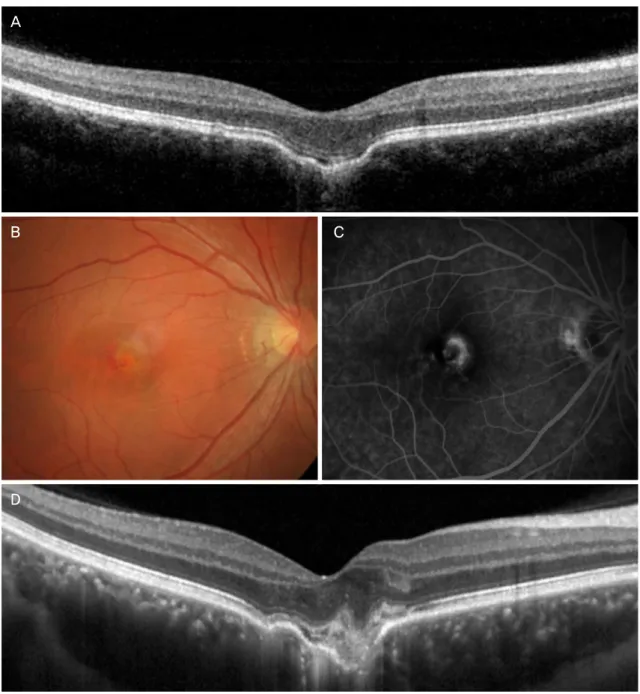
Figure 2. Clinical course of a focal choroidal excavation found in 29-year-old patient. At the diagnosis, there was no retinal disorder
(A). The patients were regularly followed-up. At 59 months (B-D), subretinal fluid and hemorrhage was noted on fundus examina- tion (B). Development of choroidal neovascularization was confirmed on fluorescein angiography (C) and optical coherence tomog- raphy (D). The patient was referred to the other hospital.24명(53.3%), 증상이 있어 본원을 방문하였거나 근처 안과 를 방문한 후 의뢰된 경우가 5명(11.1%), 다른 질환으로 진 단된 후 검사 과정에서 발견된 경우가 16명(35.6%)이었다.
16안의 경우 진단 당시 다른 망막질환을 동반하고 있었 으며, 나머지 29안의 경우 국소맥락막함몰을 제외한 다른 이상은 확인할 수 없었다. 동반된 질환은 맥락막혈관신생 8안, 중심장액맥락망막병증 4안, 망막앞막 1안, 황반원공 1안, 분지망막정맥폐쇄 1안, 포도막염 1안이었다.
동반된 질환이 없었던 29안 중 7안의 경우 최초 진단 이 후 경과관찰이 시행되지 않았다. 나머지 22안에서 평균 33.5 ± 18.2개월 동안 경과관찰하였는데, 경과관찰기간 동 안 망막질환의 발생은 3안(13.6%)에서 관찰되었다. 1안에 서 59개월에 맥락막혈관신생이 발생하였으며(Fig. 2), 2안 에서는 망막하액의 발생이 관찰되었는데, 2안 모두 망막의 출혈이나 망막내액은 없었다. 1안의 경우 17개월에 망막하 액이 발생하여 2개월만에 자연 소실되었으나 36개월에 망
A B
C D
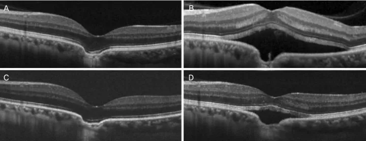
Figure 3. Clinical course of a focal choroidal excavation found in 37-year-old patient. At the diagnosis, there was no retinal disorder
(A). The patients were regularly followed-up. Development of subretinal fluid was noted at 17 months (B), but spontaneously re- solved after 2 months (C). At 28 months (D), recurrence of subretinal fluid was noted.막하액이 재발하였다(Fig. 3), 나머지 1안의 경우 28개월에 망막하액이 발생하였는데, 이후 경과관찰이 중단되었다. 망 막하액이 발생한 두 안 모두 형광안저혈관조영술이나 인도 사이아닌그린혈관조영술은 시행되지 않았으나 빛간섭단층 촬영에서 type 1 (망막색소상피하), type 2 (망막하), 혹은 type 3 (망막 내)으로 의심할 만한 고반사병변은 관찰되지 않았다. 전체 45안 중 28안(62.2%)의 경우 중심와하에 맥 락막 두께가 250 µm 이상으로 측정되었으며, 30안(66.7%) 에서 pachyvessel이 발견되었다.
고 찰
본 연구에서 국소맥락막함몰로 진단된 환자들 중 35.6%
는 다른 망막질환을 동반하고 있었으며, 나머지 64.4%는 다른 질환 없이 특발성으로 발견되었다. 최초 진단 당시 특 발성이었던 경우에도 경과관찰 과정 중 13.6%에서는 망막 질환이 발생하였다.
Jampol et al1의 최초 보고 이후 Margolis et al14은 국소맥 락막함몰 13안의 임상양상에 더불어 안저검사, 자가형광촬 영, 및 빛간섭단층촬영 소견을 정리하여 보고하였는데, 일 부 국소맥락막함몰의 경우 중심성 장액성 맥락망막병증이 나 맥락막신생혈관과 관련되어 나타날 수 있다고 하였다.
과거 Margolis et al14은 이 소견이 주로 선천적으로 발생하 는 것으로 추측하였다. 그러나 소아 환자를 대상으로 국소 맥락막함몰의 존재 여부를 확인한 Park and Oh15의 연구에 따르면 19세 미만에서는 이 소견이 관찰되지 않았는데, 이 러한 결과는 최소한 이 질환의 상당 부분이 선천적으로 발
생한 것이 아닐 수 있다는 점을 시사하였다. 또한 Lee et al2 의 연구는 맥락막혈관신생과 관련된 2차 변화로 함몰이 발 생하는 예를 보여주어 함몰이 후천적으로 발생할 수 있다 는 점을 증명하였다.
국소맥락막함몰과 동반되는 것으로 알려진 대표적인 질 환은 맥락막혈관신생3-5과 중심장액맥락망막병증6,7이다. Lee and Lee5는 국소맥락막함몰과 동반된 맥락막혈관신생 16안 의 특성을 보고하였는데, type 1의 비율(56.3%)이 type 2 (43.8%)보다 조금 높았으며, 항혈관내피성장인자 치료에 좋은 반응을 보인 것으로 나타났다. Lee et al2의 연구에서 는 전체 국소맥락막함몰의 22%에서 맥락막혈관신생을 동 반하였는데, 44%에서 type 1, 56%에서 type 2의 형태를 보 였으며, 특히 type 1 맥락막혈관신생의 경우 모든 경우에서 혈관신생의 위치가 국소맥락막함몰의 범위 안에 위치해 있 었다.2 본 연구에서는 국소맥락막함몰 진단 당시 17.8%에 서 맥락막혈관신생을 동반하여 Lee et al2의 연구와 크게 차이 나지 않는 결과를 보였다.
중심장액맥락망막병증의 경우 약 7.8%에서 국소맥락막 함몰을 동반하는 것으로 알려져 있는데, Lee et al2의 연구 에서는 전체 국소맥락막함몰환자의 24%에서 중심장액맥 락망막병증을 동반하였다. Ellabban et al6은 중심장액맥락 망막병증환자에서 맥락막 결체조직의 국소적인 반흔 형성 과 연관된 망막색소상피의 당김에 의하여 국소맥락막함몰 이 발생하는 것으로 추측하였는데, Suzuki et al7은 맥락막 순환의 이상과 함몰 부위의 망막색소상피의 위축이 중심장 액맥락망막병증의 발생과 연관된 것으로 추측하였다. 본 연구에서는 전체 환자의 8.9%에서 중심장액맥락망막병증
을 동반하였는데, 이는 Lee et al2의 연구에 비하여 상대적 으로 낮은 비율이었다. 본 연구는 국소맥락막함몰의 진단 명이 기입된 경우만을 대상으로 환자 검색을 시행하였다.
따라서 실제 국소맥락막함몰이 있었음에도 진단명이 따로 기입되지 않은 경우에는 연구에 포함되지 않았는데, 이와 같은 이유로 환자의 비율이 작게 나타났을 것으로 추측된다.
국소맥락막함몰이 관찰된 안에서는 두꺼운 맥락막, pa- chyvessel, 인도사이이난그린 혈관조영술에서의 맥락막혈 관과투과성(choroidal vascular hyperpermeability)과 같은 pachychoroid의 특성이 흔히 관찰되는데,16 Chung et al11은 이러한 특성이 국소맥락막함몰 발생의 원인과 관련된 것으 로 추측하였다. 즉, 맥락막정맥의 울혈과 과투과성에 의하 여 전염증성(proinflammatory), 전혈전성(prothrombotic) 단 백과 매개체들이 맥락막의 기질(stroma)로 유출되어 조직 의 변성, 위축을 유발하게 되며, 이러한 과정이 오래 지속 되는 경우 국소맥락막함몰이 발생할 수 있다는 것이다. 본 연구는 후향적 연구로 enhanced-depth imaging 빛간섭단층 촬영이 시행되지 않았으며, 따라서 pachychoroid의 특징을 정확하게 확인하기는 어려운 면이 있었다. 그러나 약 250 µm 두께까지는 enhanced-depth imaging 없이도 측정할 수 있 었기에 이 값을 중심으로 보다 두꺼운 경우를 확인하였으 며, pachyvessel의 경우 아래쪽(공막 쪽)의 경계가 정확하게 확인되지 않더라도 위쪽(망막 쪽)의 경계 및 전체적인 ves- sel의 모양과 주변 조직의 특성을 통하여 pachyvessel 여부 를 확인할 수 있었다. 결과적으로 전체의 62.2%에서 250 µm 보다 두꺼운 결과가 나타나 전반적으로 맥락막이 얇지 않 은 결과를 보였으며, 66.7%에서 pachyvessel이 발견되었는 데, 이는 국소맥락막함몰과 pachychoroid 관련 병태생리와 의 연관관계를 추측한 Chung et al11의 연구를 뒷받침하는 결과라 할 수 있을 것이다.
실제 환자를 진료하면서 우연히 국소맥락막함몰을 발견 할 경우 의사와 환자 모두 가장 궁금한 부분은 향후 이 눈 에 어떤 문제가 발생할 수 있을 것인지, 또한 문제가 발생 한다면 어느 정도의 비율에서 언제쯤 발생할 것인지에 관 한 내용일 것이다. 실제로 망막질환을 동반하지 않은 국소 맥락막함몰의 장기 추적관찰 결과에 대해서는 아직까지 제 한된 정보만이 알려져 있으며, 국소맥락막함몰이 진단된 경우 어느 정도 간격으로 얼마만큼의 기간 동안 경과관찰 해야 하는가에 대한 확립된 기준은 없다. 가장 많은 안을 대상으로 장기간 추적관찰한 Obata et al17의 연구에서는 망 막질환이 없던 21안(망막질환이 없었던 경우는 18안)을 대 상으로 평균 37개월간 추적관찰하였는데, 망막질환이 없던 18안 중 1안에서 중심장액맥락망막병증이 발생하였다.
본 연구에서는 최초 진단 당시 동반 질환이 없었으며, 1개
월 이상 경과관찰이 가능하였던 22안 중 3안(13.6%)에서 맥락막혈관신생 혹은 망막하액이 발생하였는데, 그 시기는 59개월, 17개월, 28개월로 다양한 분포를 보였다. 비록 경 과관찰기간이 통일되지 않았고, 환자에 따라 차이가 크다 는 제한점이 있으나 진단 59개월 이후에도 망막질환이 발 생할 수 있다는 본 연구의 결과는 환자가 이러한 부분에 대 하여 어느 정도 경각심을 가지고 있어야 한다는 점과 장기 간 정기적인 검진이 필요하다는 점을 시사한다. 맥락막혈 관신생이 발생하는 경우에도 적절한 항혈관내피성장인자 치료를 통하여 좋은 결과를 얻을 수 있으므로5 환자가 시력 저하나 변형시증이 발생하는 경우 빨리 병원에 방문할 수 있도록 교육해야 할 것이다.
경과관찰 도중 망막하액이 발생한 2안의 경우 혈관조영 술을 시행하지 않아 정확한 감별 진단은 어려웠다. 그러나 빛간섭단층촬영 결과에서 맥락막혈관신생으로 의심할 만 한 부분이 관찰되지 않았으며, 망막내액이나 출혈 없이 망 막하액만 관찰되었다는 점을 고려하였을 때 중심장액맥락 망막병증이 발생한 것으로 추측된다.
본 연구에는 다음과 같은 제한점이 있다. 본 연구는 후향 적 연구로 전자의무기록에 ‘국소맥락막함몰’이라는 진단명 이 기입된 환자만을 대상으로 시행하였다. 따라서 실제 국 소맥락막함몰이 있다 하더라도 진단명에 기입되지 않은 경 우에는 연구에 포함되지 않았다. 따라서 본 연구에 포함된 환자들의 특성은 실제 환자들의 특성과 약간의 차이를 보 일 수 있다. 이와 같은 제한점에도 불구하고 본 연구는 국 내 환자를 대상으로 한 연구들 중 가장 많은 안을 대상으로 한 연구이며, 장기간 경과관찰한 결과를 같이 보고하였다 는 점에서 그 의의가 있을 것으로 생각된다.
요약하면 국소맥락막함몰로 진단된 경우 35.6%에서 망 막질환을 동반하였는데, 빈도가 높은 질환은 맥락막혈관신 생과 중심장액맥락망막병증이었다. 최초 진단 당시 망막질 환을 동반하지 않았던 경우에도 경과관찰 과정 중 일부에 서는 맥락막혈관신생과 같은 망막질환이 발생하였는데, 이 와 같은 결과는 국소맥락막함몰로 진단된 환자를 대상으로 장기적인 경과관찰이 필요하다는 점을 시사한다.
REFERENCES
1) Jampol LM, Shankle J, Schroeder R, et al. Diagnostic and ther- apeutic challenges. Retina 2006;26:1072-6.
2) Lee CS, Woo SJ, Kim YK, et al. Clinical and spectral-domain opti- cal coherence tomography findings in patients with focal choroidal excavation. Ophthalmology 2014;121:1029-35.
3) Say EA, Jani PD, Appenzeller MF, Houghton OM. Focal choroidal excavation associated with polypoidal choroidal vasculopathy.
Ophthalmic Surg Lasers Imaging Retina 2013;44:409-11.
= 국문초록 =
국소맥락막함몰의 임상양상과 장기 추적관찰 결과
목적: 국소맥락막함몰의 임상양상을 알아보고 동반된 망막 이상이 없었던 경우의 장기 추적관찰 결과를 보고하고자 한다.
대상과 방법: 국소맥락막함몰로 진단된 환자를 대상으로 후향적 의무기록 분석을 시행하였다. 진단 당시 동반된 망막질환을 확인하였 으며, 진단 당시 망막질환을 동반하지 않은 특발성 국소맥락막함몰의 경우 경과관찰기간 동안 질환의 발생 여부를 추가적으로 조사하 였다.
결과: 전체 45안(45명)을 대상으로 연구를 시행하였다. 16안(35.6%)의 경우 진단 당시 다른 망막질환을 동반하고 있었으며, 나머지 29안의 경우 국소맥락막함몰을 제외한 다른 이상은 확인할 수 없었다. 동반된 질환은 맥락막혈관신생 8안, 중심장액맥락망막병증 4안, 망막앞막 1안, 황반원공 1안, 분지망막정맥폐쇄 1안, 포도막염 1안이었다. 동반된 질환이 없었던 29안 중 22안에서 평균 33.5
± 18.2개월 동안 경과관찰하였는데 1안에서 59개월에 맥락막혈관신생이 발생하였고, 2안에서는 각각 17개월 및 28개월에 망막하액이 발생하였다.
결론: 국소맥락막함몰로 진단된 35.6%에서 최초 진단 당시 망막질환을 동반하였다. 망막질환을 동반하지 않았던 경우에도 경과관찰 과정 중 일부에서는 망막질환이 발생하였는데, 이와 같은 결과는 국소맥락막함몰로 진단된 환자를 대상으로 장기적인 경과관찰이 필요하다는 점을 시사한다.
<대한안과학회지 2019;60(6):541-546>
이석현 / Seok Hyun Lee
건양대학교 의과대학 김안과병원 안과 Department of Ophthalmology,
Kim’s Eye Hospital, Konyang University College of Medicine 4) Xu H, Zeng F, Shi D, et al. Focal choroidal excavation complicated
by choroidal neovascularization. Ophthalmology 2014;121:246-50.
5) Lee JH, Lee WK. Choroidal neovascularization associated with fo- cal choroidal excavation. Am J Ophthalmol 2014;157:710-8.
6) Ellabban AA, Tsujikawa A, Ooto S, et al. Focal choroidal ex- cavation in eyes with central serous chorioretinopathy. Am J Ophthalmol 2013;156:673-83.
7) Suzuki M, Gomi F, Hara C, et al. Characteristics of central serous chorioretinopathy complicated by focal choroidal excavation.
Retina 2014;34:1216-22.
8) Kim H, Woo SJ, Kim YK, et al. Focal choroidal excavation in mul- tifocal choroiditis and punctate inner choroidopathy. Ophthalmology 2015;122:1534-5.
9) Roy R, Saurabh K, Chandrasekharan DP, et al. Bilateral focal cho- roidal excavation in cone dystrophy. Clin Exp Optom 2016;99:198-9.
10) Kim WJ, Cho NC, Kweon EY. A case of focal choroidal excavation associated with chronic central serous chorioretinopathy. J Korean Ophthalmol Soc 2015;56:627-31.
11) Chung CY, Li SH, Li KKW. Focal choroidal excavation-morphological
features and clinical correlation. Eye (Lond) 2017;31:1373-9.
12) Park JH, Sagong M, Chang WH. Three cases of focal choroidal ex- cavation in the macula detected by spectral-domain optical coher- ence tomography. J Korean Ophthalmol Soc 2014;55:941-6.
13) Cheung CMG, Lee WK, Koizumi H, et al. Pachychoroid disease.
Eye (Lond) 2019;33:14-33.
14) Margolis R, Mukkamala SK, Jampol LM, et al. The expanded spectrum of focal choroidal excavation. Arch Ophthalmol 2011;
129:1320-5.
15) Park KA, Oh SY. The absence of focal choroidal excavation in chil- dren and adolescents without retinal or choroidal disorders or ocu- lar trauma. Eye (Lond) 2015;29:841-2.
16) Chung H, Byeon SH, Freund KB. Focal choroidal excavation and its association with pachychoroid spectrum disorders: a review of the literature and multimodal imaging findings. Retina 2017;37:
199-221.
17) Obata R, Takahashi H, Ueta T, et al. Tomographic and angio- graphic characteristics of eyes with macular focal choroidal excavation. Retina 2013;33:1201-10.