ORIGINAL ARTICLE
건강검진 중 발견된 무증상 젊은 성인 위암의 임상병리학적 특징
문형호, 강현우, 고성준1, 김지원1, 신철민2
동국대학교 의과대학 동국대학교 일산병원 내과, 서울대학교 의과대학 서울대학교 보라매병원 내과1, 서울대학교 의과대학 분당서울대학교병원 내과2
Clinicopathological Characteristics of Asymptomatic Young Patients with Gastric Cancer Detected during a Health Checkup
Hyoung Ho Moon, Hyoun Woo Kang, Seong-Joon Koh1, Ji Won Kim1 and Cheol Min Shin2
Department of Internal Medicine, Dongguk University Ilsan Hospital, Dongguk University College of Medicine, Goyang; Department of Internal Medicine, Seoul National University Boramae Hospital, Seoul National University College of Medicine1, Seoul; Department of Internal Medicine, Seoul National University Bundang Hospital, Seoul National University College of Medicine2, Seongnam, Korea
Background/Aims: The Korean National Cancer Screening Program recommends biennial gastric cancer screening for patients aged ≥40 years. This study compared the characteristics of asymptomatic young gastric cancer patients aged <40 years, whose cancer was detected during a health checkup (screening group), with those whose disease was detected because of symptoms (diagnostic group).
Methods: Data were collected retrospectively from 84 subjects who underwent a gastroduodenoscopy before the age of 40 years and who were diagnosed with gastric cancer from January 2006 to February 2017 in three tertiary centers in Korea. The clin- icopathological characteristics, including age, sex, stage, location, pathology, and survival, were compared according to the pur- pose of endoscopy (screening group, n=23 vs. diagnostic group, n=61).
Results: The median age of the screening group was higher than that of the diagnostic group (37 vs. 35 years, p=0.027), as was the proportion of early gastric cancer cases (78.3% vs. 29.5%, p<0.01), curative endoscopic treatment or operation rate (95.7%
vs. 52.5%, p<0.01), and the overall survival (p<0.01). Poorly differentiated or signet ring cell carcinoma was less common in the screening group than in the diagnostic group (56.5% vs. 83.6%, p=0.006). The sex ratio, smoking status, family history of gastric cancer, Helicobacter pylori infection status, and tumor location were similar in the two groups.
Conclusions: Screening gastroduodenoscopy may enable the early detection of gastric cancer and prolong survival in patients
<40 years of age. (Korean J Gastroenterol 2019;74:281-290) Key Words: Gastric cancer; Screening; Endoscopy; Young adult
Received July 10, 2019. Revised October 10, 2019. Accepted October 10, 2019.
CC This is an open access article distributed under the terms of the Creative Commons Attribution Non-Commercial License (http://creativecommons.org/licenses/
by-nc/4.0) which permits unrestricted non-commercial use, distribution, and reproduction in any medium, provided the original work is properly cited.
Copyright © 2019. Korean Society of Gastroenterology.
교신저자: 강현우, 10326, 고양시 일산동구 동국로 27, 동국대학교 의과대학 동국대학교 일산병원 내과
Correspondence to: Hyoun Woo Kang, Department of Internal Medicine, Dongguk University Ilsan Hospital, Dongguk University College of Medicine, 27 Dongguk-ro, Ilsandong-gu, Goyang 10326, Korea. Tel: +82-31-961-7128, Fax: +82-31-961-9309, E-mail: gangmali@naver.com, ORCID: https://orcid.org/0000-0003-3431-0827 Financial support: This work was supported by Dongguk University Research Fund 2016.
Conflict of interest: None.
INTRODUCTION
Despite its decreasing incidence, gastric cancer is the fifth most common cancer and the third leading cause of can-
cer-related death worldwide.1 In particular, its incidence in East Asia, including Korea and Japan, is the highest in the world.2 According to the Korea Central Cancer Registry, 30,504 gastric cancer cases were diagnosed in 2016, making
Fig. 1. Flow chart of patient selection. *Means the purpose of endoscopy. F/U, follow up.
gastric cancer the most common cancer (age-adjusted in- cidence rate: 34.0 per 100,000). In men, it was the most common cancer (age-adjusted incidence rate: 49.6 per 100,000), and the incidence was high in those between the ages of 35 to 64 (age-adjusted incidence rate: 85.6 per 100,000).3 The prognosis of gastric cancer depends on many factors, including the depth of invasion, nodal involvement, and distant metastasis.4 Among them, the stage of gastric cancer is the most important prognostic factor. Early gastric cancer (EGC) usually has a good prognosis with a 5-year sur- vival rate of more than 90%. Therefore, it is very important to detect gastric cancer in the early stages to allow for earlier treatment and to cure this disease.5
Population-based screening for gastric cancer has been im- plemented in several countries with a high incidence of gastric cancer. On the other hand, the recommended screening meth- ods, age, and intervals vary.6-9 Based on the fact that gastric cancer is rare before the age of 40 years, the Korean National Gastric Cancer Screening Program recommends that upper endoscopy be performed biennially for people aged ≥40 years.9 On the other hand, the incidence of gastric cancer in patients younger than 40 years of age has been increasing in western countries over the past two decades. In particular, this tendency is prominent in patients 30 to 39 years of age.10 Several reports have suggested that younger patients are fre- quently diagnosed with advanced-stage tumors and have
poorer prognoses than older patients.11 Genetic factors and late diagnosis have been proposed as the main causes of the poorer prognoses.12-15 Therefore, proper screening and early management may be important for this age group. This study examined whether the early diagnosis of gastric cancer by screening gastroduodenoscopy in individuals <40 years of age is helpful by comparing the characteristics of gastric can- cer in young patients whose disease was detected at a health checkup with those whose disease was detected by gastro- duodenoscopy, which was undertaken because of symptoms.
SUBJECTS AND METHODS
1. Patients
For this retrospective study, this study enrolled 84 patients diagnosed with gastric cancer before 40 years of age at three tertiary centers (Dongguk University Ilsan Hospital, Seoul National University Boramae Hospital, and Seoul National University Bundang hospital) from May 2007 to February 2017. The medical records were analyzed retrospectively to evaluate the patient characteristics, including smoking status, family history of gastric cancer (at least a first-degree relative with gastric cancer), Helicobacter pylori (H. pylori) infection status, laboratory findings, stage at the time of diagnosis, can- cer location, pathologic findings, and clinical outcomes. The patients were classified into two groups according to the pur-
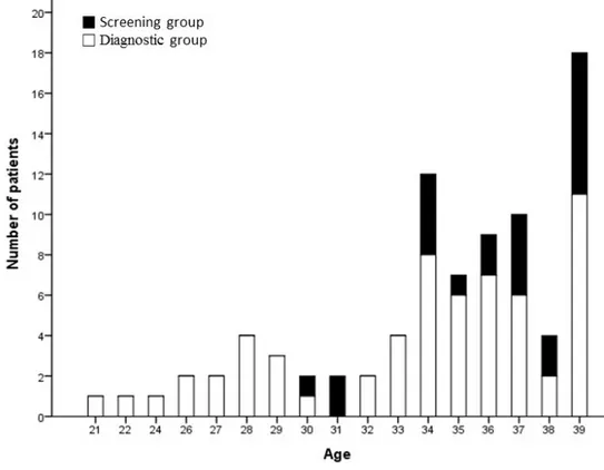
Fig. 2. Distribution of the patient’s ages study population. Median age was 38 (range 21 to 39). Forty-eight (57.1%) gastric cancer cases were detected in patients with ages ranging from 35 to 39 years. Twenty-two (26.2%) cases were detected in those from 30 to 34 years and 14 (16.7%) cases were detected in those younger than 30 years of age. All of the 14 patients diagnosed with gastric cancer under the age of 30 years were in the diagnostic group.
pose of the upper endoscopy, which is a health checkup re- gardless of symptoms (screening group) or diagnosis of symp- toms, including abdominal pain, dyspepsia, or upper gastro- intestinal bleeding (diagnostic group). The admission notes were reviewed to confirm whether the purpose of endoscopy was screening or diagnosis. Patients were classified as the screening group if they were diagnosed with gastric cancer at the outpatient department with simple dyspepsia and the purpose of endoscopy was screening. Although they were di- agnosed with gastric cancer in screening endoscopy, patients who had symptoms, including dyspepsia at that time were classified as the diagnostic group. In addition, an older patient group was formed by one- to two-stage matching to compare the characteristics of young and older patients (aged 40-50 years) diagnosed with gastric cancer by screening endoscopy in the same year. The study protocol was approved by the Institutional Review Board of each hospital (IRB no. DUIH 2014-119).
2. Classification and staging of gastric cancer
All gastric cancers were categorized pathologically as EGC or advanced gastric cancer (AGC). EGC is, by definition, con- fined to the mucosa and submucosa regardless of the tumor size or regional lymph node metastasis. In AGC, the tumor invades beyond the submucosa. The morphological classi- fication of EGC was categorized according to the Japanese classification of gastric carcinoma. AGC was classified accord- ing to the Borrmann classification.16 The location of the can- cer and the depth of invasion were also described.16 Histopathologically, gastric cancers were classified as well, moderately, and poorly differentiated tubular or papillary ad- enocarcinomas and signet ring cell carcinomas according to the Japanese classification of gastric carcinoma.16 In addition, the histological subtypes of gastric cancers were classified as intestinal and diffuse types according to Lauren’s criteria.17 Cancer was staged according to the TNM classification based on the final pathology, abdominal CT, or endoscopic ultrasound. The 8th American Joint Committee on Cancer TNM staging classification for carcinoma of the stomach was
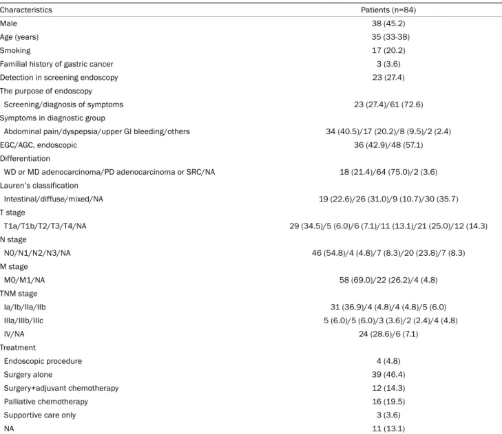
Table 1. Baseline Characteristics of Young Patients with Gastric Cancer
Characteristics Patients (n=84)
Male 38 (45.2)
Age (years) 35 (33-38)
Smoking 17 (20.2)
Familial history of gastric cancer 3 (3.6)
Detection in screening endoscopy 23 (27.4)
The purpose of endoscopy
Screening/diagnosis of symptoms 23 (27.4)/61 (72.6)
Symptoms in diagnostic group
Abdominal pain/dyspepsia/upper GI bleeding/others 34 (40.5)/17 (20.2)/8 (9.5)/2 (2.4)
EGC/AGC, endoscopic 36 (42.9)/48 (57.1)
Differentiation
WD or MD adenocarcinoma/PD adenocarcinoma or SRC/NA 18 (21.4)/64 (75.0)/2 (3.6) Lauren’s classification
Intestinal/diffuse/mixed/NA 19 (22.6)/26 (31.0)/9 (10.7)/30 (35.7)
T stage
T1a/T1b/T2/T3/T4/NA 29 (34.5)/5 (6.0)/6 (7.1)/11 (13.1)/21 (25.0)/12 (14.3)
N stage
N0/N1/N2/N3/NA 46 (54.8)/4 (4.8)/7 (8.3)/20 (23.8)/7 (8.3)
M stage
M0/M1/NA 58 (69.0)/22 (26.2)/4 (4.8)
TNM stage
Ia/Ib/IIa/IIb 31 (36.9)/4 (4.8)/4 (4.8)/5 (6.0)
IIIa/IIIb/IIIc 5 (6.0)/5 (6.0)/3 (3.6)/2 (2.4)/4 (4.8)
IV/NA 24 (28.6)/6 (7.1)
Treatment
Endoscopic procedure 4 (4.8)
Surgery alone 39 (46.4)
Surgery+adjuvant chemotherapy 12 (14.3)
Palliative chemotherapy 16 (19.5)
Supportive care only 3 (3.6)
NA 11 (13.1)
Values are presented as median (interquartile range) or n (%).
GI, gastrointestinal; EGC, early gastric cancer; AGC, advanced gastric cancer; WD, well differentiated; MD, moderately differentiated; PD, poorly differentiated; SRC, signet ring cell carcinoma; NA, not available.
used to determine the cancer staging.18 A H. pylori infection was defined as a positivity of at least one test among the rapid urease test, urea breath test, and histology for H. pylori.
3. Statistical analysis
The sequential data are expressed as the median and inter- quartile range. The continuous variables were compared using a Mann-Whitney U test or Student’s t-test as appropriate. The categorical variables were compared using a Pearson’s χ2 test or Fisher’s exact test. The survival curves were calculated using the Kaplan-Meier method. Statistical analyses were conducted
using IBM SPSS Statistics software for Windows v.22.0 (IBM Corp., Armonk, NY, USA). All tests were two-sided and statistical significance was considered when the p-value was <0.05.
RESULTS
1. Baseline characteristics of the study population In this study, 84 patients were enrolled. Twenty-three pa- tients <40 years of age with gastric cancer were classified into the screening group. Fourteen patients were diagnosed with gastric cancer in three tertiary health checkup centers.
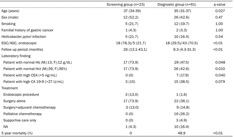
Table 2. Clinical Characteristics of Gastric Cancer according to the Purpose of Endoscopy
Screening group (n=23) Diagnostic group (n=61) p-value
Age (years) 37 (34-39) 35 (31-37) 0.027
Sex (male) 12 (52.2) 26 (42.6) 0.47
Smoking 5 (21.7) 12 (19.7) 1.00
Familial history of gastric cancer 1 (4.3) 2 (3.3) 1.00
Helicobacter pylori infection 5 (21.7) 10 (16.4) 0.54
EGC/AGC, endoscopic 18 (78.3)/5 (21.7) 18 (29.5)/43 (70.5) <0.01
Follow up period (months) 26 (13.1-43.1) 9.3 (4.3-31.3) <0.01
Laboratory finding
Patient with normal Hb (M≥13, F≥12 g/dL) 17 (73.9) 29 (47.5) 0.048
Patient with normal Hct (M≥39, F≥36%) 17 (73.9) 26 (42.6) 0.010
Patient with high CEA (>5 ng/mL) 0 (0) 7 (17.9) 0.040
Patient with high CA 19-9 (>27 U/mL) 3 (15) 15 (38.5) 0.079
Treatment
Endoscopic procedure 3 (13.0) 1 (1.6)
Surgery alone 17 (73.9) 22 (36.1)
Surgery+adjuvant chemotherapy 3 (13.0) 9 (14.8)
Palliative chemotherapy 0 (0) 16 (26.2)
Supportive care only 0 (0) 3 (4.9)
NA 1 (4.3) 10 (16.4)
5 year mortality (%) 0 48.9 <0.01
Values are presented as median (interquartile range) or n (%).
EGC, early gastric cancer; AGC, advanced gastric cancer; Hb, hemoglobin; M, male; F, female; Hct, hematocrit; CEA, carcinoembryonic antigen; CA 19-9, carbohydrate antigen 19-9; NA, not available.
The estimated prevalence of gastric cancer in patients younger than 40 years in screening endoscopy was 7.2 per 100,000 (14/195,711). One patient was transferred to another hospital by the patient’s direction. In outpatient clinics, 12 patients diagnosed with gastric cancer during screening endoscopy.
Among them, two patients were transferred to other hospitals.
As a result, 23 were included in the screening group. An addi- tional 61 patients who had visited the hospital because of symptoms were also diagnosed with gastric cancer (Fig. 1).
The male to female sex ratio was 0.83, and the median age of the study population was 35 years (range, 21 to 39 years) (Fig. 2). In total, 48 (57.1%) gastric cancer patients had ages ranging from 35 to 39, and 22 (26.2%) patients were 30 to 34 years of age. Fourteen (16.7%) patients were younger than 30 years of age; all of these patients belonged to the diagnostic group. The study population was comprised of 36 cases of EGC and 48 cases of AGC. The median follow-up period was 16.1 months (interquartile range 6.5-38.4).
Pathologically, there were 40 cases of adenocarcinoma and 42 cases of signet ring cell carcinoma (well differentiated carci-
noma, 5; moderately differentiated carcinoma, 13; poorly differ- entiated adenocarcinoma, 22; signet ring cell carcinoma, 42).
The most common stage was stage 1A (31 cases, 36.9%), followed by stage 4 (24 cases, 28.6%) (Table 1).
2. Comparison of the clinical characteristics of the screening and diagnostic groups
Abdominal pain was the most common symptom in the diagnostic group (34 cases, 40.5%). No patients had under- lying diseases except for two patients who had type 2 diabetes. Three patients had a familial history of gastric can- cer (one case in the screening group, and two cases in the diagnostic group). The median age of the screening group was significantly higher than that of the diagnostic group (37 vs. 35 years, p=0.03). The ratios of patients with normal he- moglobin and hematocrit were significantly lower in the diag- nostic group (47.5% vs. 73.9%, p=0.048, 42.6% vs. 73.9%, p=0.010). The ratio of patients with a high CEA level was higher in the diagnostic group (17.9% vs. 0%, p=0.040). No significant differences were found in the other laboratory
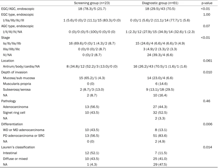
Table 3. Pathologic Characteristics of Gastric Cancer according to the Purpose of Endoscopy
Screening group (n=23) Diagnostic group (n=61) p-value
EGC/AGC, endoscopic 18 (78.3)/5 (21.7) 18 (29.5)/43 (70.5) <0.01
EGC type, endoscopic 1.00
I/IIa/IIb/IIc/III 1 (5.6)/0 (0)/2 (11.1)/15 (83.3)/0 (0) 0 (0)/1 (5.6)/2 (11.1)/14 (77.7)/1 (5.6)
AGC type, endoscopic 0.07
I/II/III/IV/NA 0 (0)/0 (0)/5 (100)/0 (0)/0 (0) 1 (2.3)/12 (27.9)/15 (34.9)/14 (32.6)/1 (2.3)
Stage <0.01
Ia/Ib/IIa/IIb 16 (69.6)/0 (0)/1 (4.3)/2 (8.7) 15 (24.6)/4 (6.6)/4 (6.6)/3 (4.9)
IIIa/IIIb/IIIc 0 (0)/0 (0)/2 (8.7) 3 (4.9)/2 (3.3)/2 (3.3)
IV/NA 0 (0)/2 (8.7) 24 (39.3)/4 (6.6)
Location 0.061
Antrum/body/cardia/NA 8 (34.8)/12 (52.2)/3 (13.0)/0 (0) 16 (26.2)/43 (70.5)/1 (1.6)/1 (1.6)
Depth of invasion 0.010
Mucosa/sub mucosa 15 (65.2)/1 (4.3) 14 (23.0)/4 (6.6)
Muscularis propria 0 (0) 6 (14.6)
Subserosa/serosa 2 (8.7)/3 (13.0) 9 (13.1)/18 (29.5)
NA 2 (8.7) 10 (16.4)
Pathology 0.46
Adenocarcinoma 13 (56.5) 27 (44.3)
Signet ring cell 10 (43.5) 32 (52.5)
NA 2 (3.3)
Differentiation 0.006
WD or MD adenocarcinoma 10 (43.5) 8 (13.1)
PD adenocarcinoma or SRC 13 (56.5) 51 (83.6)
NA 0 (0) 2 (4.9)
Lauren’s classification 0.014
Intestinal 12 (52.1) 7 (11.5)
Diffuse or mixed 10 (43.5) 25 (41.0)
NA 1 (4.3) 29 (47.5)
Values are presented as n (%).
EGC, early gastric cancer; AGC, advanced gastric cancer; NA, not available; WD, well differentiated; MD, moderately differentiated; PD, poorly differentiated; SRC, signet ring cell carcinoma.
findings. The proportion of EGC in the screening group was significantly higher than that of the diagnostic group (78.3%
vs. 29.5%, p<0.01). The number of patients who received che- motherapy was significantly higher in the diagnostic group (13.0% vs. 41.0%, p<0.01). The proportion of patients who underwent a curative resection was significantly higher in the screening group (95.7% vs. 52.5%, p<0.01). During the fol- low-up period, 21 deaths were observed, which all occurred in the diagnostic group (Table 2). All 21 deaths were due to gastric cancer. The 5-year estimated mortality rate was sig- nificantly lower in the screening group than in the diagnostic group (0% vs. 48.9%, p<0.01).
3. Comparison of the pathologic characteristics of the screening and diagnostic groups
Among the 36 cases of EGC, the depressed type (IIc) was the most common type, regardless of the presence of symptoms. Among the 48 cases of AGC, the ulceroinfiltrative type (Borrmann type 3) was most common. The stage at the time of diagnosis was significantly earlier in the screening group than in the diagnostic group (p<0.01), and the pro- portion of EGC was significantly higher (78.3% vs. 29.5%, p<0.01). The depth of invasion of primary cancer in the diag- nostic group was significantly deeper than that in the screen- ing group (p=0.010), and poorly differentiated carcinoma was more common (56.5% vs. 83.6%, p=0.006). The location and pathological type were similar in the two groups (Table 3).
Table 4. Comparisons of the Characteristics between Young and Older Patients Diagnosed with Gastric Cancer with a Screening Endoscopy Young patient group (n=23) Older patient group (n=46) p-value
Age (years) 37 (34-39) 47.5 (45-49)
Sex (male) 12 (52.2) 19 (41.3) 0.61
Smoking 5 (21.7) 9 (37.0) 0.20
Familial history of gastric cancer 1 (4.3) 1 (2.2) 0.61
EGC/AGC, endoscopic 18 (78.3)/5 (21.7) 36 (78.3)/10 (21.7) 1.00
Pathology 0.072
WD or MD adenocarcinoma 10 (43.5) 31 (67.4)
PD adenocarcinoma or SRC 13 (56.5) 15 (32.6)
NA 0 (0) 0 (0)
Lauren’s classification 0.002
Intestinal 7 (30.4) 27 (58.7)
Diffuse or mixed 16 (69.6) 11 (23.9)
NA 0 (0) 8 (17.4)
5 year mortality (%) 0 6.7 0.265
Values are presented as median (interquartile range) or n (%). Young patient group: young patients whose gastric cancer was detected at a health checkup; Older patient group: 1 to 2 stage-matched older (41-50 years old) patients whose gastric cancer was detected at a health checkup in the same year.
EGC, early gastric cancer; AGC, advanced gastric cancer; WD, well differentiated; MD, moderately differentiated; PD, poorly differentiated; SRC, signet ring cell carcinoma; NA, not available.
Fig. 3. Kaplan-Meier survival curve between the two groups. Kaplan-Meier curves illustrating the overall survival of the screening and diagnostic groups. Among the patients in the diagnostic group, there were no deaths. A total of 23 deaths occurred in the diagnostic group. Overall the survival was significantly higher in the screening group than in the diagnostic group.
4. Comparison of the characteristics of young and older patients diagnosed with gastric cancer with a screen- ing endoscopy
A one to two stage-matched older patient group (aged 40-49 years) was formed to compare the characteristics be- tween young and older patients diagnosed with gastric cancer with a screening endoscopy. The young and older groups showed a similar smoking status, family history of gastric can- cer, and survival. The ratio of patients with poorly differ- entiated adenocarcinoma or signet ring cell carcinoma was higher in the young patients (56.5% vs. 32.6%, p=0.072).
Diffuse type cancer was more prevalent in the young screen- ing group (69.6% vs. 23.9%, p=0.002) (Table 4).
5. Comparison of the overall survival in the screening and diagnostic groups
Fig. 3 presents a schematic diagram of the overall survival of patients according to the purpose of their endoscopy. The overall 1-, 2-, 3-, and 5-year survival rates for the screening group were higher than those of the diagnostic group (100%
vs. 77.1%, 100% vs. 72.1%, 100% vs. 67.2%, and 100% vs.
51.1% respectively, p<0.01).
DISCUSSION
This retrospective study evaluated the role of screening gastroduodenoscopies in young patients indirectly by compar- ing the screening and diagnostic groups, which differed in the purpose of endoscopy. To the best of the authors’ knowl- edge, this is the first report of gastric cancer in young patients detected during a health checkup. These results show that young patients aged <40 years, who were diagnosed with gas- tric cancer after a screening gastroduodenoscopy, tended to be diagnosed in the earlier stages than those who underwent upper endoscopy because of symptoms. In addition, this group showed a significantly longer survival than the sympto- matic group.
Previous studies have investigated the features of gastric cancer in young patients with gastric cancer. The definition of young onset gastric cancer is controversial, but it is usually defined as gastric cancer diagnosed in patients <40-45 years of age.19-22 A Mexican study that evaluated gastric cancer in patients under 30 years of age from 1985 to 2006 reported a tendency towards a late diagnosis of gastric cancer in young
patients.12 Almost all cancers were already at an advanced stage at the time of diagnosis, and 25 patients (83%) were diagnosed with stage 4 disease.12 On the other hand, this study enrolled only symptomatic patients. Therefore, it is diffi- cult to apply this result to asymptomatic young patients. In the present study, the survival rate of asymptomatic patients was significantly higher than that of symptomatic patients.
A European study comparing the clinicopathologic features of young gastric cancer patients with older patients >45 years old, showed that young-onset gastric cancer tends to be more advanced, but the prognosis is similar to that of older patients.23 In the younger age group, diffuse type carcinoma (73%), lymph node metastasis (59%), and stage IV disease (49%), and non-curative treatment (36%) are more prevalent than in older patients. These results are similar to the results of a diagnostic group in the present study.
In a retrospective Korean study, the prognosis of young pa- tients (<40 years old) with early gastric carcinoma was similar to that of older patients with EGC. They insisted that the belief that the prognosis of young gastric cancer patients is poorer than that of aged patients should be abandoned and that early detection is critical to achieve a good prognosis.24 Another study reported that young gastric cancer patients (<40 years old) present with poorly differentiated cancer and with more metastatic and advanced disease.25 In addition, they concluded that the patient’s health status, tumor local- ization, smoking status did not confer a significant difference in the disease state, which coincides with the present data showing a poorly differentiated histology in 52% of the study population. Another Korean study revealed the predominance of female patients and histologically undifferentiated carcinoma.20 In the present study, there was no difference in sex and location of the primary cancer between the screen- ing and diagnostic groups, and adenocarcinoma and signet ring cell carcinoma showed a similar incidence. On the other hand, poorly differentiated adenocarcinoma was more com- mon in the diagnostic group than the screening group (p<0.01).
In terms of screening an upper endoscopy for gastric can- cer, a Korean study previously reported the characteristics of gastric cancer diagnosed at a health screening.26 EGC rep- resented 81 out of 111 cases (73%) in this study. The mean overall survival was 103 months and the cumulative proba- bility of survival at 5 and 10 years was 82.7% and 67%,
respectively. Five patients (4.5%) under the age of 40 years were diagnosed with gastric cancer. Another study showed that annual endoscopic screening in a region with a high in- cidence of gastric cancer improved the detection of ear- ly-stage and endoscopically treatable gastric cancer.27 A re- cent large-scale Korean study, using nationwide screening pro- gram results, reported that gastric cancer screening with up- per endoscopy reduced gastric cancer mortality.28 A recent meta-analysis of observational studies of gastric cancer mor- tality reduction after endoscopic screening suggested that pa- tients might achieve a 40% reduction in gastric cancer mortal- ity after endoscopic screening but screening did not reduce incidence in Asian countries, including South Korea.29 These results support the role of screening endoscopy for gastric cancer in individuals aged ≥40 years.
A comparison of the characteristics of a young screening group in this study with stage-matched gastric cancer patients aged 40 to 49 years diagnosed by screening endoscopy re- vealed similar characteristics, including the proportion of EGC and survival, except for the Lauren’s classification. Previous studies reported that gastric cancer at a young age is more frequently associated with diffuse and undifferentiated histol- ogy types.30-33 In the present study, diffuse type cancer was more prevalent in the young screening group. The ratio of patients with poorly differentiated adenocarcinoma or signet ring cell carcinoma tended to be higher in the young patients, which was not statistically different. The lack of significant differences might be due to the relatively small number of patients in the study population.
A higher ratio of patients with lower serum hemoglobin or hematocrit levels in the diagnostic group may be related to their advanced disease. When this study was designed, it is believed that the serum CEA and CA 19-9 levels in the screen- ing group would be different from those of the diagnostic group. Patients with abnormal CEA levels were more prevalent in the diagnostic group and the CA 19-9 levels tended to be higher in the diagnostic group.
This study had several limitations. First, this study was con- ducted as an observational, retrospective study. Therefore, biases, such as length-time bias and lead-time bias, could not be controlled. Slow progressive cancers are more likely to be detected in screening endoscopy. Therefore, these are likely to be classified as the screening group, and show a better prognosis. In contrast, rapidly progressive cancer tends
to cause symptoms rapidly and is unlikely to be found during screening endoscopy; these were classified as a diagnostic group. In the present study, the ratios of patients with poorly differentiated carcinoma or signet ring cell carcinoma and dif- fuse type cancer, which are characterized by rapid progression and a poorer prognosis,34 were significantly lower in the screening group than in the diagnostic group. Regarding the lead-time bias, patients diagnosed at the early stage during screening endoscopy may show increased survival without af- fecting the course of the disease. Therefore, a well-designed, randomized controlled trial is needed to prove the effective- ness of screening endoscopy for survival gain in young patients. Second, this was a retrospective study using medical records. Therefore, data, such as the underlying disease, symptoms at the time of diagnosis, and familial history could be inaccurate. In addition, the patients were classified into two groups according to the purpose of endoscopy based on the medical records. Hence, misclassification bias could occur. Third, only a small number of patients were included in the screening group. In addition, this study included pa- tients who were referred to the three territory centers from other clinics with abnormal endoscopy finding suggesting gas- tric cancer. Therefore, it was impossible to calculate the exact incidence of gastric cancer. Future large-scale, prospective studies should resolve these limitations.
In conclusion, asymptomatic patients aged <40 diagnosed with gastric cancer by screening endoscopy may allow a down- ward migration of the stage, and in some populations, screen- ing endoscopy could result in a better prognosis compared to those by diagnostic endoscopy for symptoms. A well de- signed, multi-center, large-scale study for the effects of screening endoscopy in individuals aged <40 will be needed.
REFERENCES
1. Bray F, Ferlay J, Soerjomataram I, Siegel RL, Torre LA, Jemal A.
Global cancer statistics 2018: GLOBOCAN estimates of in- cidence and mortality worldwide for 36 cancers in 185 countries.
CA Cancer J Clin 2018;68:394-424.
2. Lee HJ, Yang HK, Ahn YO. Gastric cancer in Korea. Gastric Cancer 2002;5:177-182.
3. Jung KW, Won YJ, Kong HJ, Lee ES. Cancer statistics in Korea: in- cidence, mortality, survival, and prevalence in 2016. Cancer Res Treat 2019;51:417-430.
4. Miwa K. Evaluation of the TNM classification of stomach cancer and proposal for its rational stage-grouping. Jpn J Clin Oncol 1984;14:385-410.
5. Park IS, Lee YC, Kim WH, Noh SH, Lee KS, Kim H. Clinicopathologic characteristics of early gastric cancer in Korea. Yonsei Med J 2000;41:607-614.
6. Mizoue T, Yoshimura T, Tokui N, et al. Prospective study of screen- ing for stomach cancer in Japan. Int J Cancer 2003;106:103-107.
7. Llorens P. Gastric cancer mass survey in Chile. Semin Surg Oncol 1991;7:339-343.
8. Pisani P, Oliver WE, Parkin DM, Alvarez N, Vivas J. Case-control study of gastric cancer screening in Venezuela. Br J Cancer 1994;69:1102-1105.
9. Park HA, Nam SY, Kim SK, et al. The Korean guideline for gastric cancer screening. J Korean Med Assoc 2015;58:373-384.
10. Anderson WF, Camargo MC, Fraumeni JF Jr, Correa P, Rosenberg PS, Rabkin CS. Age-specific trends in incidence of noncardia gas- tric cancer in US adults. JAMA 2010;303:1723-1728.
11. Leung WK, Wu MS, Kakugawa Y, et al. Screening for gastric can- cer in Asia: current evidence and practice. Lancet Oncol 2008;9:279-287.
12. López-Basave HN, Morales-Vásquez F, Ruiz-Molina JM, et al.
Gastric cancer in young people under 30 years of age: worse prognosis, or delay in diagnosis? Cancer Manag Res 2013;
5:31-36.
13. Smith BR, Stabile BE. Extreme aggressiveness and lethality of gastric adenocarcinoma in the very young. Arch Surg 2009;144:
506-510.
14. Buffart TE, Carvalho B, Hopmans E, et al. Gastric cancers in young and elderly patients show different genomic profiles. J Pathol 2007;211:45-51.
15. Bedikian AY, Khankhanian N, Heilbrun LK, Bodey GP, Stroehlein JR, Valdivieso M. Gastric carcinoma in young adults. South Med J 1979;72:654-656.
16. Japanese Gastric Cancer Association. Japanese classification of gastric carcinoma: 3rd English edition. Gastric Cancer 2011;14:
101-112.
17. Lauren P. The two histological main types of gastric carcinoma:
diffuse and so-called intestinal-type carcinoma. An attempt at a histo-clinical classification. Acta Pathol Microbiol Scand 1965;
64:31-49.
18. Brierley JD, Gospodarowicz MK, Wittekinde C. TNM classification of malignant tumours. 8th ed. Hoboken (NJ): Wiley-Blackwell, 2017.
19. Lee J, Lee MA, Kim IH, Roh SY. Clinical characteristics of young-age onset gastric cancer in Korea. BMC Gastroenterol 2016;16:110.
20. Lai JF, Kim S, Li C, et al. Clinicopathologic characteristics and prognosis for young gastric adenocarcinoma patients after cura- tive resection. Ann Surg Oncol 2008;15:1464-1469.
21. Takatsu Y, Hiki N, Nunobe S, et al. Clinicopathological features of gastric cancer in young patients. Gastric Cancer 2016;19:
472-478.
22. Kono Y, Kanzaki H, Tsuzuki T, et al. A multicenter observational study on the clinicopathological features of gastric cancer in young patients. J Gastroenterol 2019;54:419-426.
23. Santoro R, Carboni F, Lepiane P, Ettorre GM, Santoro E.
Clinicopathological features and prognosis of gastric cancer in young European adults. Br J Surg 2007;94:737-742.
24. Park YK, Kim JC, Koh YS, et al. Early gastric carcinoma in young patients. Int Surg 2006;91:316-319.
25. Isik M, Caner S, Metin Seker M, et al. Gastric adenocarcinoma under the age of 40; more metastatic, less differentiated. J BUON 2011;16:253-256.
26. Lee HJ, Chung JM, Seo EH, Jeon SW. Clinicopathologic character- istics of gastric cancer diagnosed at health screening. Korean J Med 2008;75:665-672.
27. Chung SJ, Park MJ, Kang SJ, et al. Effect of annual endoscopic screening on clinicopathologic characteristics and treatment modality of gastric cancer in a high-incidence region of Korea. Int J Cancer 2012;131:2376-2384.
28. Jun JK, Choi KS, Lee HY, et al. Effectiveness of the Korean na- tional cancer screening program in reducing gastric cancer mortality. Gastroenterology 2017;152:1319-1328.e7.
29. Zhang X, Li M, Chen S, et al. Endoscopic screening in Asian coun- tries is associated with reduced gastric cancer mortality: a meta-analysis and systematic review. Gastroenterology 2018;
155:347-354.e9.
30. Theuer CP, de Virgilio C, Keese G, et al. Gastric adenocarcinoma in patients 40 years of age or younger. Am J Surg 1996;172:
473-477.
31. Park HJ, Ahn JY, Jung HY, et al. Clinical characteristics and out- comes for gastric cancer patients aged 18-30 years. Gastric Cancer 2014;17:649-660.
32. Park JC, Lee YC, Kim JH, et al. Clinicopathological aspects and prognostic value with respect to age: an analysis of 3,362 con- secutive gastric cancer patients. J Surg Oncol 2009;99:
395-401.
33. Kong X, Wang JL, Chen HM, Fang JY. Comparison of the clin- icopathological characteristics of young and elderly patients with gastric carcinoma: a meta analysis. J Surg Oncol 2012;
106:346-352.
34. Chon HJ, Hyung WJ, Kim C, et al. Differential prognostic im- plications of gastric signet ring cell carcinoma: stage adjusted analysis from a single high-volume center in Asia. Ann Surg 2017;265:946-953.