65 http://www.jchestsurg.org
JCSJournal of Chest Surgery
CaseReport
Successful Dual-Patch Closure of a Fistula between the Right Pulmonary Artery and the Left Atrium
Abhishek Shrinivas Joshi, Balaji Dattatray Aironi
Department of Cardiovascular and Thoracic Surgery, Seth Gordhandas Sunderdas Medical College and King Edward VII Memorial Hospital, Mumbai, India
ARTICLE INFO Received May 6, 2020 Revised June 28, 2020 Accepted July 1, 2020 Corresponding author Abhishek Shrinivas Joshi
Tel 91-9820326126; 91-224107429 E-mail [email protected] ORCID
https://orcid.org/0000-0001-9893-1556
A fistula between the right pulmonary artery and the left atrium is a very rare congenital anomaly, for which there is no definitive embryogenetic explanation. Patients present with cyanosis or clubbing, and the treatment strategy is to close the fistula, which can be done by an open surgical technique or by percutaneous intervention. Although rare, this con- dition should be considered in the differential diagnosis when evaluating a patient with central cyanosis.
Keywords: Anomaly, Cyanosis, Fistula
Copyright©The Korean Society for Thoracic and Cardiovascular Surgery. 2021. All right reserved.
This is an Open Access article distributed under the terms of the Creative Commons Attribution Non-Commercial License (http://creativecommons.org/licenses/
by-nc/4.0) which permits unrestricted non-commercial use, distribution, and reproduction in any medium, provided the original work is properly cited.
Case report
A fistulous connection between the right pulmonary ar- tery (RPA) and the left atrium (LA), although rare, is a condition that cannot be neglected [1]. The largest collec- tive case review by Chowdhury et al. [2] included 59 cases.
This entity is also hardly mentioned in current textbooks of cardiology and cardiac surgery. Hence, the exact inci- dence of this rare condition has not been determined. The majority of cases are congenital, although traumatic etiolo- gy has been implied in a couple of cases. The usual pre- senting complaints and signs in patients with this condi- tion are dyspnea on exertion, central cyanosis, decreased arterial oxygen saturation, and clubbing. The modalities used to diagnose this condition include 2-dimensional echocardiography, computed tomography (CT) scans, and angiography [3]. This anomaly has been classified into 4 types, and the type of fistula dictates the treatment ap- proach. Although catheter-guided closure of the fistula has been recently proposed as a less invasive technique, nu- merous other factors are relevant when deciding upon the treatment approach for this condition [4]. It is always best to adopt a case-directed, individualized approach for the treatment of RPA-LA fistula. The mortality rate of this condition has decreased in recent years. Herein, we de- scribe a case of RPA-to-LA fistula in a 32-year-old male
patient.
A 32-year-old male farmer presented with complaints of grade II dyspnea on exertion for the last 8 years and pedal edema. He also gave a history of hospital admission for symptoms of severe headache and fever with chills. On ex- amination, the patient’s vital parameters were normal; he had grade 3 clubbing, central symmetrical cyanosis, and arterial oxygen saturation of 70%. An ejection systolic murmur could be heard over the left second and third parasternal areas. A chest X-ray showed cardiomegaly and a wide carinal angle, and an electrocardiogram was sug- gestive of left atrial enlargement. There was early contrast filling of the LA on contrast echocardiography. The patient underwent a cardiac CT scan, which showed a fistula be- tween the RPA and the LA (Fig. 1A). Aneurysmal dilata- tion of the left atrial appendage was also noted. Angiogra- phy was done to denote the course of the fistula from the RPA to the LA (Fig. 1B). Contrast injection clearly showed the opening in the LA. Individual pressures were not cal- culated; however, the patient’s oxygen saturation increased to 96% upon complete occlusion of the right pulmonary ostium of the fistula. The patient was classified as having a type 1 communication and surgery was planned. The pa- tient underwent surgery under cardiopulmonary bypass with moderate hypothermia using Del NIdo cardioplegia.
Intraoperatively, the 3-cm-long fistula was found to arise
https://doi.org/10.5090/kjtcs.20.034 pISSN: 2765-1606 eISSN: 2765-1614 J Chest Surg. 2021;54(1):65-67
66
https://doi.org/10.5090/kjtcs.20.034
http://www.jchestsurg.org
JCS
from the RPA and communicate with the roof of the LA.
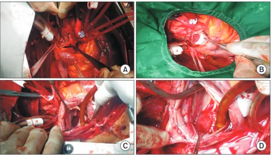
The RPA was opened and the opening of the fistulous communication, which measured around 1.5 cm×1.5 cm, could be easily seen at the inferior side of the RPA (Fig.
2A). The LA was entered via a trans-septal approach through the right atrium (RA). The opening in the roof of the LA was readily noted, and measured around 2 cm×2 cm (Fig. 2C). We decided to perform a double-patch clo- sure of the fistula instead of the more routine ligation or single-patch closure, as the fistula was thin-walled. Using 2 different patches also served as a safety mechanism for preventing air embolism during the postoperative period.
The RPA opening was closed with a Dacron patch (Sauvage Filamentous Dacron; Bard, Tempe, AZ, USA) using inter- rupted polypropylene 6-0 sutures, and the LA opening was closed with a pericardial patch using continuous polypro-
pylene 5-0 sutures after allowing the fistula to fill with blood (Fig. 2B, D). The interatrial septum and RA were closed and the patient was weaned off bypass. The total bypass time was 156 minutes and the cross-clamp time for the procedure was 124 minutes. The chest was closed pri- marily. Postoperatively, arterial oxygen saturation was 100%. The patient had an uneventful postoperative course and was discharged on postoperative day 6. After 2 months, another CT scan and 2-dimensional echocardiog- raphy showed no evidence of any fistulous communication.
The patient is currently symptom-free.
The patient provided written informed consent for pub- lication of the clinical details and images.
Discussion
A fistula between the RPA and the LA is a rare cause of cyanosis in adult patients. It is difficult to diagnose this condition based only on a clinical history and routine 2-di- mensional echocardiography. Additional investigations, namely contrast echocardiography and CT, are needed to delineate such fistulous tracts precisely. This condition was classified into 3 types by de Souza e Silva et al. [5], and a fourth type was later added by Ohara et al. [6]. Hence, there are 4 major types of these fistulae and every type has a distinct line of management. Type 1 is an anomalous connection connecting the posterior aspect of the proximal RPA into the LA with normal pulmonary venous connec- tion. Type 2 is a persistent and supernumerary branch of the pulmonary artery, which establishes an abnormal con- nection with the LA. In type 3, all pulmonary veins drain into an aneurysmal pouch between the RPA and the LA.
A B
Fig. 1. (A) Cardiac computed tomography demonstrating filling of the left atrium and pulmonary bifurcation. (B) Catheter course from the inferior vena cava to the right atrium, right ventricle, pulmo- nary artery, right pulmonary artery, left atrium, and left ventricle.
Fig. 2. (A) Opening in the RPA. (B) Dacron patch closure of the RPA opening. (C) Opening in the LA.
(D) Pericardial patch closure of the LA opening. RPA, right pulmonary artery; LA, left atrium.
A
C D
B
67
Abhishek Shrinivas Joshi and Balaji Dattatray Aironi. Right Pulmonary Artery to Left Atrial Fistula
http://www.jchestsurg.org
JCS
In type 4, a very proximal pulmonary arteriovenous fistula joins the LA, such that the right pulmonary veins drain into the fistula and the left pulmonary veins drain into the LA directly. Even though this condition can be tackled by either vascular intervention or open surgery, it is best to plan the treatment on a patient-specific basis. It is neces- sary to treat this condition as early as possible upon diag- nosis. Median sternotomy is a safe approach and closure of both ends using a patch is a safe method for the treatment of an RPA-LA fistula, as is simple division and ligation.
In conclusion, this report highlights a rare case of cyano- sis in an adult patient and describes the pathway of diag- nosing this condition. Dual closure of the fistula from both ends by using a Dacron and pericardial patch under car- diopulmonary bypass is a safe and simple procedure to treat this condition that led to a good outcome.
Conflict of interest
No potential conflict of interest relevant to this article was reported.
ORCID
Abhishek Shrinivas Joshi: https://orcid.org/0000-0001-9893-1556
Balaji Dattatray Aironi: https://orcid.org/0000-0002-6223-5011
References
1. Muthialu N, Joshi S, Hoskote A, Bhole V. Rare cause of central cya- nosis: right pulmonary artery to left atrial fistula. Interact Cardiovasc Thorac Surg 2016;23:839-40.
2. Chowdhury UK, Kothari SS, Airan B, Subramaniam KG, Venugopal P. Right pulmonary artery to left atrium communication. Ann Thorac Surg 2005;80:365-70.
3. Narula J, Choudhury M, Chowdhary UK, Kiran U. Pulmonary artery- pulmonary vein fistula: contrast echocardiography using agitated sa- line for indirect evaluation of adequacy of surgical repair. J Perioper Echocardiogr 2014;2:61-4.
4. Benz DC, Burkhardt B, Quandt D, Stambach D, Knirsch W, Kretsch- mar O. Interventional closure of RPA-to-LA communication in an oligosymptomatic neonate. Eur J Pediatr 2014;173:1703-5.
5. De Souza e Silva NA, Giuliani ER, Ritter DG, Davis GD, Pluth JR.
Communication between right pulmonary artery and left atrium. Am J Cardiol 1974;34:857-63.
6. Ohara H, Ito K, Kohguchi N, et al. Direct communication between the right pulmonary artery and the left atrium: a case report and re- view of the literature. J Thorac Cardiovasc Surg 1979;77:742-7.