INTRODUCTION
The key updates in the revised 2016 WHO criteria for the recat- egorization of acute myeloid leukemia (AML) are as follows [1-3].
1) Two new provisional entities, AML with BCR-ABL1 and AML with mutated RUNX1 were added to one of the AML subtypes with recurrent genetic abnormalities (abbreviated as RGA in the following text). 2) AML diagnosis with mutated CEBPA required biallelic mutations instead of a single mutation. 3) The diagnostic precedence of AML with myelodysplasia-related changes (abbre- viated as MRC in the following text) over AML with NPM1 or bial- lelic CEBPA (CEBPA
bi) mutations was clarified for patients show- ing myelodysplasia-associated cytogenetic abnormalities. 4)
급성골수성백혈병의 개정된 세계보건기구 기준에 의한 대규모 환자군의 분류: 재분류된 사례의 빈도 및 특성
Revised World Health Organization Criteria-Defined Acute Myeloid Leukemia in a Large Cohort: Highlighting the Frequency and Characterization of Recategorized Cases
김시환1·이민영2·조영욱1·황상현1·장성수1·서을주1·박찬정1
Sihwan Kim, M.D.
1, Min-Young Lee, M.D.
2, Young-Uk Cho, M.D.
1, Sang-Hyun Hwang, M.D.
1, Seongsoo Jang, M.D.
1, Eul-Ju Seo, M.D.
1, Chan-Jeoung Park, M.D.
1울산대학교 의과대학 서울아산병원 진단검사의학과1, 경희대학교 의과대학 강동경희대학교병원 진단검사의학과2
Department of Laboratory Medicine
1, University of Ulsan, College of Medicine and Asan Medical Center, Seoul; Department of Laboratory Medicine
2, Kyung Hee University School of Medicine, Kyung Hee University Hospital at Gangdong, Seoul, Korea
Vol. 11, No. 2: 115-123, April 2021
https://doi.org/10.47429/lmo.2021.11.2.115
진단혈액학
Corresponding author: Young-Uk Cho, M.D., Ph.D.
https://orcid.org/0000-0002-4403-8989
Department of Laboratory Medicine, Asan Medical Center, University of Ulsan College of Medicine, 88 Olympic-ro 43-gil, Songpa-gu, Seoul 05505, Korea
Tel.: +82-2-3010-4501, Fax: +82-2-478-0884, E-mail: [email protected] Received: July 6, 2020
Revision received: September 9, 2020 Accepted: September 10, 2020
This article is available from https://www.labmedonline.org 2021, Laboratory Medicine Online
This is an Open Access article distributed under the terms of the Creative Commons Attribution Non-Commercial License (https://creativecommons.org/licenses/by-nc/4.0/) which permits unrestricted non-commercial use, distribution, and reproduction in any medium, provided the original work is properly cited.
Background: Application of the 2016 revised WHO criteria for categorization of acute myeloid leukemia (AML) highlights certain discrepancies from that of the 2008 WHO criteria. We thus analyzed the frequency, categorization patterns, and features of discrepant cases, and characterized the AML subtypes that had undergone major changes under the revised criteria.
Methods: We divided the patients into the following seven categories based on the previous and the revised WHO criteria: AML with recurrent genetic abnormalities (RGA), AML with myelodysplasia-related changes (MRC), therapy-related AML, AML not otherwise specified (NOS), AML as- sociated with Down syndrome, AML with subcategory not determined, and myelodysplastic syndrome (MDS).
Results: In total, 1,185 AML cases were reviewed. The concordance rate in categorization between the two criteria was 93.4%. Among 78 dis- crepant cases, the three most common discrepancy patterns were for the RGA to NOS, MRC to MDS, and MRC to RGA, representing cases with a single mutation in CEBPA, erythroleukemia, and recurrent genetic abnormalities showing myelodysplasia, respectively. We identified three cases of erythroleukemia harboring an NPM1 mutation, who might clinically benefit from chemotherapy rather than MDS-oriented treatment; we also found AML with del(9q) in 3% of patients, which might contribute to leukemogenesis either via haploinsufficiency of deleted genes or gene-to- gene interaction.
Conclusions: This study revealed that approximately 7% of patients with AML were reclassified into a different category due to the introduction of new entities, changed definitions, and refined subcategorization. Therefore, further refinement should be considered during the next revision.
Key Words: Acute myeloid leukemia, WHO classification, Revised, Recategorization, Myelodysplastic syndrome
2017-03-16 https://crossmark-cdn.crossref.org/widget/v2.0/logos/CROSSMARK_Color_square.svg
del(9q) was removed from the definition of cytogenetic abnor- mality for MRC. 5) Erythroleukemia, defined as
≥50% bone mar- row (BM) erythroid cells and
≥20% myeloblasts among non-ery- throid cells, was removed from the AML category.
Since the publication of the revised 2016 WHO criteria, several studies have been undertaken on AML reclassification [4-9]. Half of these studies have investigated erythroleukemia or erythroid- dominant AML, the subtype that underwent critical changes in the revised criteria [4, 6, 7]. The remaining studies have focused on cases harboring a RUNX1 mutation [5] or cases with NPM1 and CEBPA mutations [8, 9]. Unfortunately, there are no real- world data on the systemic comparison of AML categorization based on the two classification systems with a large number of patients. From the viewpoint of hematopathologists, it is impor- tant to ensure the best course of treatment for patients with AML by providing the most up-to-date and refined diagnostic informa- tion to the treating physician. Considering this, in this study, we categorized AML cases based on the previous and revised WHO criteria and identified discrepant cases in a large patient cohort.
We analyzed the frequency, categorization patterns, and features of discrepant cases and characterized the AML subtypes that have undergone major changes under the revised criteria.
MATERIALS AND METHODS
1. Patients
The BM archive of our laboratory was searched for newly diag- nosed patients with AML from January 2009 to December 2018.
We reviewed patient data including karyotype, presence of multi- lineage dysplasia (MLD), mutational status (NPM1 and CEBPA), as well as a prior history of myelodysplastic syndrome (MDS), my- elodysplastic/myeloproliferative neoplasm (MDS/MPN), cytotoxic chemotherapy, or radiotherapy. MLD was defined when dysplasia was present in 50% or more cells in at least two hematopoietic cell lines [1]. Of the 1,195 consecutive patients with AML in our BM archive, 10 were excluded owing to an absence of cytogenetic data, and a total of 1,185 cases were analyzed in the present study.
We divided the patients into the following seven categories based on the WHO criteria; RGA, MRC, therapy-related AML (abbrevi- ated as TR in the following text), AML not otherwise specified (abbreviated as NOS in the following text), AML associated with Down syndrome (abbreviated as DS in the following text), AML
with subcategory not determined (abbreviated as ND in the fol- lowing text), and MDS. Patients with TR who had recurrent cyto- genetic or genetic abnormalities remained in the TR subcategory [1]. We omitted the detailed characteristics of the patients for each category because we focused on their categorization based on the previous and revised WHO criteria and the recognition of re- categorized cases. This study was approved by the institutional review board of Asan Medical Center (2019-1274) and was per- formed in accordance with the Declaration of Helsinki. Informed consent was waived because of the retrospective nature of the study and the analysis used anonymous clinical data.
2. Mutational analyses and cytogenetics
FLT3-internal tandem duplication (ITD) was detected using PCR and fragment analysis as described previously [10]. Until De- cember 2017, NPM1 and CEBPA mutations were detected using bidirectional Sanger sequencing as reported previously [11, 12].
Thereafter, mutations were primarily analyzed using a custom- ized hybridization capture-based next-generation sequencing (NGS) platform that targeted 141 genes including FLT3, NPM1, CEBPA, and RUNX1 that are involved in the pathogenesis of he- matologic malignancies. However, we continued to use FLT3-ITD fragment analysis to calculate the FLT3-ITD allelic ratio, which has a prognostic significance in AML with mutated NPM1 [13].
The subtype of AML with mutated RUNX1 was not considered in the present study because of inadequate data regarding RUNX1 mutation. As genetic profiling based on NGS data is beyond the scope of the present study, we omitted these details for the NGS assay. Recurrent fusion genes were detected using the HemaVi- sion multiplex reverse-transcriptase (RT)-PCR kit (Bio-Rad Labo- ratories, Hercules, CA, USA). Cytogenetic analysis was performed using the conventional G-banding technique applied to unstimu- lated diagnostic BM or blood samples. At least 20 metaphase cells were analyzed, if possible. Cytogenetic abnormalities were de- scribed following the procedures described by the 2016 Interna- tional System for Human Cytogenomic Nomenclature [14].
3. Statistical analyses
Descriptive statistics including frequency and distribution were
calculated. A Sankey diagram was drawn using R and an R pack-
age, ‘ggplot2,’ was used to visualize the pattern of recategoriza-
tion. Overall survival (OS) was calculated from the date of diag-
nosis to the date of death (uncensored) or the last follow-up (cen- sored). OS was compared using a log-rank test and plotted using Kaplan-Meier curves. Results with P values less than 0.05 were considered significant. The MedCalc program (version 19.0.3, MedCalc Software, Acacialaan, Belgium) was used to perform the statistical analyses.
RESULTS
1. Frequency of categorization in patients with AML
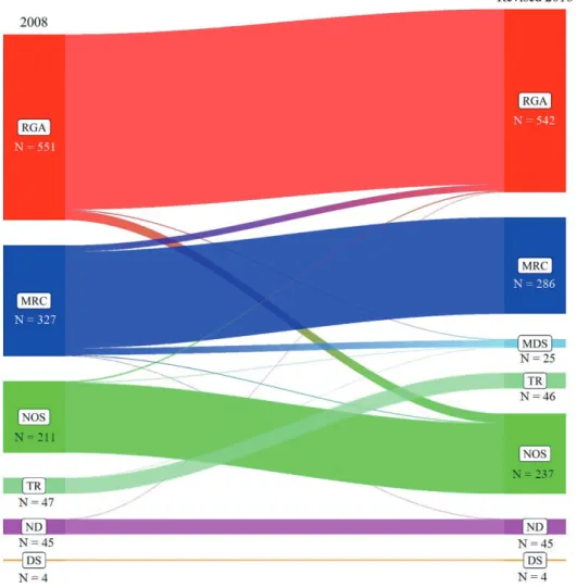
Based on the 2008 WHO criteria, the number of patients in each category was as follows: RGA 551 (46.5%), MRC 327 (27.6%), NOS 211 (17.8%), TR 47 (4.0%), DS 4 (0.3%), and ND 45 (3.8%).
Based on the revised criteria, the number of patients in each cate- gory was as follows: RGA 542 (45.7%), MRC 286 (24.1%), NOS 237 (20.0%), TR 46 (3.9%), DS 4 (0.3%), MDS 25 (2.1%), and ND 45 (3.8%). The ND category was obtained using the incomplete mu- tation (NPM1 and/or CEBPA) data. Application of the revised cri- teria redistributed a subset of each category into another category.
Overall, this redistribution resulted in a slight increase in the NOS category and a slight decrease in the RGA and MRC categories un- der the revised criteria. Generation of the MDS category repre- sented a relocation of erythroleukemia cases with blasts
<20% of the total marrow cells (Fig. 1). The overall concordance rate of cat- egorization between the two criteria was 93.4% (1107/1185) and 78 discrepant cases (6.6%) were identified.
Fig. 1. Comparison of categorization based on the previous and revised WHO criteria. Categories based on the previous 2008 WHO criteria are
shown on the left, and categories based on the revised 2016 WHO criteria are shown on the right. The gradient colors represent the diagnostic categories and the widths of the bands are proportional to the counts of cases in each category based on the previous and revised criteria.Abbreviations: RGA, AML with recurrent genetic abnormalities; MRC, AML with myelodysplasia-related changes; NOS, AML not otherwise speci- fied; TR, therapy-related AML; ND, AML with subcategory not determined; DS, AML associated with Down syndrome.
2. Features and categorization patterns of discrepant cases
Table 1 summarizes the recategorization patterns of discrepant cases and their details. Among the discrepancies, RGA to NOS
Table 1. Summary of discrepant cases between the previous and re-
vised WHO criteria and the basis for recategorization2008 WHO Revised 2016 WHO Rationale for recategorization RGA (N
=31) NOS (N
=28)
CEBPAmonoMDS (N
=3) Erythroleukemia and mutated NPM1 MRC (N
=41) MDS (N
=20) Erythroleukemia
RGA (N
=18) Mutated NPM1 (N
=11)/CEBPA
bi(N
=4)/
BCR-ABL1 (N=
3) NOS (N
=2) del(9q)
ND (N
=1) del(9q) and CEBPA
NANOS (N
=4) RGA (N
=3)
BCR-ABL1MDS (N
=1) Erythroleukemia TR (N
=1) MDS (N
=1) Erythroleukemia ND (N
=1) RGA (N
=1)
BCR-ABL1Abbreviations: RGA, AML with recurrent genetic abnormalities; MRC, AML with myelodysplasia-related changes; TR, therapy-related AML; NOS, AML not other- wise specified; ND, AML with subcategory not determined; NA, not available.
(N
=28, 35.9%) was the most common recategorization and in- cluded cases with a single mutation of CEBPA. The recategoriza- tion of MRC to MDS (N
=20, 25.6%) was the second most com- mon and encompassed cases of erythroleukemia. The MRC to RGA (N
=18, 23.1%) was the third most common recategorization and included AML with mutated NPM1 (N
=11), AML with mu- tated CEBPA
bi(N
=4), and AML with BCR-ABL1 (N
=3). Other re- categorizations were RGA to MDS and NOS to RGA (N
=3, 3.8%
each), and these included erythroleukemia with mutated NPM1, and AML with BCR-ABL1, respectively. Miscellaneous recategori- zations were MRC to NOS (N
=2, 2.6%), MRC to ND, NOS to MDS, TR to MDS, and ND to RGA (N
=1, 1.3% each).
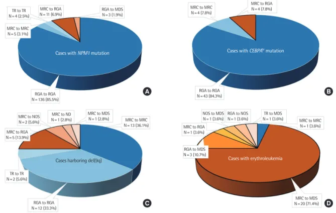
3. Characterization of specific subtypes
We further analyzed the categorization patterns in patients with specific subtypes that underwent critical changes in the revised 2016 criteria. Patients with NPM1 mutation (N
=159, 13.4% of all patients) revealed 145 (91.2%) agreements and 14 (8.8%) discrep-
Fig. 2. Pattern and frequency of categorization in AML patients with (A) NPM1 mutation, (B) CEBPA
bi mutation, (C) del(9q), and (D) erythroleuke- mia. The black-brimmed pieces indicate a discrepancy and non-brimmed pieces indicate an agreement. There may be overlapping cases in each patient group.Abbreviations: RGA, AML with recurrent genetic abnormalities; MRC, AML with myelodysplasia-related changes; NOS, AML not otherwise speci- fied; TR, therapy-related AML; ND, AML with subcategory not determined; CEBPAbi, biallelic CEBPA.
A B
C D
TR to TR N=4 (2.5%)
MRC to RGA N=11 (6.9%)
MRC to MRC N=4 (7.8%)
MRC to RGA N=4 (7.8%)
RGA to RGA N=43 (84.3%)
NOS to MDS N=1 (3.6%)
RGA to NOS N=1 (3.6%)
TR to MDS
N=1 (3.6%) MRC to MRC N=1 (3.6%)
MRC to MDS N=20 (71.4%) MRC to RGA
N=1 (3.6%)
RGA to MDS N=3 (10.7%) RGA to MDS
N=3 (1.9%)
MRC to MRC N=5 (3.1%)
RGA to RGA N=136 (85.5%)
MRC to NOS N=2 (5.6%) MRC to RGA N=5 (13.9%)
TR to TR N=2 (5.6%)
RGA to RGA N=12 (33.3%)
MRC to ND N=1 (2.8%)
MRC to MDS
N=1 (2.8%) MRC to MRC N=13 (36.1%)
Cases with NPM1 mutation Cases with CEBPAbi mutation
Cases with erythroleukemia Cases harboring del(9q)
ancies. The majority (85.5%) of cases were categorized as RGA. A subset of patients (N
=16, 10.1%) was categorized as MRC using the previous criteria and was split into MRC (N
=5, 3.1%) and RGA (N
=11, 6.9%) using the revised criteria. Another subset of patients was subjected to TR categorization (N
=4, 2.5%) and RGA to MDS recategorization (N
=3, 1.9%), which included erythroleukemia with mutated NPM1 (Table 1, Fig. 2A). Patients with CEBPA
bimu- tation (N
=51, 4.3% of all patients) revealed 47 (92.2%) agreements and 4 (7.8%) discrepancies. Similar to patients with NPM1 muta- tion, the majority (84.3%) of cases were categorized as RGA. A subset of patients (N
=8, 15.7%) was categorized as MRC using the previous criteria and was split into MRC (N
=4, 7.8%) and RGA (N
=4, 7.8%) using the revised criteria (Table 1, Fig. 2B). In total, 15 cases with NPM1 or CEBPA
bimutations were identified within the MRC to RGA recategorization. Among these, 11 cases with mutated NPM1 were previously categorized as MRC owing to a prior history of MDS or MDS/MPN (N
=6), MLD (N
=4), and del(9q) (N
=1). The remaining four cases with CEBPA
bimutation were previously categorized as MRC owing to the presence of del(9q) in all cases (Table 2). Patients with a prior history of MDS or MDS/
MPN showed shorter OS compared to those with MLD or del(9q) (P
=0.026; Fig. 3).
Patients harboring del(9q) (N
=36, 3.0% of all patients) revealed 27 (75.0%) agreements and 9 (25.0%) discrepancies. In total, 22
patients (61.1%) were categorized as MRC using the previous crite- ria and were split into MRC (N
=13, 36.1%), RGA (N
=5, 13.9%), NOS (N
=2, 5.6%), ND (N
=1, 2.8%), and MDS (N
=1, 2.8%) using the revised criteria. Among these, five cases in the MRC to RGA recategorization included those with CEBPA
bi(N
=4) or NPM1
Table 2. Characteristics and clinical outcomes of patients with mutated NPM1 or CEBPA
bi included in the MRC to RGA recategorizationCase No. Sex/Age FAB Cytogenetic abnormality FLT3-ITD
(allelic ratio)/
NPM1/CEBPAbi
Prior
disease MLD FU time (m)
Clinical outcome
1 M/76 M2 80-84, XY,inc [7]/46, XY [20] -/+/- - + 1.6 Dead
2 F/68 M2 46, XX [20] +(1.04)/+/- - + 6.3 Dead
3 F/44 M1 46, XX [20] -/+/- - + 43.9 Dead
4 M/71 M2 46, XY [30] -/+/- - + 3.3 FU loss
5 F/40 M1 46, XX, del (9) (q21q34) [4]/46, XX [16] +(NA)/+/- - - 10.8 Dead
6 M/45 M2 46, XY [25] -/+/- MDS + 6.8 Dead
7 F/75 M2 47, XX, +8 [1]/46, XX [7] +(11.31)/+/- MDS - 3.0 Dead
8 M/49 M1 46, XY [27]//46, XX [3] +(0.52)/+/- MDS - 3.5 Dead
9 F/53 M5 46, XX [20] -/+/- CMML - 5.7 Dead
10 F/60 M1 46, XX [30] -/+/- MDS - 39.6 Dead
11 F/64 M1 46, XX, inv (9) (p12q13) c [20] +(0.30)/+/- MDS - 0.5 FU loss
12 M/56 M1 46, XY, del (9) (p13) [6]/46, XY, del (9) (q21) [6]/46, XY [8] -/-/+ - - 51.5 Alive
13 F/18 M1 46, XX, del (9) (q13q32) [16]/46, XX [4] +(0.03)/-/+ - - 50.4 Alive
14 M/39 M1 45, X, -Y, del (9) (q22q34) [2]/46, XY [18] -/-/+ - - 43.6 Alive
15 F/63 M1 46, XX, del (9) (q13q22) [9]/46, XX, del (9) (q12) [2]/46, XX [9] -/-/+ - - 30.6 Alive
Abbreviations: CEBPA
bi, biallelic CEBPA; MRC, AML with myelodysplasia-related changes; RGA, AML with recurrent genetic abnormalities; M, male; F, female; FAB, French- American-British classification; ITD, internal tandem duplication; NA, not available; CMML, chronic myelomonocytic leukemia; MLD, multilineage dysplasia; FU, follow-up; m, month.
Fig. 3. Influence of MDS history on the outcomes of patients with
AML harboring gene mutations identified in the MRC to RGA recate- gorization. Kaplan-Meier plots for overall survival according to the defining criteria for the MRC category using the 2008 WHO criteria, including multilineage dysplasia or del(9q) (N=9) and a prior history of MDS or MDS/MPN (N=6).Abbreviations: MRC, AML with myelodysplasia-related changes; RGA, AML with recurrent genetic abnormalities; MDS/MPN, myelodysplas- tic/myeloproliferative neoplasm.
100
80
60
40
20
0
Overall survival (%)
Prior history of MDS or MDS/MPN
P=0.026
Month
Multilineage dysplasia or del(9q)
0 10 20 30 40 50 60
mutation (N
=1). The 12 cases in the RGA categorization included those with nine t(8;21), two t(15;17), and one inv(16). Thus, a total of 17 cases (47.2%) with recurrent (cyto)genetic abnormalities were included in the del(9q) subtype (Table 1, Fig. 2C). The OS of patients with del(9q) alone tended to be shorter compared to that of patients with concurrent t(8;21) or CEBPA
bi(P
=0.158; Supple- mental Data Fig. S1). Patients with erythroleukemia (N
=28, 2.4%
of all patients) revealed only one (3.6%) agreement and 27 (96.4%) discrepancies. A total of 22 patients (78.6%) were catego- rized under MRC using the previous criteria and were split into MDS (N
=20, 71.4%), RGA (N
=1, 3.6%), and MRC (N
=1, 3.6%) us- ing the revised criteria. Overall, 25 cases (89.3%) of erythroleuke- mia were reclassified as MDS, whereas the remaining three cases (10.7%) remained classified as the AML entity (one case each in the MRC, RGA, and NOS categories). The RGA to MDS recategori- zation (N
=3, 10.7%) represented erythroleukemia cases harbor- ing the NPM1 mutation (Table 1, Fig. 2D). Finally, patients with AML harboring BCR-ABL1 (N
=7, 0.6% of all patients) were char- acterized by male predominance (85.7%), high incidence of ane- mia (71.4%) and thrombocytopenia (71.4%), and isoform p210 dominance (66.7%). Three cases (42.9%) showed a complex karyotype and were categorized as MRC according to the previ- ous criteria. Recurrent mutations were not detected in any of the patients tested. Four (57.1%) patients have survived for more than 3 years after allogeneic hematopoietic stem cell transplantation (Table 3).
DISCUSSION
According to the revised 2016 criteria, the 2008 criteria-defined patients with AML were most commonly categorized as RGA (45.7%), followed by MRC (24.1%), NOS (20.0%), TR (3.9%), ND (3.8%), MDS (2.1%), and DS (0.3%). The discrepancy rate between the two criteria was 6.6%. The RGA to NOS (35.9% of discrep- ancy) recategorization was the most common and represented cases with CEBPA
monomutation. The MRC to MDS (25.6%) recate- gorization represented erythroleukemia cases and the MRC to RGA (23.1%) recategorization represented recurrent genetic ab- normalities showing myelodysplasia features. Patients with NPM1 or CEBPA
bimutations within the MRC to RGA recategorization were characterized by a high agreement rate, categorization un- der MRC by the previous criteria in some cases, and identification of cases with a prior history of MDS or MDS/MPN. We demon- strated that patients with secondary AML and gene mutations had a worse prognosis than those presenting MLD or del(9q). Patients harboring del(9q) were characterized by three quarters of the agreement rate and enrichment in core binding factor AML or cases with CEBPA
bimutations. The vast majority of erythroleuke- mia was recategorized as MDS, with only 10.7% of cases remain- ing as an AML entity.
The frequency of discrepancy (approximately 7%) was not high between the two classification systems because the revision largely inherited the concept of the previous criteria and incorpo- Table 3. Characteristics and clinical outcomes of cases of AML harboring BCR-ABL1 according to the revised WHO criteria
Case No. Sex/Age FAB WBC (×10
9/L) Hb
(g/dL) PLT
(×10
9/L) Cytogenetic abnormality Isoform
FLT3-ITD/NPM1/CEBPAbi
FU time
(m) Clinical outcome 1* M/41 M6 3.6 9.4 30 42,XY,-2,-5,-7,der(17)t(4;17)(q12;p13),der(18)t(7;18)
(q11.2;q21.1),dic(19;21)(p13.1;p11.2)[11]/42,idem,t(9;22) (q34;q11.2),-15,+mar[5]/43,XY,-2,-5,-7,i(8)(q10),-17, der(17)t(4;17)(q12;p13),+18,der(18)add(18)(p11.2) t(7;18)(q11.2;q21.1)[4]
NA -/-/NA 7.7 Dead
§2* F/42 M0 23.8 6.1 162 46,XX,?add(5)(q35),t(9;22)(q34;q11.2),t(21;19;?17)
(q22;q13.1;?q25)[20] e1a2 -/-/NA 89.4 Alive
§3* M/28 M1 77.5 10.4 22 49,XY,+X,der(9)t(9;22)(q34;q11.2),+11,+12,ider(22)(q10) t(9;22)[13]/50,XY,+X,t(9;22)(q34;q11.2)+11,+12,+der(22) t(9;22)[6]/46,XY[1]
b3a2 -/NA/NA 62.5 Alive
§4
†M/38 M7 5.2 7.5 86 46,XY,inv(9)(p12q13)c,t(9;22)(q34;q11.2)[20] b3a2 -/-/- 62.3 Alive
§5
†M/54 M4 268.1 9.1 84 46,XY,t(9;22)(q34;q11.2)[20] b3a2 -/-/- 33.4 Alive
§6
†M/70 M2 71.4 12.7 100 46,XY,t(9;22)(q34;q11.2)[20] b3a2 -/-/- 2.7 FU loss
7
‡M/71 M0 2.0 9.1 158 46,XY,inv(9)(p12q13)c[20] e1a2 -/NA/NA 14.3 FU loss
Categorized as MRC*, RGA
†, and ND
‡, respectively, based on the 2008 WHO criteria;
§Cases that underwent allogeneic hematopoietic stem cell transplantation.
Abbreviations: M, male; F, female; FAB, French-American-British; WBC, white blood cell; PLT, platelet; NA, not available; ITD, internal tandem duplication; CEBPA
bi, biallelic
CEBPA; FU, follow-up; m, month; MRC, AML with myelodysplasia-related changes; RGA, AML with recurrent genetic abnormalities; ND, AML with subcategory not deter-mined.
rated clinical features, morphology, immunophenotyping, cytoge- netics, and molecular genetics to define the disease entities of clinical significance. This frequency is largely in line with the dis- crepancy frequency (8.9%) of a recent study that performed cate- gorization using two classification systems in 610 patients [8]. The RGA to NOS and MRC to MDS recategorizations were anticipated consequences. However, further discussion is required regarding the MRC to RGA recategorization. This is partly due to the result of classifying the newly-introduced subtype, AML with BCR- ABL1 in the revised criteria. This was added to recognize this dis- ease as distinct from blast-phase chronic myeloid leukemia that benefits from tyrosine kinase inhibitors and hematopoietic stem cell transplantation [15, 16]. However, categorization of cases with gene mutations (NPM1 or CEBPA
bi) using the revised criteria is not straightforward. MLD and del(9q) have no adverse prognostic significance in these patients and their presence does not exclude a case from AML with gene mutations, whereas cases co-occur- ring with myelodysplasia-associated cytogenetic abnormalities should be diagnosed under the MRC category. In the present study, a subset of patients with gene mutations was classified in the MRC category using the previous criteria. However, rather than remaining in the MRC category, a larger number of these pa- tients were recategorized into other categories by the revised cri- teria. This is an example of an effort to minimize diagnostic ambi- guity based on clinical significance. Despite refinement, the MRC to RGA recategorization can be complicated by a complexity of priority rules in patients with gene mutations. In particular, there are no clear guidelines regarding the categorization of patients with recurrent gene mutations as well as a history of MDS or MDS/MPN. Schnittger et al. [17] observed that NPM1 mutation was identified in 13.1% of 283 patients with secondary AML.
These authors argued that this mutation might contribute to the transformation of MDS to AML; however, they found no survival benefit in their secondary AML cohort [17]. These data suggest that the NPM1 mutation could reflect differently in de novo and secondary AML. This is in line with our observations that, within the MRC to RGA recategorization, patients with a prior history of MDS or MDS/MPN showed worse outcomes than those with MLD or del(9q). In this context, a recent expert review suggested that the diagnosis of MRC is appropriate in AML patients with a his- tory of MDS and NPM1 mutation, particularly when supported with molecular genetic findings [18]. Nonetheless, large studies
are required to confirm the prognostic influence of NPM1 or CEBPA
bimutations in secondary AML cohorts.
Del(9q) is a recurrent cytogenetic abnormality in AML and ac- counts for approximately 2% of unselected cases [19]. It is en- riched in core binding factor AML, particularly in t(8;21), or pa- tients with CEBPA mutation [19, 20]. These findings are consistent with our observations that 3.0% of cases harbored del(9q) and nearly half of them had recurrent (cyto)genetic abnormalities, particularly t(8;21) and CEBPA
bimutations. A previous study de- lineated many genes in the commonly deleted region of del(9q) and the leukemogenic role of haploinsufficiency of TLE1 and TLE4 adjacent to this region by overcoming the negative survival and anti-proliferative effects of AML1-ETO fusion protein on my- eloid progenitors and allowing preleukemic stem cells to expand into AML [21, 22]. Other investigators confirmed the low expres- sion of TLE4 in AML with del(9q) and showed strong associations with DNMT3A and NPM1 mutations [23]. Naarmann-de Vries et al. [24] recently reported that the mRNA expression of the HNRNPK gene in the 9q21.32–9q21.33 region was enhanced to a normal karyotype level in a group of del(9q)/CEBPA-mutated pa- tients, indicating a molecular relationship of CEBPA and the del(9q) CDR genes. Thus, del(9q) might contribute to leukemo- genesis by the haploinsufficiency of tumor suppressor genes in CDR or a regulatory interaction between CEBPA and HNRNPK.
Thus, AML with del(9q) could be a candidate for an anti-leuke- mogenic strategy by blocking the effect of HNRNPK or other CDR gene products on CEBPA expression, which could improve the outcomes of del(9q) AML.
A vast majority of erythroleukemia cases were recategorized as
MDS in the present study. This finding is consistent with a recent
study in which 30 (88.2%) of 34 de novo erythroleukemia cases
were reclassified as MDS [7]. The present study found that ap-
proximately 10% of erythroleukemia cases had an NPM1 muta-
tion and these cases were all recategorized as MDS using the re-
vised criteria. Montalban-Bravo et al. [25] recently demonstrated
that the frequency of NPM1 mutations among 1,900 patients with
newly diagnosed MDS or MDS/MPN was 1.6%. Most cases (61%)
were classified as MDS with excess blasts, and patients treated
with chemotherapy had better clinical outcomes than those
treated with hypomethylating agents [25]. Thus, NPM1 mutations
are rarely detected in MDS and these patients might benefit from
chemotherapy compared to MDS-based treatment approaches.
As RUNX1 mutation had not been systematically tested before the use of NGS in our laboratory, we only considered mutations in NPM1 and CEBPA
bias the defining drivers of AML in the pres- ent study. Exclusion of RUNX1 mutation data from the analysis re- sulted in a slight increase in the NOS category during recategori- zation. We have previously demonstrated that RUNX1 mutations were detected in approximately 15% of patients in the NOS cate- gory [26]. Thus, integration of RUNX1 mutation data is expected to shift some cases from the NOS category to the RGA category.
Our actual data showed that RUNX1 mutation was detected in 11 (11.2%) of 98 patients with NGS results. Among these, 2 (2.0%) were diagnosed with AML with mutated RUNX1 according to the revised criteria (Supplemental Data Fig. S2). Comprehensive and high-throughput mutational analysis using NGS has replaced the Sanger sequencing-based stepwise approach of the mutational as- say in patients with AML. Future studies should thus focus on mu- tation profiling and genotype-phenotype correlation, and their clinical relevance in myeloid neoplasms and acute leukemias in the context of categorization using the revised WHO criteria.
In conclusion, the present study highlights that applying the re- vised 2016 WHO criteria identified discrepancies in categorization using the previous criteria in approximately 7% of patients with AML. The study demonstrated that these discrepancies were due to the introduction of new entities, changed definitions, and re- fined subcategorization. Further refinement could be considered for the next version of the WHO criteria, including the prece- dence of a history of MDS over NPM1 mutation as a criterion for classifying the MRC category as well as potential new subtypes such as AML with del(9q), or MDS and MDS-related disorders with mutated NPM1.
요 약
배경: 2016년 개정된 WHO 기준과 2008년 WHO 기준에 따라 AML을 분류하면 일부 불일치하는 경우가 부각된다. 본 연구에서 는 불일치 사례의 빈도, 분류 패턴, 특성을 분석하고, 개정된 기준 에 의해 주요 변화를 보인 AML의 아형들을 특성화하였다.
방법: 기존 및 개정된 WHO 기준에 따라 환자군을 다음 7가지 범 주로 나누었다: 반복유전자이상 AML (AML with recurrent genetic abnormalities, RGA), 골수형성이상관련 AML (AML with myelo- dysplasia-related changes, MRC), 치료관련 AML, 상세불명 AML (AML not otherwise specified, NOS), 다운증후군관련 AML, 아형
미결정 AML, 그리고 MDS.
결과: 총 1,185예의 AML 증례를 검토하였다. 두 기준 사이의 일치 율은 93.4%이었다. 78예의 불일치 사례 중 가장 흔한 패턴 3개는 RGA에서 NOS, MRC에서 MDS, 그리고 MRC에서 RGA이었으며, 각 각 CEBPA 단일 돌연변이, 적백혈병(erythroleukemia), 그리고 골 수형성이상을 보이는 반복유전자이상으로 인한 불일치였다. MDS 위주 치료보다 항암화학요법으로 임상적 이득을 취할 수 있는 NPM1 돌연변이 동반 적백혈병이 3예에서 확인되었다. 그리고 결 실된 유전자의 홑배수부전(haploinsufficiency) 혹은 유전자 사이 의 상호작용을 통해 백혈병을 유발할 수 있는 9번 염색체 장완의 결실이 환자의 3%에서 확인되었다.
결론: 본 연구를 통해 AML 환자의 약 7%에서 새로운 질환명의 도 입, 변경된 정의, 그리고 정밀한 하위범주화로 인해 다른 범주로 재 분류됨을 확인하였다. 이에 따라 다음 개정 시에는 추가적인 세밀 한 개선이 고려되어야 할 것이다.
Conflicts of Interest
None declared.
REFERENCES
1. Swerdlow SH, Campo E, et al. eds. WHO classification of tumours of haematopoietic and lymphoid tissues. Revised 4th ed. Lyon, France:
International Agency for Research on Cancer, 2017:129-71.
2. Leonard JP, Martin P, Roboz GJ. Practical implications of the 2016 re- vision of the World Health Organization classification of lymphoid and myeloid neoplasms and acute leukemia. J Clin Oncol 2017;35:2708-15.
3. Arber DA, Orazi A, Hasserjian R, Thiele J, Borowitz MJ, Le Beau MM, et al. The 2016 revision to the World Health Organization classification of myeloid neoplasms and acute leukemia. Blood 2016;127:2391-405.
4. Chen Y, Pourabdollah M, Atenafu EG, Tierens A, Schimmer A, Chang H. Re-evaluation of acute erythroid leukemia according to the 2016 WHO classification. Leuk Res 2017;61:39-43.
5. Toth LN, de Abreu FB, Peterson JD, Loo EY. Impact of molecular se- quencing information as related to 2008 and 2016 World Health Orga- nization classification of acute myeloid leukemia and myelodysplasia.
Arch Pathol Lab Med 2018;142:1017.
6. Margolskee E, Mikita G, Rea B, Bagg A, Zuo Z, Sun Y, et al. A reevalu- ation of erythroid predominance in acute myeloid leukemia using the updated WHO 2016 criteria. Mod Pathol 2018;31:873-80.
7. Ryu S, Park HS, Kim SM, Im K, Kim JA, Hwang SM, et al. Shifting of
erythroleukemia to myelodysplastic syndrome according to the re- vised WHO classification: biologic and cytogenetic features of shifted erythroleukemia. Leuk Res 2018;70:13-9.
8. Jung J, Cho BS, Kim HJ, Han E, Jang W, Han K, et al. Reclassification of acute myeloid leukemia according to the 2016 WHO classification.
Ann Lab Med 2019;39:311-6.
9. Kansal R. Classification of acute myeloid leukemia by the revised fourth edition World Health Organization criteria: a retrospective sin- gle-institution study with appraisal of the new entities of acute my- eloid leukemia with gene mutations in NPM1 and biallelic CEBPA.
Hum Pathol 2019;90:80-96.
10. Huang Q, Chen W, Gaal KK, Slovak ML, Stein A, Weiss LM. A rapid, one step assay for simultaneous detection of FLT3/ITD and NPM1 mutations in AML with normal cytogenetics. Br J Haematol 2008;142:
489-92.
11. Falini B, Mecucci C, Tiacci E, Alcalay M, Rosati R, Pasqualucci L, et al.
Cytoplasmic nucleophosmin in acute myelogenous leukemia with a normal karyotype. N Engl J Med 2005;352:254-66.
12. Wouters BJ, Löwenberg B, Erpelinck-Verschueren CA, van Putten WL, Valk PJ, Delwel R. Double CEBPA mutations, but not single CEBPA mutations, define a subgroup of acute myeloid leukemia with a dis- tinctive gene expression profile that is uniquely associated with a fa- vorable outcome. Blood 2009;113:3088-91.
13. Döhner H, Estey E, Grimwade D, Amadori S, Appelbaum FR, Büchner T, et al. Diagnosis and management of AML in adults: 2017 ELN rec- ommendations from an international expert panel. Blood 2017;129:
424-47.
14. McGowan-Jordan J, Simons A, eds. An international system for human cytogenomic nomenclature (2016). Unionville, CT: S. Karger Publish- ers, Inc., 2016.
15. Bhatt VR, Akhtari M, Bociek RG, Sanmann JN, Yuan J, Dave BJ, et al.
Allogeneic stem cell transplantation for Philadelphia chromosome- positive acute myeloid leukemia. J Natl Compr Canc Netw 2014;
12:963-8.
16. Konoplev S, Yin CC, Kornblau SM, Kantarjian HM, Konopleva M, An- dreeff M, et al. Molecular characterization of de novo Philadelphia chromosome-positive acute myeloid leukemia. Leuk Lymphoma 2013;
54:138-44.
17. Schnittger S, Bacher U, Haferlach C, Alpermann T, Dicker F, Sunder- mann J, et al. Characterization of NPM1-mutated AML with a history of myelodysplastic syndromes or myeloproliferative neoplasms. Leuke- mia 2011;25:615-21.
18. Lin P and Falini B. Acute myeloid leukemia with recurrent genetic ab- normalities other than translocations. Am J Clin Pathol 2015;144:19-28.
19. Peniket A, Wainscoat J, Side L, Daly S, Kusec R, Buck G, et al. Del (9q) AML: clinical and cytological characteristics and prognostic implica- tions. Br J Haematol 2005;129:210-20.
20. Fröhling S, Schlenk RF, Krauter J, Thiede C, Ehninger G, Haase D, et al. Acute myeloid leukemia with deletion 9q within a noncomplex karyotype is associated with CEBPA loss-of-function mutations. Genes Chromosomes Cancer 2005;42:427-32.
21. Sweetser DA, Peniket AJ, Haaland C, Blomberg AA, Zhang Y, Zaidi ST, et al. Delineation of the minimal commonly deleted segment and identification of candidate tumor-suppressor genes in del(9q) acute myeloid leukemia. Genes Chromosomes Cancer 2005;44:279-91.
22. Dayyani F, Wang J, Yeh JR, Ahn EY, Tobey E, Zhang DE, et al. Loss of TLE1 and TLE4 from the del(9q) commonly deleted region in AML co- operates with AML1-ETO to affect myeloid cell proliferation and sur- vival. Blood 2008;111:4338-47.
23. Herold T, Metzeler KH, Vosberg S, Hartmann L, Jurinovic V, Opatz S, et al. Acute myeloid leukemia with del(9q) is characterized by fre- quent mutations of NPM1, DNMT3A, WT1 and low expression of
TLE4. Genes Chromosomes Cancer 2017;56:75-86.
24. Naarmann-de Vries IS, Sackmann Y, Klein F, Ostareck-Lederer A, Os- tareck DH, Jost E, et al. Characterization of acute myeloid leukemia with del(9q) - impact of the genes in the minimally deleted region.
Leuk Res 2019;76:15-23.
25. Montalban-Bravo G, Kanagal-Shamanna R, Sasaki K, Patel K, Ganan- Gomez I, Jabbour E, et al. NPM1 mutations define a specific subgroup of MDS and MDS/MPN patients with favorable outcomes with inten- sive chemotherapy. Blood Adv 2019;3:922-33.
26. You E, Cho YU, Jang S, Seo EJ, Lee JH, Lee JH, et al. Frequency and clinicopathologic features of RUNX1 mutations in patients with acute myeloid leukemia not otherwise specified. Am J Clin Pathol 2017;
148:64-72.
Supplemental Data Fig. S1. Influence of other (cyto)genetic abnor-
malities on overall survival in cases of AML harboring del (9q). Kaplan- Meier plots for the overall survival of patients with del (9q) alone (N=5), in combination with biallelic CEBPA mutation (N=5) or denovo t(8;21) (N=9).
100 90 80 70 60 50 40 30 20 10
Overall survival (%)
del(9q) with t(8;21) del(9q) with bi-CEBPA
sole del(9q)
P=0.158
Month
0 10 20 30 40 50 60 70
Supplemental Data Fig. S2. Pattern and frequency of categorization
for patients with AML harboring a RUNX1 mutation. The NOS to NOS represented patients with AML harboring mutated RUNX1 when con- sidering the application of RUNX1 mutation in recategorization ac- cording to the revised criteria. The RGA case was based on the pres- ence of t(3;3) (q21;q26).Abbreviations: MRC, AML with myelodysplasia-related changes; NOS, AML not otherwise specified; RGA, AML with recurrent genetic ab- normalities.