Original Article
J
Intra- and Inter-rater Reliabilities of Infrasternal Angle Measurement
Moon-Hwan Kim1, Jong-Hyuck Weon2
1Department of Physical Therapy, The Graduate School, Yonsei University, Wonju; 2Department of Physical Therapy, College of Tourism & Health Science, Joongbu University, Geumsan-gun, Chungnam, Korea
Purpose: The purpose of this study was to propose a new reference point for measurement of the infrasternal angle and to investigate the intra- and inter-rater reliabilities of infrasternal angle measurement using photographs.
Methods: Twenty-four healthy male college students participated as subjects in this study. Photographs were taken of subjects in two postures, one standing with the shoulders relaxed and one standing with the shoulders at 150° abduction. All photographs were ana- lyzed using Image J software. Raters used the photographs to measure the infrasternal angle between the xiphoid process, the medial margin of rib and navel on the right and left sides. The reliability of the infrasternal angle measurement was assessed by means of intra- class correlation coefficients [ICC (3,1)]. The level of statistical significance was set at p< 0.05.
Results: The intra- and inter-rater reliabilities of the infrasternal angle measurement for the right side at rest were excellent (ICC= 0.866 and 0.813, respectively), as were those for the left side at rest (ICC= 0.919 and 0.846, respectively). At 150° shoulder abduction, the in- tra- and inter-rater reliabilities for measurement of the infrasternal angle on the right side were excellent (ICC= 0.972 and 0.778, re- spectively), as were those for the left side (ICC= 0.914 and 0.826, respectively).
Conclusion: These findings suggest that this technique can be successfully used to measure the infrasternal angle, thus suggesting a new reference point for determining the length of the internal oblique and external oblique muscles in clinical situations.
Keywords: Thoracic rotation syndrome, Infrasternal angle, Photograph
INTRODUCTION
The thorax is composed of stable structures including the thoracic vertebrae, ribs, and sternum.1 These stable structures aid in main- taining breathing, protect internal organs such as the heart and lungs, and support attachments of trunk muscles.2-4 The thoracic vertebrae are able to move in several directions; especially notable is their degree of rotation (40-55°) compared with the lumbar verte- brae.2,5 Rotation of the thoracic vertebrae occurs when the rib cage is rotated, and repetitive and sustained rotation of the thoracic ver- tebrae or scoliosis associated with the thoracic vertebrae can have an effect on the movement or alignment of the rib cage.6-9
Thoracic rotation syndrome is typified by pain related to repeti-
tive and sustained rotational movement and rib cage asymmetry associated with thoracic rotation in the sagittal plane.6 Asymmetry of the rib cage affects recruitment of the abdominal muscles and in- fluences the length of the internal oblique and external oblique muscles bilaterally.10-12 In a previous study, asymmetrical length of the internal and external oblique muscles between the right and left sides resulted in limitations to trunk rotation.13 Asymmetry of oblique muscle may be occurred asymmetric activation of oblique muscle, these asymmetric activation can led to pain, causing micro- trauma-related tissue inflammation around the facet joints.14 There- fore, assessment of rib cage differences between the right and left sides is important to identify subjects with thoracic rotation syn- drome.6
Vol. 27, No. 3, June 2015 pISSN 1229-0475 eISSN 2287-156X
JKPT
Received May 28, 2015 Revised Jun 11, 2015 Accepted Jun 18, 2015
Corresponding author Jong-Hyuck Weon E-mail [email protected]
Copylight © 2015 The Korea Society of Physical Therapy
This is an Open Access article distribute under the terms of the Creative Commons Attribution Non-commercial License (Http:// creativecommons.org/license/by-nc/3.0.) which permits unrestricted non-commercial use, distribution,and reproduction in any medium, provided the original work is properly cited.
J Kor Phys Ther 2015:27(3):154-158
http://dx.doi.org/10.18857/jkpt.2015.27.3.154
angle between the xiphoid process and the medial margin of the rib on each side.6 A previous study reported that the ideal infrasternal angle was 90°, as that value reflected a balance between the internal oblique and external oblique muscles.6 If the internal oblique muscle is contracted or shortened bilaterally, the infrasternal angle becomes wider than this ideal, whereas if the external oblique muscle is con- tracted or shortened bilaterally, the infrasternal angle is narrower.6,15 Furthermore, in individuals with perpetual rotation toward one side, the infrasternal angles of the right and left side differ due to length changes in the internal and external oblique muscles.6
Thus, measurement of the infrasternal angle is a useful method for identifying the length of the internal and external oblique mus- cles. However, the inter-rater reliability of infrasternal angle was re- ported to be low, with testers apparently obtaining differing values due to the curved shape of the medial margin of the rib.6 The low inter-rater reliability of infrasternal angle measurement has ren- dered it inadequate as a ‘gold standard’ for identifying thoracic rota- tion syndrome and determining the length of the internal and ex- ternal oblique muscles. Therefore, the purpose of this study was to suggest a reference point for the infrasternal angle and to investigate the intra- and inter-rater reliability of measurements using this ref- erence point.
METHODS
1. Subjects
In this study, 24 healthy subjects (all male) were recruited from among students at Y University, Wonju-si. Exclusion criteria were any neuromuscular disorder, cardiopulmonary disorder, congenital disorder of the trunk or lower extremities, scoliosis, fracture of the ribs or xiphoid process, subject with thoracic or lumbar rotation syndrome, and limitation of bilateral shoulder flexion. All subjects participated voluntarily in this study. Before the study, the investiga- tor explained the experimental procedures to the subjects. Charac- teristics of the subjects are presented in Table 1.
2. Experimental methods
The study was conducted by two testers and one assistant. Tester 1 was a physical therapist who has worked in a general hospital for 10
years, tester 2 was a sophomore student in the Department of Physi- cal Therapy, and the assistant was a graduate student in the Depart- ment of Physical Therapy. Prior to the study, the testers and assistant became acquainted with the technique for measuring the infraster- nal angle and with the experimental procedures.
The infrasternal angle was measured with the subject in two po- sitions. Subjects were measured first in a relaxed standing position and then in a standing position with 150° shoulder abduction. Sub- jects were instructed to breathe comfortably during measurement.
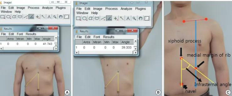
For the infrasternal angle measurement, first, we suggested the ref- erence point of medial margin (a perpendicular line was drawn from the mid clavicle to the medial margin of the rib cage). Markers were attached on the xiphoid process, navel, and the reference point of medial margin. An angle formed between two lines were mea- sured. The two lines formed an angle for measurement. (One line:
the line connecting the xiphoid process and the navel, the other line:
the line connecting the xiphoid process and medial margin of rib cage) (Figure 1).
After the markers were attached, the assistant took photographs from a position 1.5 m in front of the subject using a camera (450 D, Cannon, Inc., Japan). The tester measured each subject twice in each position; the order of subjects and positions were randomized.
The assistant then analyzed each photograph using Image J soft- ware (National Institutes of Health, Bethesda, MD, USA). To do this, the assistant drew a line on the image from the xiphoid process to the navel, and then drew lines from the xiphoid process to the markers indicating the medial margin of the 10th rib on both sides.
The right and left sides of the infrasternal angle were calculated as the angle formed by the line connecting the markers on the xiphoid process and the navel and the line connecting the markers on the xiphoid process and medial margin of the 10th rib. Intra-rater reli- ability was calculated based on the measurement values obtained by tester 1, and the inter-rater reliability was calculated by comparing the first trial values of tester 1 and tester 2.
Mean± SD Range
Age (year) 22.9± 1.8 20.0-22.0
Height (cm) 173.5± 1.4 152.0-183.0
Weight (kg) 70.6± 2.7 46.0-100.0
SD, standard deviation.
Moon-Hwan Kim and Jong-Hyuck Weon
JKPT The Journal of Korean Physical Therapy
3. Statistical analysis
The subject`s characteristics and the infrasternal angle of the right and left sides are presented as means and standard deviations (SD).
A repeated-measures analysis of variance (ANOVA) with a post hoc t-test was used to test for systematic bias between the first and sec- ond measurements. Intraclass correlation coefficients [ICC (3,1)]
were used to determine the inter- and intra-rater reliabilities of the infrasternal angle measurement on the right and left sides. The standard error of measurement (SEM) was calculated for each mea- surement to assess absolute consistency.16 Statistical analyses were performed using the Statistical Package for the Social Sciences ver- sion 21.0 for Windows (SPSS, Inc., Chicago, IL, USA). The signifi-
cance level was set at p< 0.05.
RESULTS
The measurement values of the infrasternal angle for testers 1 and 2 was shown the mean±SD (Table 2). The intra-rater reliabilities for measurement of this angle on the right and left sides were 0.866 and 0.919, respectively, in the resting position, and 0.972 and 0.914, re- spectively, with 150° shoulder flexion (Table 3).
Inter-rater reliabilities for measurement of the infrasternal angle on the right and left sides were 0.813 and 0.846, respectively, in the resting position, and 0.778 and 0.826, respectively, with 150° shoul- der flexion (Table 4).
DISCUSSION
Although measurement of the infrasternal angle has been a com- mon method for identifying thoracic rotation syndrome and assess-
A B C
Figure 1. Measurement of infrasternal angle using image J software program. (A) In resting position, (B) In shoulder abduction 150°, (C) Landmark of measurement.
Table 2. Mean and standard deviation of infrasternal angle (N= 24) In rest standing posture In standing posture
with 150° shoulder abduction Right side Left side Right side Left side Tester 1 35.57± 5.83 40.00± 5.55 34.76± 5.86 39.64± 5.26 Tester 2 38.07± 6.01 39.81± 5.88 34.85± 5.99 36.90± 5.96
Table 3. Intra-rater reliability of infrasternal angle (N= 24) In rest standing posture In standing posture
with 150° shoulder abduction Right side Left side Right side Left side
ICC (3,1) 0.866 0.919 0.972 0.914
(95% CI) (-4.62-2.32) (-5.24-1.41) (-4.16-3.58) (-3.72-2.72)
SEM 2.14 1.58 0.98 1.54
ICC, intraclass correlation coefficients; CI, confidence interval; SEM, standard er- ror of measurement.
Table 4. Inter-rater reliability of infrasternal angle (N= 24) In rest standing posture In standing posture
with 150° shoulder abduction Right side Left side Right side Left side
ICC (3,1) 0.813 0.846 0.778 0.826
(95% CI) (-6.10-1.10) (-3.29-3.37) (-3.70-3.52) (-0.67-6.16)
SEM 2.52 2.18 2.76 2.19
ICC, intraclass correlation coefficients; CI, confidence interval; SEM, standard er- ror of measurement.
for measuring the infrasternal angle and calculated intra- and inter- rater reliability by analyzing photographs.
The present study demonstrated excellent intra- and inter-rater reliability (0.866 and 0.778 or higher, respectively) for measurements with and without 150° shoulder flexion. Tester 1 was a physical therapist, and tester 2 was sophomore student. Although tester 2 lacked measurement experience, we nevertheless found excellent inter-rater reliability following training in the measurement meth- od. Thus, if physical therapists working in clinical settings are taught this method of measuring the infrasternal angle, they should be able to perform the assessment with comparative ease.
For tester 1, the mean infrasternal angles on the right and left sides were 35.57°± 5.83° and 40.00°± 5.55°, respectively, and in tester 2, they were 38.07°± 6.01° and 39.81°± 5.88°, respectively. The mean differences between tester 1 and tester 2 were 2.50° and 0.19° for the right and left sides, respectively, without shoulder abduction. With 150° shoulder abduction, the differences in mean values between testers 1 and 2 were 0.09° and 2.74° for the right and left sides, re- spectively (Table 2). The mean difference between measurements with and without shoulder abduction was less than 3°; this value was not significantly different between testers 1 and 2. If an infra- sternal angle of 90° is considered ideal,6 then the infrasternal angle of subjects in the present study was narrower than ideal, suggesting a dominant external oblique relative to the internal oblique muscle.
Also, measurements in this study indicated that 150° shoulder ab- duction decreased the right and left infrasternal angles slightly (by 0.81 and 0.36, respectively). Shirmann (2011) reported no difference in the infrasternal angle between a standing position with and one without shoulder abduction,6 similar to the results presented here.
Little research has been carried out on the infrasternal angle. Be- cause X-rays do not show the infrasternal angle, no study has yet evaluated the validity of infrasternal angle measurement. Because X-rays can’t use for measuring infrasternal angle, we take advantage of photography in this study. In previous study, intra- and interrater reliability are showed high correlation between radiographic and photographic measurement.17 Also, other researcher suggested that photographic measurement used when it was difficult to use the X- ray measurement.18 Therefore, this study evaluating the reliability of such measurement is important. Limitations of the present study
muscular or musculoskeletal problems. Further research, preferably using electromyography, will be helpful in determining differences in muscle activity between the internal and external oblique mus- cles in subjects with asymmetrical infrasternal angles.
REFERENCES
1. Koumbourlis AC. Chest Wall Abnormalities and their Clinical Signifi- cance in Childhood. Paediatr Respir Rev. 2014;15(3):246-54.
2. Neumann DA. KINESIOLOGY of the MUSCULOSKELETAL SYS- TEM. St. Louis, Mosby, 2002:369-70.
3. Magee DJ. Orthopedic Physical Assessment. 4th edition. Saunders. Phil- adelphia. 2002:425-44.
4. De Troyer A, Wilson TA. Mechanism of the increased rib cage expan- sion produced by the diaphragm with abdominal support. J Appl Physi- ol (1985). 2015;118(8):989-95.
5. Johnson KD, Kim KM, Yu BK, et al. Reliability of thoracic spine rotation range-of-motion measurements in healthy adults. J Athl Train. 2012;
47(1):52-60.
6. Shirmann S. MOVEMENT SYSTEM IMPAIRMENT SYNDROMES of the Extremities, Cervical and Thoracic Spines. St. Louis, Mosby, 2011:114-37.
7. Campbell RM Jr, Smith MD, Mayes TC, et al. The characteristics of tho- racic insufficiency syndrome associated with fused ribs and congenital scoliosis. J Bone Joint Surg Am. 2003;85(3):399-408.
8. Kang JI, Jung DG, Park SK, et al. Forced expiratory volume in one sec- ond, Chest resistance exercise, Proprioceptive neuromuscular facilita- tion. J Kor Phys Ther. 2011;23(2):37-43.
9. Linek P, Saulicz E, Wolny T, et al. Ultrasound evaluation of the symmetry of abdominal muscles in mild adolescent idiopathic scoliosis. J Phys Ther Sci. 2015;27(2):465-68.
10. Kim BK, Lee MH, Kim GC. Comparison of Abdominal Muscle Activity during Exercises Using a Sling and Swiss-ball. J Kor Phys Ther. 2012;
25(3):149-54.
11. Lim JH. Correlations of Symmetry of the Trunk Muscle Thickness by Gender with the Spinal Alignment in Healthy Adults. J Kor Phys Ther.
2013;25(6):405-10.
12. Kang HK, Chang SK. The Effect of Robo-horseback Riding Exercise on Trunk Muscle Activity Ratios in Patients with Low Back Pain. J Kor Phys Ther. 2012;24(6):393-7.
13. Richardson C, Hodges PW, Hides J. Therapeutic Exercise for Lumbopel- vic Stabilization: A Motor Control Approach for the Treatment and Pre- vention of Low Back Pain. 2nd ed. London. Churchill Livingstone. 2004.
14. Vera-Garcia FJ, Brown SHM, Gray JR, et al. Effects of different levels of torso coactivation on trunk muscular and kinematic responses to poste- riorly applied sudden loads. Clinical Biomechanics 2006;21(5):443-55.
15. Kendall FP, McCreary EK, Provance PG, et al. Muscles: Testing and function with posture and pain. 5th ed. Baltimore, MD, Williams &
Wilkins,2005:200-1.
Moon-Hwan Kim and Jong-Hyuck Weon
JKPT The Journal of Korean Physical Therapy
16. Shrout PE, Fleiss JL. Intraclass correlations: Uses in assessing rater reli- ability. Psychol Bull. 1979;86(2):420-8.
17. Nix S, Russell T, Vicenzino B, et al. Validity and reliability of hallux valgus angle measured on digital photographs. J Orthop Sports Phys Ther.
2012;42(7):642-8.
18. Choung SD, Kang SY, Kim MH, et al. Reliability and validity of the goni- ometer for hallux valgus angle measurement. Phys Ther Kor. 2013;
20(2):46-51.