Bite Force, Occlusal Contact Area and Occlusal Pressure of Patients with Temporomandibular Joint Internal
Derangement
Ki-Seo Kim, D.D.S.,M.S.D., Jong-Hoon Choi, D.D.S.,M.S.D.,Ph.D., Seong-Taek Kim, D.D.S.,M.S.D., Chong-Youl Kim, D.D.S.,M.S.D.,Ph.D.,
Hyung-Joon Ahn, D.D.S.,M.S.D.,Ph.D.
Department of Oral Medicine, College of Dentistry, Yonsei University
Temporomandibular joint (TMJ) internal derangement, especially disc displacement with reduction (DDwR) is the most common TMJ arthropathy and has been thought to do some effects on masticatory performance. Measuring of maximal bite force has been widely used as objective and quantitative method of evaluating masticatory performance, but previous studies showed various results due to various characteristics of subjects and different measuring devices and techniques.
In a few studies about the correlation of bite force and temporomandibular disorders (TMD), some authors reported that bite force and masticatory performance would be reduced in patients with TMD because of pain. But the correlation of changes in structure of articular disc and masticatory performance has not been well investigated yet. In this study, to investigate the influences of non-painful disc change on the masticatory performance, we measured the value of maximal bite force, occlusal contact area and occlusal pressure of 39 patients with non-painful DDwR of the TMJ using pressure sensitive film, and compared it with that of 59 controls. The results are summarized as follows:
1. The maximal bite force (P < 0.01) and the occlusal contact area (P < 0.05) of the DDwR patients were greater than the controls.
2. There was no significant difference in occlusal pressure between the DDwR patients and the controls (P > 0.05).
3. The maximal bite force of the male group was greater than that of the female group (P < 0.05). However, the occlusal contact area and the occlusal pressure between the male and the female group didn't show significant difference (P > 0.05).
From the results above, we can suggest that DDwR could be a factor of changing bite force, but more controlled, large scaled and EMG related further study is needed.
Key words : Bite force, Occlusal contact area, Occlusal pressure, Pressure sensitive film, Disc displacement with reduction
Corresponding Author: Prof. Hyung-Joon Ahn
Department of Oral Medicine, College of Dentistry, Yonsei University, 134 Shinchon-Dong, Seodaemun-Ku, Seoul 120-752, Korea
E-mail: [email protected] received: 2006-07-20
accepted: 2006-09-08
I. INTRODUCTION
Mastication, or chewing, is one of the main
functions of the stomatognathic system
1).
Masticatory performance can be influenced by
many changes on the stomatognathic system such
as orthognathic surgery
2), muscle problems
3),
disorders of the temporomandibular joint
4), loss of occlusal support
5), different types of dentures
6), age
7)and sex
8).
Temporomandibular disorders(TMD) are divided into temporomandibular joint(TMJ) disorders and masticatory muscle disorders. TMJ disorders include internal derangement(or articular disc displacement) and it is the most common TMJ arthropathy and is characterized by several stages of clinical dysfunction that involve the condyle-disc relation. It is subdivided into disc displacement with reduction(DDwR) and disc displacement without reduction(DDw/oR)
9).
Masticatory function has been evaluated using both subjective and objective methods. Bite force is generally evaluated as objective method. Some investigators have reported the comparison of bite force between denture wearers and normal dentate subjects in the dental clinics
10,11). But, until now, the effects of temporomandibular disorders(TMD) on masticatory performance have not frequently been investigated. A few studies have assessed bite force of patients with TMD
12,13).
Although Helkimo
12)found an increasing bite force correlated with decreasing TMD symptoms, this observation could not be confirmed in a more recent study
13). Patients with DDw/oR had decreased bite force endurance in static chewing, caused by increasing pain in the joints
14). Electromyographic studies of patients with DDw/oR
15)revealed a significant reduction in bite force when patients were compared with controls.
In addition to their studies of bite force and occlusal contact area, the same authors also evaluated the masticatory function of patients with DDw/oR using photometric devices
16).
All parameters were reduced in patients compared with controls and reductions in occlusal contact area and/or impaired chewing were discussed as sources of reduced masticatory function.
Many studies describe reduced chewing ability caused by pain among patients with TMD, especially DDw/oR
12,14), but influence of structural
change of disc-condyle relation such as non-painful DDwR to masticatory function has not been well described.
A number of appliances for bite force measurement have been reported including strain gauge transducer
17-25), biting fork
26-28)and bite force dynamometers
29,30). But these methods were time consuming and unstable because of various level of vertical occlusal height.
Recently, an occlusal diagnostic system called the Dental Prescale System(Fuji Film Co., Tokyo, Japan.) was developed. As compared with the appliances described above, the latter system is more flexible, permits natural occlusion and prevents mandibular displacement during clenching
31)
. The reproducibility of this system has been confirmed for use in complete dentitions
32,33)and complete denture patients
31)as well as in implant supported fixed cantilever prosthesis
34)and hemi-maxillectomy patients
35).
The aim of this study is to investigate the influences of non-painful disc change on the masticatory performance by means of comparing the maximal bite force, the occlusal contact area and the biting pressure.
Ⅱ. MATERIALS AND METHODS 1. Subjects
39 patients(18 males, 21 females) diagnosed with DDwR of the TMJ served as the experimental group. Their ages ranged from 21 to 38 years, with an average of 25.9 years. They were fully informed of the nature of the study and agreed to participate.
Reciprocal clicking was a clinical sign of DDwR.
The clicking can occur at various stages during opening and closing. Patients who have pain on mandibular movement were ruled out. Bony change on the condylar head area of the TMJ was evaluated by radiographic examination to rule out osteoarthritis and osteoarthrosis.
59 individuals were selected as controls(30 males,
29 females). The controls had no past history or
Subjects
Control group Experimental group
n 59 39
Male/Female 30/29 18/21
Age 25.9 ± 3.1(21~38) 25.6 ± 3.5(19~34) Table 1. Classification of subject group
present symptoms related to the TMD, joint sounds or pain, muscle pain, deviation on opening or limited mouth opening. Their ages ranged from 19 to 34 years, with an average of 25.6 years. In both the patients with DDwR and the controls, the upper and lower dentition was complete. They had neither malocclusion nor dento-facial deformity. Third molars were either absent or unerupted, uncontacted(Table 1).
2. Methods
1) Measurement of maximal bite force, occlusal contact area, occlusal pressure
Maximal bite force, occlusal contact area, occlusal pressure was measured by a Dental Prescale System (Fuji Film Co.), consisting of pressure sensitive sheets (Dental Prescale
®, Fuji Film Co., Fig. 1) and an analyzing computer
Fig. 1. Dental Prescale
®film (50H type-R, large size)
(Occluzer
®; GC company Co., Fig. 2).
The Dental Prescale
®consists of two papersheets and numerous microcapsules containing a red dye between them. When the teeth are brought into occlusion, these microcapsules rupture and discharge the dye, staining one of the papers red.
The density of the color is in proportion to the degree of pressure applied. The occlusal contact area was measured, and the occlusal pressure and the bite force calculated for each subject. All measurements were made with the subject seated with the head upright, looking forward, and in an unsupported natural head position. The subject was instructed to bite as forcefully as possible. Tooth contact area and the density of the color for the occlusal pressure recorded on the Dental Prescale
®were measured by an Occluzer
®computer(FPD707) and the bite force was also calculated using this apparatus. In this study, the value of the occlusal contact area and the value of the bite force were explained, respectively, as the total contact areas and the total force calculated on contact area between the upper and lower teeth in occlusion for each subject. The value of the occlusal pressure was represented by the mean value of pressures measured on the contact area between the upper and lower teeth in occlusion for each subject.
Before recording, each subject was seated at the
Fig. 2. Occluzer
®(GC company Co.)
upright position, in parallel with the Frankfort plane and instructed to bite so as to exert and produce maximal clenching. Dental Prescale
®sheet(50H, type R) was placed between the upper and lower dentitions and the subjects were instructed to bite at the intercuspal position for 2~3 seconds. On occlusion, the midline of the Dental Prescale
®was matched with that of the subject's dental arch.
After testing, the sheets were stored in a lightproof box for at least 3 hours to stabilize the intensity of color.
2) Statistical analysis
Differences in maximal bite force, occlusal contact area, occlusal pressure between patients with DDwR and the controls, and between male and female group were evaluated with paired t-test.
Ⅲ. RESULTS 1. Maximal bite force
The maximal bite force of experimental group(patients with DDwR) were greater than that of control group in both of male(989.0±309.7 versus 818.6±227.50 N) and female group(813.8±186.3 versus 700.2±178.2 N). Standard deviation were relative small in female group in both of control and experimental group. The mean and standard deviation(S.D.) of maximal bite force of each subject groups are listed below(Table 2).
The value of bite force measured in patients with DDwR was significantly greater than that in the controls (P < 0.01).
Group Maximal bite force (N)
Control group Experimental group P value
Male 818.6 ± 227.50 989.0 ± 309.7 0.0336*
Female 700.2 ± 178.2 813.8 ± 186.3 0.0339*
Total 760.4 ± 211.6 894.2 ± 262.7 0.0064**
* : P < 0.05 ** : P < 0.01 Table 2. Maximal bite force(Mean±S.D.)
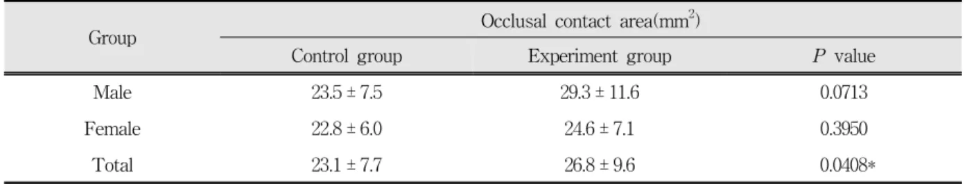
2. Occlusal contact area
The occlusal contact area of experimental group(26.8±9.6 mm
2) (patients with DDwR) were greater than the controls (23.1±7.7 mm
2). The mean and standard deviation(S.D.) of occlusal contact area of each subject groups are listed below(Table 3).
The value of occlusal contact area measured in patients with DDwR was greater than that in the controls(P < 0.05).
3. Occlusal pressure
There was no significant difference in occlusal pressure between the DDwR patients(34.0±4.7 Mpa) and the controls(33.9±5.8 Mpa). The mean and standard deviation(S.D.) of occlusal pressure of each subject groups are listed below(Table 4).
4. Comparison of male and female group
In both of control and experiment group, only the value of the bite force measured in males was significantly greater than that in females(P < 0.05).
The maximal bite force of the male
group(818.6±227.50 N in control, 989.0±309.7 N in
experimental group) was greater than that of the
female group(700.2±178.2 N in control, 813.8±186.3
N in experimental group) in both of control and
experimental group(P < 0.05). However, the
occlusal contact area and the occlusal pressure
between the male and the female group didn't show
significant difference(P > 0.05). The mean and
Group Occlusal contact area(mm
2)
Control group Experiment group P value
Male 23.5 ± 7.5 29.3 ± 11.6 0.0713
Female 22.8 ± 6.0 24.6 ± 7.1 0.3950
Total 23.1 ± 7.7 26.8 ± 9.6 0.0408*
* : P < 0.05 ** : P < 0.01 Table 3. Occlusal contact area(Mean±S.D.)
Group Occlusal pressure(Mpa)
Control group Experimental group P value
Male 35.4 ± 5.1 34.5 ± 4.7 0.5700
Female 32.4 ± 6.2 33.5 ± 4.8 0.5066
Total 33.9 ± 5.8 34.0 ± 4.7 0.9615
* : P < 0.05 ** : P < 0.01 Table 4. Occlusal pressure(Mean±S.D.)
Group Control group
Male Female P value
Maximal bite force(N) 818.6 ± 227.50 700.2 ± 178.2 0.0303*
Occlusal contact area(mm
2) 23.5 ± 7.5 22.8 ± 6.0 0.7084
Occlusal pressure (Mpa) 35.4 ± 5.1 32.4 ± 6.2 0.0504
* : P < 0.05 ** : P < 0.01
Table 5. Comparison between male and female control group
Group Experimental group(DDwR)
Male Female P value
Maximal bite force (N) 989.0 ± 309.7 813.8 ± 186.3 0.0455*
Occlusal contact area (mm
2) 29.3 ± 11.6 24.6 ± 7.1 0.1513
Occlusal pressure (Mpa) 34.5 ± 4.7 33.5 ± 4.8 0.5023
* : P < 0.05 ** : P < 0.01
Table 6. Comparison between male and female experiment group
standard deviation(S.D.) of each evaluated values of control and experiment group are listed below (Table 5, 6).
Ⅳ. DISCUSSION
Most of previous studies provided variable results on maximal bite force, and it maybe due to a lack in the control of such variables to affect bite force measurements as age
17,26,28,36), sex
17,19,21,22,36), physical characteristics of subjects including height and weight
19,26,21,22), the number of teeth present
37,36), diverse population and different measuring devices and techniques
29,30,38).
In the present study, age, sex, the number of tooth present and measuring device were considered except physical characteristics including body height and weight. Several authors found positive correlation between bite force and growth variables such as age and body height and weight.
It is assumed that persons with larger body build, size and/or weight may exhibit a greater bite force.
Kiliaridis et al.
27)noticed a correlation between body height and the mean amplitude of the maximal bite force. Ringqvist
30)found significant correlation between body height and maximal molar bite force in female, whereas Linderholm and Wennstörm
29)and Braun et al.
21,22)did not. In this study, the standard deviation of maximal bite force ranged from 178.2 to 309.2, and it is consistent to that of the previous studies
36,16,39). Threrfore, a group of similar body height and weight could be an important factor in future studies.
In the previous studies, maximal bite force was measured using a metal material such as transducer, biting fork or bite force dynamometer of various sizes and thicknesses. The thickness(4~16 mm) is one of the reasons for unstable positions.
Moreover, subjects might be reluctant to bite maximally because of fear of tooth damage or pain.
In this study, maximal bite force was measured using the Dental Prescale System, which consists of pressure sensitive sheet(Dental Prescale
®, Fuji Film Co., Tokyo, Japan) and a computer for
analysis(Occluzer
®, GC company Co., Japan).
Suzuki et al.
31)reported that Dental Prescale System has some advantages as follows: (i) the thin material induces only a small change in the occlusal vertical dimension(98 ㎛ thickness), and makes it available to measure at a position near the intercuspal position, (ii) it is not necessary to prepare special measurement equipment, (iii) many patients may be examined for a short time, (iv) recording storage, even for an extended period, is simplified and (v) it is easy to explain the treatment to patients by using dental images. In addition, the clinical usefulness of this system was documented in terms of quantitative evaluation for occlusal load distribution
35,31).
In the present study, the mean value of maximal bite force was greater than that obtained by previous studies. Bakke et al.
40)examined a large sample of subjects(63 females and 59 males), and found a mean bite force of 522 N in males and 441 N in females. Dean et al.
41)found the mean values at the molar region to be 490 N in males and 402 N in females. Braun et al.
21,22)reported a mean value of 814 N for males and 615 N for females. This variability might be due to the differences of measuring point and appliances used.
There are few studies which have examined
occlusal pressure, contact area and bite force in
patients with TMD or anterior disc displacement of
TMJ. Takenoshita et al.
42)observed no difference in
occlusal contact area between the patients with
TMJ dysfunction and the controls without TMJ
dysfunction. Our results show that the value of the
occlusal contact area measured in patients with
DDwR of the TMJ was slightly greater than that in
the controls(P < 0.05), and are thus not consistent
with the results of Sato et al.
16), who reported
smaller occlusal contact area in patients with
anterior disc displacement of the TMJ. It remains
obscure why the value of occlusal contact area
measured in patients with DDwR was greater than
that in the controls. But we can guess increased
masticatory muscle activity, as in bruxism patients,
could make increased tooth wear, thus occlusal
contact area could be increased. Further EMG related studies could be valuable. Our results also show that the bite force in patients with DDwR of the TMJ was greater than that in the controls, although there was no difference in the occlusal pressure. From these results we can think that occlusal contact area should have more important role in masticatory performance than occlusal pressure. In previous studies, the bite force and occlusal contact area in patients with DDwR of the TMJ was smaller than that in the controls and it was thought because the patients' symptoms - such as TMJ pain or masticatory muscle pain, fear to TMJ noise etc. - could disturb a stronger bite force. Sinn DP et al.
43)examined the bite force in patients with TMJ dysfunction. They also found that the maximal bite force in patients was smaller than that in the controls without TMJ dysfunction.
However, most of previous studies included various types of TMJ dysfunction, including myofascial pain, internal derangement of the TMJ(DD with or without reduction), osteoarthritis etc. In our study, we examined only in patients with non-painful DDwR of the TMJ, therefore, influence of disc displacement could be reflected more critically. In other words, increased bite force and occlusal contact area could be a cause of disc displacement.
Both of the control and the DDwR group, maximal bite force of male group was greater than that of female group. This result is consistent with the results of previous studies
8,40,36).
In conclusion, we can suggest that DDwR could be a factor of changing bite force regardless of pain, but more controlled, large scaled and EMG related further study is needed.
Ⅴ. CONCLUSIONS
To investigate the influences of non-painful disc change on the masticatory performance, we measured the value of maximal bite force, occlusal contact area and occlusal pressure of 39 patients with non-painful DDwR of the TMJ using pressure sensitive film, and compared it with that of 59
controls. the results are summarized as follows:
1. The maximal bite force (P < 0.01) and the occlusal contact area (P < 0.05) of the DDwR patients were greater than those of the controls.
2. There was no significant difference in the occlusal pressure between the DDwR patients and the controls (P > 0.05).
3. The maximal bite force of the male group was greater than that of the female group (P < 0.05).
However, there was no significant difference in the occlusal contact area and the occlusal pressure between the male and the female group (P > 0.05).
From the results above, we can suggest that DDwR could be a factor of changing bite force, but more controlled, large scaled and EMG related further study is needed.
REFERENCES
1. Peroz I, Tai S. Masticatory performance in patients with anterior disk displacement without reduction in comparison with reduction in comparison with symptom-free volunteers. Eur J Oral Sci 2002;
110:341-344.
2. Zarrinkelk HM, Throckmorton GS, Ellis E, Sinn DP.
A longitudinal study of changes in masticatory performance of patients undergoing orthognatic surgery. J Oral Maxillofac Surg 1995;53:777-782.
3. Liu ZJ, Yamagata K, Kasahara Y, Ito G:
Electromyographic examination of jaw muscles in relation to symptoms and occlusion of patients with temporomandibular joint disorders. J Oral Rehabil 1999;26:33-47.
4. Harper RP, Brown CM, Triplett MM, Villasenor A, Gatchel RJ: Masticatory function in patients with juvenile rheumatoid arthritis. Pediatr Dent 2000;22:200-206.
5. van der Bilt A, Olthoff LW, Bosman F, Oosterhaven SP: Chewing performance before and after rehabilitation of postcanine teeth in man. J Dent Res 1994;73:1677-1683.
6. Fontijn-Tekamp FA, Slagter AP, Van Der Bilt A, Van 'T Hof MA, Witter DJ, Kalk W, Jansen JA:
Biting and chewing in overdentures, full dentures,
and natural dentitions. J Dent Res 2000;79:1519-1524.
7. Hirai T, Ishijima T, Koshino H, Anzai T: Age-related change of masticatory function in complete denture wearers: evaluation by a sieving method with peanuts and a food intake questionnaire method. Int J Prosthodont 1994;7:454-460.
8. Julien KC, Buschang PH, Throckmorton GS, Dechow PC: Normal masticatory performance in young adults and children. Arch Oral Biol 1996;41:69-75.
9. Okeson J: Orofacial pain : guidelines for classification, assessment, and management. 3rd ed. 1996, pp.
127-141, Quintessence Pub Co Inc. Chicago.
10. Atkinson HF, Ralph WJ: Tooth loss and biting force in man. J Dent Res 1973;52:225-228.
11. Haraldson T, Karlsson U, Carlsson GE: Bite force and oral function in complete denture wearers. J Oral Rehabil 1979;6:41-48.
12. Helkimo E, Carlsson GE, Carmeli Y: Bite force in patients with functional disturbances of the masticatory system. J Oral Rehabil 1975;2:397-406.
13. Rudy TE, Greco CM, Yap GA, Zaki HS, Leader JK, Boston JR: The association between research diagnostic criteria for temporomandibular disorder findings and biting force and endurance in patients with temporomandibular disorders. Pain Med 2001;2:35-45.
14. Stegenga B, Broekhuijsen ML, De Bont LG, van Willigen JD: Bite-force endurance in patients with temporomandibular joint osteoarthrosis and internal derangement. J Oral Rehabilfac Surg 1992;19:639-647.
15. Sato S, Ohta M, Goto S, Kawamura H, Motegi K:
Electromyography during chewing movement in patients with anterior disc displacement of the temporomandibular joint. Int J Oral Maxillofac Surg 1998;27:274-277.
16. Sato S, Ohta M, Sawatari M, Kawamura H, Motegi K: Occlusal contact area, occlusal pressure, bite force, and masticatory efficiency in patients with anterior disc displacement of the temporomandibular joint. J Oral Rehabil 1999;26:906-911.
17. Garner LD, Kotwal NS: Correlation study of incisive biting forces with age, sex and anterior occlusion. J Dent Res 1973;52:698-702.
18. Proffit WR, Fields HW, Nixon WL: Occlusal forces in normal and long-face adults. J Dent Res 1983;62:566-570.
19. Fields HW, Proffit WR, Case JC, Vig KWL: Variables affecting measurements of vertical force. J Dent Res 1986;65:135-138.
20. Waltimo A, Nyström M, Könönen M: Bite force and dentofacial morphology in men with severe dental attrition. Scand J Dent Res 1994;102:92-96.
21. Braun S, Bantleon HP, Hnat WP, Freudenthaler JW, Marcotte MR, Johnson BE: A study of bite force, part 1. Relationship to various physical characteristics.
Angle Orthod 1995;65:367-372.
22. Braun S, Bantleon HP, Hnat WP, Freudenthaler JW, Marcotte MR, Johnson BE: A study of bite force, part 2. Relationship to various cephalometric measurements. Angle Orthod 1995;65:373-377.
23. Kikuchi M, Korioth TWP, Hannam AG: The association among occlusal contacts, clenching effort, and bite force distribution in man. J Dent Res 1997;76:13-16.
24. Raadsheer MC, van Eijden TM, van Ginkel FC, Prahl-Andersen B: Contribution of jaw muscle size and craniofacial morphology to human bite force magnitude. J Dent Res 1999;78:31-42.
25. Throckmorton GS, Ellis E, Buschang PH: Morpho- logic and biomechanical correlates with maximum bite forces in orthognathic surgery patients. J Oral Maxillofac Surg 2000;58:515-524.
26. Helkimo E, Carlsson GE, Helkimo M: Bite force and state dentition. Acta Odontol Scand 1977;35:297-303.
27. Kiliaridis S, Kjellberg H, Wenneberg B, Engström C:
The relationship between maximal bite force, bite force endurance and facial morphology during growth. A cross sectional study. Acta Odontol Scand 1993;51:323-331.
28. Fogle LL, Glaros AG: Contribution of facial morphology, age and gender to EMG activity under biting and resting condition. A canonical correlation analysis. J Dent Res 1995;74:1496-1500.
29. Linderholm H, Wennstörm A: Isometric bite force and its relation to general muscle force and body build.
Acta Odontol Scand 1970;28:679-689.
30. Ringqvist M: Isometric bite force and its relation to dimensions of the facial skeleton. Acta Odontol Scand 1973;31:35-42.
31. Suzuki T, Kumagai H, Watanabe T, Uchida T, Nagao M: Evaluation of complete denture occlusal contacts using pressure-sensitive sheets. Int J Prosthod 1997;10:386-391.
32. Watanabe M, Hattori Y, Satoh C: Bite force distribution on the dental arch in normal dentitions.
In Brain and Oral Functions, oral motor function and
dysfunction. Morimoto T, Matsuya T, Takada K,
eds., 1995, pp. 399-403, Elsevier Science, Oxford.
33. Kumagai H, Suzuki T, Hamada T, Sondang P, Fujitani M, Nikawa H: Occlusal force distribution on the dental arch during various levels of clenching. J Oral Rehabil 1999;26:932-935.
34. Suzuki T, Kumagai H, Yoshitomi N, McGlumphy EA:
Occlusal contacts of edentulous patients with mandibular hybrid dentures opposing maxillary complete dentures. Int J Maxillofac Impl 1999;14:
504-509.
35. Matsui Y, Ohno K, Michi K, Suzuki Y, Yamagata K:
A computerized method for evaluating balance of occlusal load. J Oral Rehabil 1996;23:530-535.
36. Miyaura K, Matsuka Y, Morita M, Yamashita A, Watanabe T: Comparison of biting forces in different age and sex groups: a study of biting efficiency with mobile and non-mobile teeth. J Oral Rehabil 1999;26:223-227.
37. Hidaka O, Iwasaki M, Saito M, Morimoto T: Influence of clenching intensity on bite force balance, occlusal contact area, and average bite pressure. J Dent Res 1999;78:1336-1344.
38. Tortopidis D, Lyons MF, Baxendale RH, Gilmour WH:
The variability of bite force measurement between sessions, in different positions within the dental arch.
J Oral Rehab 1998;25:681-686.
국문요약
측두하악관절 내장증 환자의 교합력, 교합 접촉 면적 및 교합압