Multiple sclerosis (MS) is a chronic in- flammatory disease occurring in the brain and spinal cord, and which manifests as various neu- rological symptoms. MS mainly occurs at a young age when individuals are socially active and the physical disabilities create a socioeconomic and individual burden.1 Because there are no charac- teristic clinical symptoms or tests with high diag- nostic specificity, the diagnosis of MS is based on excluding MS-like diseases, and finding imag- ing evidence of time and spatially diffuse lesions in the central nervous system. Over the last deca- des, disease modifying treatment (DMT) has been applied at the onset of MS based on several stud- ies, and it is known that starting DMT early slows down the disease progression and prevents the disability. The first step for rapid treatment is cor-
rect diagnosis, and this review looks at MS diag- nostic criteria newly published in 2016 and high- lights the importance of differential diagnosis and early treatment.
There is no single test to confirm an MS diagnosis. For this reason, researchers have changed and applied various diagnostic criteria for MS over the past two decades based on new evidence and expert recommendations. MS diag- nosis, including clinical approaches, should ex- clude other explanatory diseases and the demyeli- nated lesion of the central nervous system should be identified in many places (dissemination in space; DIS) and several times (dissemination in time; DIT). Although the diagnosis is based on clinical evidence, magnetic resonance imaging
Review Article
The early diagnosis and treatments in multiple sclerosis
So Young Huh
Department of Neurology, College of Medicine, Kosin University, Busan, Korea
Multiple sclerosis (MS) is a chronic inflammatory demyelinating disorder of the central nervous system that leads to neurological disability. The diagnosis of MS relies on the MRI criteria, which can demonstrate dissemination in space and time. Exclusion of other demyelinating mimics is essential because there are no specific biomarker for MS and MRI criteria are still have imperfect. There is incremental improvements in MS treatment option that have contributed to the delay of disease progression. The early initiation of DMT may ameliorate the neurological disability. In this review, we discusses the new diagnostic MS criteria and summarize the evidences supporting the early treatment in the course of MS.
Key Words: Diagnosis, Multiple sclerosis
Corresponding Author: So Young Huh, Department of Neurology, College of Medicine, Kosin University, 262, Gamcheon-ro, Seo-gu, Busan 49267, Korea
Tel: +82-51-990-6461 Fax: +82-51-990-3077 E-mail: [email protected]
Received:
Revised:
Accepted:
Apr. 06, 2017 Apr. 17, 2017 Apr. 19, 2017
(MRI) should support the clinical diagnosis. With the development of MRI and the application of new diagnostic criteria, accurate diagnosis of ear- ly stage MS has recently become possible.
Nevertheless, correct consideration of clinical in- formation is critical for excluding MS-like dis- eases that may cause white matter degeneration.
1. Clinical Symptoms
Clinical symptoms are manifested as symptoms impacting movement, sensory, visual and auto- nomic nervous systems. Additionally, most relaps- ing-remitting MS (RRMS) patients experience central nervous system symptoms such as optic neuritis, incomplete transverse neuritis and acute brain stem syndrome that lasts for more than 24 hours. At this time, the central nervous system symptoms that are experienced first are called CIS features typically seen in MS Less common CIS features which
may be seen in MS
Atypical CIS features not expected in MS
Optic nerve
Unilateral optic neuritis Pain on eye movement
Partial and mainly central visual blur- ring
Normal disc or mild disc swelling
Bilateral simultaneous optic neuritis No pain
No light perception
Moderate to severe disc swellingwith- no hemorrhages
Uveitis (mild, posterior)
Progressive optic neuropathy Severe, continuous orbital pain Persistent complete loss of vision Neuroretinitis (optic disc swelling with macular star)
Uveitis (severe, anterior) Brain stem/cerebellum
Bilateral internuclear ophthalmoplegia Ataxia and multidirectional nystagmus Sixth nerve palsy
Facial numbness
Unilateral internuclear
ophthalmoplegia, facial palsy, facial myokymia
Deafness
One-and-a-half syndrome Trigeminal neuralgia Paroxysmal tonic spasms
Complete external ophthalmoplegia;
verticalgaze palsies
Vascular territory syndrome, e.g., lat- eramedullary
Third nerve palsy
Progressive trigeminal sensory neuro- pathy
Focal dystonia, torticollis Spinal cord
Partial myelopathy Lhermitte’s symptom Deafferented hand Numbness
Urinary urgency, incontinence, erectile dysfunction
Progressive spastic paraplegia (asymmetrical)
Complete transverse myelitis Radiculopathy, areflexia
Segmental loss of pain andtemper- ature sensation
Partial Brown-Sequard syndrome (sparing posterior columns)
Faecal incontinence
Progressive spastic paraplegia (symmetrical)
Anterior spinal artery territory lesion (sparing posterior columns only) Cauda equina syndrome
Sharp sensory level to all modalities andlocalized spinal pain
Complete Brown-Sequard syndrome Acute urinary retention
Progressive sensory ataxia (posterior columns)
Cerebral hemispheres
Mild subcortical cognitive impairment Hemiparesis
Epilepsy Hemianopia
Encephalopathy (obtundation, con- fusion, drowsiness)
Cortical blindness Table 1. CIS clinical features and likelihood of signaling an MS diagnosis4
clinically isolated syndrome (CIS).2 After CIS, many patients experience symptoms of the cen- tral nervous system again, and when the second episode occurs, MS can be diagnosed as clinically definite multiple sclerosis (CDMS). About 85% or more of CIS cases will develop into MS that sat- isfies both DIS and DIT together.2,3 The clinical features of CIS suggesting MS are summarized in (Table 1).4
2. 2016 MAGNIMS Diagnostic Criteria
The diagnostic criteria were determined by McDonald in 2001 and were revised in 2005 and again in 2010. New diagnostic criteria were sug-
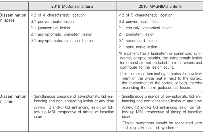
gested in 2016 as “The magnetic resonance imag- ing in multiple sclerosis (MAGNIMS)” (Table 2).5-8 Diagnosis of MS is possible when DIS and DIT are simultaneously satisfied in CIS patients by apply- ing these diagnostic criteria.
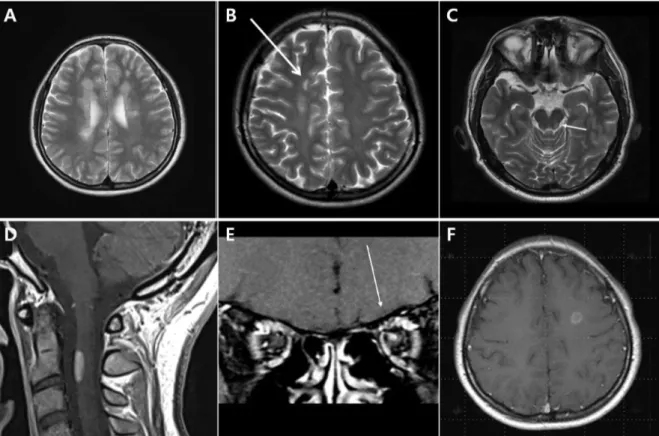
According to the 2016 MAGNIMS diagnostic cri- teria, among 5 lesions including ≥ 3 periventricle lesions, juxtacortical lesions, posterior fossa le- sions, spinal cord lesions, and optic nerve lesions (Fig. 1A-E) if 2 or more lesions are invaded, it sat- isfies the DIS.8 DIT is satisfied if a con- trast-enhanced lesion and non-contrast-enhanced lesion are present at the same time or a new T2 lesion or contrast-enhanced lesion is observed in the MRI regardless of the follow-up period. In the
Fig. 1. Example of lesion for multiple sclerosis MRI Criteria of dissemination in space (A) periventricular lesions; (B) juxtacortical lesions; (C) infratentorial lesions; (D) spinal cord lesion; (E) optic nerve lesion.
Gadolinium-enhancing lesion in T1 gadolinium weighted image; (F) are visible in the patient with MS.
2010 McDonald diagnostic criteria, brain stem or spinal cord lesions that caused symptoms were ex- cluded,7 but the 2016 MAGNIMS diagnostic criteria reflects the lesions regardless of symptoms.8 Spinal nerve MRI is recommended for DIS evaluation pur- poses but it has limitations for DIT identification.9,10 Abnormal findings of optical coherence tomog- raphy and visual evoked potential are recognized to support optic nerve lesions as well as clinical symptoms and MRI findings.8 These changes in the diagnostic criteria are believed to increase the sen- sitivity and thus promote early diagnosis.
With the development of imaging technology and the increased accessibility of MRI, there are cases in which a lesion suggestive of MS is found in a person who does not have typical MS clinical
symptoms or signs. Such a case is named as radio- logically isolated syndrome (RIS).11 However, even RIS that satisfies the DIS should not be diagnosed as MS based on MRI. However, MS can be diag- nosed if at least one episode that is appropriate for demyelinated central nervous system symptom has been confirmed.8
3. MRI Findings of MS
There is no specific MS lesion, but lesions of early MS patients are mainly identified around the cerebral ventricle, and are observed as either a oval-shaped lesion with high-intensity signal on T2-weighted MRI or a “Dawson Finger” lesion ar- ranged perpendicular to the lateral cerebral ven- tricle (Fig. 1-A).12
2010 McDonald criteria 2016 MAGNIMS criteria
Dissemination in space
≥2 of 4 characteristic location
≥1 periventricular lesion
≥1 juxtacortical lesion
≥1 asymptomatic brainstem lesion
≥1 asymptomatic spinal cord lesion
≥2 of 5 characteristic location
≥3 periventricular lesion
≥1 cortical/juxtacortical lesion
≥1 brainstem lesion
≥1 spinal cord lesion
≥1 optic nerve lesion
*If a patient has a brainstem or spinal cord syn- drome, or optic neuritis, the symptomatic lesion (or lesions) are not excluded from the criteria and contribute to the lesion count.
†This combined terminology indicates the involve- ment of the white matter next to the cortex, the involvement of the cortex, or both, thereby expanding the term juxtacortical lesion.
Dissemination in time
- Simultaneous presence of asymptomatic Gd-en- hancing and non-enhancing lesion at any time - A new T2 and/or Gd-enhancing lesion on fol- low-up MRI irrespective of timing of baseline scan
- Simultaneous presence of asymptomatic Gd-en- hancing and non-enhancing lesion at any time - A new T2 and/or Gd-enhancing lesion on fol- low-up MRI irrespective of timing of baseline scan
- Clinical symptoms should be associated with radiologically isolated syndrome
Table 2. 2010 McDonald criteria and 2016 MAGNIMS Diagnosis criteria8
An increase of the contrast enhancement re- lated to damage of the cerebral vascular barrier in the form of a complete or incomplete ring en- hancement may be seen in the border of MS le- sions (Fig. 1F). However, these findings can also be observed in brain abscess, brain tumors and sarcoidosis, and therefore requires caution in diagnosis. In MS, spinal lesions are localized in the axial region of the MRI not invading the whole, and 90% of the lesions have a short length of less than 2 spinal segments (Fig. 1D).13
Cortical lesions (Fig. 1-B) are classified as leu- kocortical, intracortical, and subpial lesion pathologically. Approximately 38% of cortical le- sions are found in early MS patients,14 and autops- ies reveal the demyelinated lesions in the local cortex in approximately 90% of MS patients.
However, it is difficult to find these cortical lesions with conventional clinical MRI protocols. Recently, double inversion recovery, phase-sensitive inversion recovery, and MP-RAGE (magnetization-prepared rapid acquisition with gradient echo sequence) were reported to have high sensitivity for detecting cortical lesions.8 It is reported that about 30% of CIS patients have cortical lesions when using dou- ble inversion recovery images.15,16
Recently, with the development of 7T MRI neu- roimaging, a central vein sign has been reported in MS patients. Kilsdonk et al. analyzed 1004 cases of brain lesions in 33 MS patients and found a central vessel in around 78% of the lesions.17 These central vein signs are not observed in ischemic brain white matter degeneration and are likely to
be helpful in differential diagnosis.18
4. Differential Diagnosis
Infections, neoplasms, congenital diseases, metabolic diseases, vascular diseases and idio- pathic inflammatory demyelinated diseases can also meet the MS diagnostic criteria by displaying clinical forms or MRI findings similar to those of MS.4,19
Therefore, MS cannot be diagnosed based only on the location of the lesion according to the MRI diagnosis criteria, so efforts should first be con- centrated on differential diagnosis. Differential diagnosis is based on clinical observation, blood test results and imaging. In 2008, Miller et al. re- ported 79 clinical and imaging red flags that should differentiate between MS and non-MS patients.4 If clinical features such as lung, kidney and bone invading lesions, peripheral neuro- pathy, myopathy, rash, and arthritis are observed, they are clinical red flags that indicate non-MS diagnoses should be considered (Table 3).
Differential diagnosis of neuromyelitis optica spectrum disorder (NMOSD) or acute dis- seminated encephalomyelitis (ADEM) is especially important in patients suspected of having idio- pathic inflammatory demyelinated disease.
Patients with ADEM may not need prophylactic immunotherapy because of their low possibility of recurrence, and it is important to note that if interferon-beta (IFN-b), the primary treatment for MS, is applied to NMOSD, recurrence cannot be effectively prevented.20 Particularly, since
Red flag Type Examples of alternative diagnosis Bone lesions Clinical Histiocytosis; Erdheim Chester disease Lung involvement Clinical Sarcoidosis; Lymphomatoid granulomatosis Multiple cranial neuropathies or pol-
yradiculopathy
Clinical Chronic meningitis, including sarcoidosis and tuberculosis; Lyme disease Peripheral neuropathy Clinical B12 deficiency; adrenoleukodystrophy; metachromatic leukodystrophy,
Lyme disease
Tendon xanthomas Clinical Cerebrotendinous xanthomatosis
Cerebral venous sinus thrombosis MRI Behçet’s disease; vasculitis; chronic meningitis, antiphospholipid or anti- cardiolipin antibody syndromes
Cardiac disease Clinical Multiple cerebral infarcts; brain abscesses with endocarditis or right to left cardiac shunting
Myopathy Clinical Mitochondrial encephalomyopathy (e.g.,MELAS); Sjögren’s syndrome Cortical infarcts MRI Embolic disease; thrombotic thrombocytopenic purpura; vasculitis Hemorrhages/ microhemorrhages MRI Amyloid angiopathy; Moya Moya disease; CADASIL; vasculitis Meningeal enhancemen MRI Chronic meningitis; sarcoidosis; lymphomatosis; CNS vasculitis Extrapyramidal features Clinical Whipple’s disease; multisystem atrophy; Wilson’s disease
Livedo reticularis Clinical Antiphospholipid antibody syndrome; systemic lupus erythematosus;
Sneddon’s syndrome
Retinopathy Clinical Mitochondrial encephalomyopathy; Susac, and other vasculitides (retinal infarction); neuronal ceroid lipofuscinosis
Calcifications on CT scans MRI Cysticercosis; toxoplasmosis, mitochondrial disorders Diabetes insipidus Clinical Sarcoidosis; histiocytosis
Increase serum lactate level Clinical Mitochondrial disease Selective involvement of the ante-
rior temporal and inferior frontal lobe
MRI CADASIL
Hematological manifestations Clinical Thrombotic thrombocytopenic purpura; vitamin B12 deficiency; Wilson’s disease (hemolytic anemia); copper deficiency
Lacunar infarcts MRI Hypertensive ischemic disease; CADASIL; Susac syndrome Persistent Gd-enhancement and
continued enlargement of lesions
MRI Lymphoma; glioma; vasculitis; sarcoidosis
Mucosal ulcers Clinical Behçet’s disease
Myorhythmia Clinical Whipple’s disease
Hypothalamic disturbance Clinical Sarcoidosis; neuromyelitis optica; histiocytosis Recurrent spontaneous abortion or
thrombotic events
Clinical Antiphospholipid antibody syndrome; thrombotic thrombocytopenic purpura; metastatic cancer with hypercoagulable state
Simultaneous enhancement of all lesions
MRI Vasculitis; lymphoma; sarcoidosis
Rash Clinical Systemic lupus erythematosus; T-cell lymphoma; Lyme disease, Fabry disease
T2-hyperintensity in the dentate nuclei
MRI Cerebrotendinous xanthomatosis
Arthritis, polyarthalgias, myalgias Clinical Systemic lupus erythematosus; Lyme disease; fibromyalgia Amyotrophy Clinical Amyotrophic lateral sclerosis; syringomyelia; polyradiculpathy Table 3. Clinical and MRI major red flags suggestive of alternative diagnosis to multiple sclerosis4
there is more NMOSD than MS in the Asian pop- ulation, it is necessary to check whether aqua- porin-4 antibody is detected and if there are any characteristic MRI findings suggesting NMOSD.
The 2015 International consensus diagnostic cri- teria21 presented characteristic MRI findings sug- gestive of NMOSD as follows: lesions invading cor- ticospinal tract, prolonged invasion of the optic nerve or invasion of the posterior optic nerve in- cluding the optic chiasm, especially, when con- firming the invasion of both optic nerves and si- multaneous increase of T2 high shading or con- trast enhancement in optic chiasm, lesions around the third cerebral ventricle or the thala- mus and hypothalamus, medulla oblongata around the fourth cerebral ventricle, large sub- cortical or deep white matter lesions (tumefative lesion), long lesions of corpus callosum (arch bridge pattern), long contrast-enhanced lesions around the ependyma (pencil-thin lesion), and long myelitis more than 3 spinal segments (longitudinally extensive transverse myelitis).
Mendelian or mitochondrial genetic disorder is also differentiated from MS.22 There are various
diseases that can cause white matter degeneration due to inherent factors, and leukodystrophy which occurs as an adult may appear similar to MS (Table 3). However, there are many cases in which leukodystrophy appears as a bilateral, sym- metric white matter lesion in MRI and clinical manifestations are also accompanied by mental symptoms such as cognitive dysfunction and be- havioral abnormalities, which can be a lead to the differential diagnosis.
Leber hereditary optic neuropathy (LHON) is an inherited disease caused by mitochondrial genetic mutation, which causes loss of central vision in one or both eyes and causes severe vision loss.
It occurs mainly between the ages of 25 to 35 years. RRMS was diagnosed in MS patients by ge- netic testing and detection of a mitochondrial ge- netic mutation (LHON-MS, Harding disease), therefore, for MS patients with a family history of LHON or severe visual impairment, the possi- bility of accompanying LHON disease should be considered.23
5. Need for Early Treatment
Headache or meningismus Clinical Venous sinus thrombosis; chronic meningitis; lymphoma or glioma, vasculitis, systemic lupus erythematosus
T1-hyperintensity of the pulvinar MRI Fabry disease; hepatic encephalopathy; manganese toxicity Persistently monofocal manifes-
tations
Clinical Structural lesion (e.g., Chiari malformation); cerebal neoplasm Large and infiltrating brainstem le-
sions
MRI Behçet’s disease; pontine glioma Predominance of lesions at the
cortical/subcortical junction
MRI Embolic infarction; vasculitis; progressive multifocal leukoencephalop- athy
CADASIL; cerebral autosomal dominant arteriopathy with subcortical infarcts and leukoencephalopathy,MELAS;
mitochondrial myopathy, encephalopathy, lactic acidosis, stroke-like episodes syndrome
Axonal damage starts at the beginning of the MS, but does not lead to significant clinical symp- toms, initially due to the complementary elements of the central nervous system; but over time, ex- tensive axonal damage causes irreversible clinical symptoms.24 Encephalatrophy is also observed in clinically isolated syndrome (CIS), the period when the first symptoms of MS occur. According to a study on brain volume in 263 CIS patients, brain volume reduction measured at the first symptom and at 1 or 2 years after the onset of symptoms was greater in patients who were con- verted from CIS to CDMS than in CIS patients who did not experience recurrence. In addition, the number of newly developed T2 lesions during the first year of the disease after symptoms occurred was found to be related to the brain volume change rate in the second year, indicating that damage to brain parenchyma progresses very rap- idly in the early stages of the disease.25 Decreased cognitive function is also observed in advanced MS as well as in CIS. In a study on CIS patients and RRMS patients, within two years of onset, about 19.6% of patients showed abnormalities in more than four cognitive measures and showed a concentration of disorders, performance abnor- malities, and a significant decrease in memory and learning abilities compared to the control group.26
T2 lesion load seen in early MRI lesions is re- lated to the long-term prognosis of the patient.
Filippi et al. analyzed the initial MRI results in MS patients and the expanded disability status
scale (EDSS) of disease severity after 5 years. In early MRI results, 52% of patients with a lesion load of more than 1.23 ㎤ had an EDSS score of 6 or greater and 23% of patients with a lesion load less than 1.23 ㎤ showed an EDSS score of below 6.27 Based on these studies, the need for ther- apeutic intervention at the beginning of MS has become apparent.
Large clinical studies (CHAMPS, ETOMS, BENEFIT, Precise, and TOPIC) were conducted in CIS, the first symptomatic stage of MS, and dem- onstrated that preventing recurrence and delaying the conversion to CDMS could reduce the risk of disease progression.
The CHAMPS study28 was a 3-year follow-up study of 383 patients with CIS by dividing them into an IFN-b-1a 30 mg (Avonex®) treatment group and placebo group. The incidence of CDMS was significantly lower in the IFN-b-1a group than in the placebo group (P = 0.002). At Month 18, the volume of brain lesions in the treatment group was significantly smaller (P < 0.001), and newly developed T2 lesions and contrast-enhanced le- sions were also significantly less than the placebo group (P < 0.001). Conversion from CIS to CDMS was approximately 66% lower in the IFN-b-1a treatment group than in the placebo group. In a 10-year follow-up study,29 an early treatment group that started treatment within one month after CIS diagnosis showed lower CDMS con- version (hazard ratio = 0.64) and lower annual re- currence rate (P = 0.03) than the delayed treatment group which started treatment after an average
of 30 months from diagnosis.
The ETOMS study30 was a 2-year follow-up study on 308 patients with CIS by dividing them into an IFN-b-1a 22 mg (Rebif®) treatment group and a placebo group. During the follow-up period of 2 years, the IFN-b-1a treatment group (34%) showed lower CDMS conversion than the placebo group (45%) (P = 0.047). CDMS conversion was significantly prolonged in the IFN-b-1a treatment group (569 days) compared to the placebo group (252 days) (P = 0.034). The annual recurrence rate was also significantly lower in the IFN-b-1a treat- ment group (0.33) than the placebo group (0.43) (P = 0.045). In MRI, the lesion load was sig- nificantly lower in the treatment group, demon- strating the effectiveness of IFN-b-1a treatment using brain imaging as well as clinical effects in early MS patients.
The PreCISe study31 was a 3-year study compar- ing 481 CIS patients with more than two T2 lesions greater than 6 mm in size, which divided the pa- tients into a glatiramer acetate 20 mg (Copaxone®) treatment group and a placebo group. CDMS con- version was significantly lower in the glatiramer acetate treatment group (25%) than the placebo group (43%) (P = 0.0001) and the time when CDMS conversion occurred was also significantly pro- longed in the glatiramer acetate treatment group (722 days) than the placebo group (336 days) (P
= 0.0005). In the last observation, the number of newly developed T2 lesions was significantly less in the glatiramer acetate treatment group (0.7 le- sions) compared to the placebo group (1.8 lesions)
(P = 0.0001).
The BENEFIT study32 was a study comparing 468 CIS patients by dividing them into an IFN-b-1b 250 mg (Betaferon®) treatment group and a pla- cebo group. Two years later, 45% patients in the placebo group were converted into CDMS, how- ever, only 28% of patients in the IFN-b-1b treat- ment group were converted into CDMS, reducing the risk of CDMS conversion by about 50% when administering IFN-b-1b. Since then, an 11-year extended follow-up study33 showed that early treatment within 60 days after CIS occurrence re- duced the risk of the first recurrence by 34.5%
compared to a delayed treatment group (start of treatment after the second clinical recurrence or 2 years after CIS occurrence) and reduced the an- nual recurrence rate by 19.1%. There was no sig- nificant change in EDSS between the early treat- ment group and the delayed treatment group, but after 11 years the EDSS score remained low and the conversion of SPMS was low in both groups.
The TOPIC study34 compared 618 patients with CIS for more than 108 weeks by dividing them into two treatment groups administering 7 mg or 14 mg of teriflunomide (Aubagio®) daily and a placebo group. Compared with the placebo group (28%), CDMS conversion in the 14 mg (18%) and 7 mg (19%) teriflunomide treatment groups was significantly reduced (P = 0.0087, P = 0.0271). The risk of recurrence or new MRI lesions was also significantly reduced in the 14 mg treatment group compared to the placebo group (HR 0.651, P = 0.0003).
In addition to large-scale clinical studies of sin- gle agents, there was a study demonstrating that factors affecting cumulative impairment are re- lated to the onset of treatment with various DMTs (IFN-b-1a, IFN-b-1b, glatiramer acetate, natali- zumab, fingolimod, and rituximab).35 In 639 MS patients, by applying a regression analysis model for about 8 years, the risk of reaching 4 points of EDSS was investigated by dividing the patients into three DMT treatment groups that started treatment after either 1 year, between 2 and 3 years, or more than 3 years after the first clinical symptoms developed. As the time of treatment was delayed, the risk increased by 7.4% per year, and patients who started treatment 3 years after the first clinical symptoms developed had a rela- tive risk of 2.63 until reaching 4 points of EDSS, compared to patients who started treatment with- in 1 year after the first symptoms. Therefore, early treatment may be a factor that affects the occur- rence of long-term disability.
It has been reported that various anti-in- flammatory drugs and DMTs (dimethyl fumarate, fingolimod, alemtuzumab, rituximab, laquini- mode, daclizumab, and ocelizumab) reduce the frequency and severity of new demyelinated epi- sodes in RRMS as well as CIS.36,37 However, when the disease has progressed to secondary pro- gressive MS (SPMS), there is no effective drug. As for the pathological features, the cerebrovascular barrier is compromised and lymphocytes infiltrate into the central nervous system, so that the active inflammatory lesions can be identified at the time
of RRMS. On the other hand, once the disease has progressed to SPMS, the effect of DMT or an- ti-inflammatory drugs is reduced because in- flammatory changes in the central nervous system are reduced, and it is compartmentalized within the cerebrovascular barrier.38 Whether newly in- troduced drugs can effectively prevent SPMS or not requires more research, but there is no doubt that aggressive treatment is needed at the time of RRMS where therapeutic effects can be expected.
CONCLUSION
In MS, axonal damage and encephalatrophy are initiated prior to progression to CDMS, which is related to the patient's severity of impairment.
Many studies have shown that since DMT treat- ment reduces the frequency and severity of de- myelinated episodes in MS patients, it is prudent to start DMT treatment at the early stage of dis- ease, which is the period of the highest in- flammatory activity. The most important thing for treatment of patients and their prognosis is to ac- curately diagnose CIS and MS as early as possible.
Therefore, it is necessary to understand various differential diseases, to make an accurate diag- nosis by collecting clinical symptoms and MRI findings and to perform frequent follow-ups.
REFERENCES
1. Compston A, Coles A. Multiple sclerosis. Lancet 2008;372:1502-17.
2. Miller DH, Chard DT, Ciccarelli O. Clinically iso- lated syndromes. Lancet Neurol 2012;11:157–69.
3. Miller D, Barkhof F, Montalban X, Thompson A, Filippi M. Clinically isolated syndromes sugges- tive of multiple sclerosis, part I: natural history, pathogenesis, diagnosis, and prognosis. Lancet Neurol 2005;4:281–8.
4. Miller DH, Weinshenker BG, Filippi M, Banwell BL, Cohen JA, Freedman MS, et al. Differential diagnosis of suspected multiple sclerosis: a con- sensus approach. Mult Scler 2008;14:1157–74.
5. McDonald WI, Compston A, Edan G, Goodkin D, Hartung HP, Lublin FD, et al. Recommended diag- nostic criteria for multiple sclerosis: guidelines from the International Panel on the diagnosis of multiple sclerosis. Ann Neurol 2001;50:121–7.
6. Polman CH, Reingold SC, Edan G, Filippi M, Hartung HP, Kappos L, et al. Diagnostic criteria for multiple sclerosis: 2005 revisions to the
“McDonald Criteria”. Ann Neurol 2005;58:840–6.
7. Polman CH, Reingold SC, Banwell B, Clanet M, Cohen JA, Filippi M, et al. Diagnostic criteria for multiple sclerosis: 2010 revisions to the McDonald criteria. Ann Neurol 2011;69:292–302.
8. Filippi M, Rocca MA, Ciccarelli O, De Stefano N, Evangelou N, Kappos L, et al. MRI criteria for the diagnosis of multiple sclerosis: MAGNIMS consensus guidelines. Lancet Neurol 2016;15:292 –303.
9. Sombekke MH, Wattjes MP, Balk LJ, Nielson JM, Vrenken H, Uitdehaag BM, et al. Spinal cord le-
sions in patients with clinically isolated syndrome:
a powerful tool in diagnosis and prognosis.
Neurology 2013;80:69–75.
10. Weier K, Mazraeh J, Naegelin Y, Thoeni A, Hirsch JG, Fabbro T, et al. Biplanar MRI for the assess- ment of the spinal cord in multiple sclerosis. Mult Scler 2012;18:1560–9.
11. Okuda DT, Mowry EM, Beheshtian A, Waubant E, Baranzini SE, Goodin DS, et al. Incidental MRI anomalies suggestive of multiple sclerosis: the radiologically isolated syndrome. Neurology 2009;72;800–5.
12. Horowitz AL, Kaplan RD, Grewe G, White RT, Salberg LM. The ovoid lesion: a new MR ob- servation in patients with multiple sclerosis. AJNR Am J Neuroradiol. 1989;10:303–5.
13. Grossman RI, Barkhof F, Filippi M. Assessment of spinal cord damage in MS using MRI. J Neurol Sci. 2000;15;172 Suppl 1:S36-9.
14. Lucchinetti CF, Popescu BF, Bunyan RF, Moll NM, Roemer SF, Lassmann H, et al. Inflammatory cort- ical demyelination in early multiple sclerosis. N Engl J Med 2011;365:2188-97
15. Calabrese M, De Stefano N, Atzori M, Bernardi V, Mattisi I, Barachino L, et al. Detection of cort- ical inflammatory lesions by double inversion re- covery magnetic resonance imaging in patients with multiple sclerosis. Arch Neurol 2007;64:
1416-22.
16. Filippi M, Rocca MA, Calabrese M, Sormani MP, Rinaldi F, Perini P, et al. Intracortical lesions:
relevance for new MRI diagnostic criteria for mul- tiple sclerosis. Neurology 2010;75:1988-94.
17. Kilsdonk ID, Lopez-Soriano A, Kuijer JP, de Graaf WL, Castelijns JA, Polman CH, et al. Morphological features of MS lesions on FLAIR* at 7 T and their relation to patient characteristics. J Neurol 2014;261:1356-64.
18. Tallantyre EC, Dixon JE, Donaldson I, Owens T, Morgan PS, Morris PG, et al. Ultra-high-field imaging distinguishes MS lesions from asympto- matic white matter lesions. Neurology 2011;76:
534-9.
19. Charil A, Yousry TA, Rovaris M, Barkhof F, De Stefano N, Fazekas F, et al. MRI and the diagnosis of multiple sclerosis: expanding the concept of
“no better explanation”. Lancet Neurol 2006;5:
841–52.
20. Kim SH, Kim W, Li XF, Jung IJ, Kim HJ. Does interferon beta treatment exacerbate neuro- myelitis optica spectrum disorder? Mult Scler 2012;18:1480-3.
21. Wingerchuk DM, Banwell B, Bennett JL, Cabre P, Carroll W, Chitnis T, et al. International con- sensus diagnostic criteria for neuromyelitis op- tica spectrum disorders. Neurology 2015;85:177–
89.
22. Weisfeld-Adams JD, Katz Sand IB, Honce JM, Lublin FD. Differential diagnosis of Mendelian and mitochondrial disorders in patients with sus- pected multiple sclerosis. Brain 2015;138:517-39.
23. Pfeffer G, Burke A, Yu-Wai-Man P, Compston DA, Chinnery PF. Clinical features of MS asso- ciated with Leber hereditary optic neuropathy mtDNA mutations. Neurology 2013;81:2073-81.
24. Rovaris M, Gambini A, Gallo A, Falini A, Ghezzi
A, Benedetti B, et al. Axonal injury in early multiple sclerosis is irreversible and independent of the short-term disease evolution. Neurology 2005;65:
1626–30.
25. Filippi M, Rovaris M, Inglese M, Barkhof F, De Stefano N, Smith S, et al. Interferon beta-1a for brain tissue loss in patients at presentation with syndromes suggestive of multiple sclerosis: a randomised, double-blind, placebo-controlled trial. Lancet 2004;364:1489–96.
26. Baysal Kıraç L, Ekmekçi Ö, Yüceyar N, Sağduyu Kocaman A. Assessment of early cognitive im- pairment in patients with clinically isolated syn- dromes and multiple sclerosis. Behav Neurol 2014;2014:637694.
27. Filippi M, Horsfield MA, Morrissey SP, MacManus DG, Rudge P, McDonald WI, et al. Quantitative brain MRI lesion load predicts the course of clin- ically isolated syndromes suggestive of multiple sclerosis. Neurology 1994;44:635-41.
28. Jacobs LD, Beck RW, Simon JH, Kinkel RP, Brownscheidle CM, Murray TJ, et al.
Intramuscular interferon beta-1a therapy ini- tiated during a first demyelinating event in multi- ple sclerosis CHAMPS Study Group. N Engl J Med 2000;343:898-904.
29. Kinkel RP, Dontchev M, Kollman C, Skaramagas TT, O'Connor PW, Simon JH. Association between immediate initiation of intramuscular interferon beta-1a at the time of a clinically isolated syn- drome and long-term outcomes: a 10-year fol- low-up of the Controlled High-Risk Avonex Multiple Sclerosis Prevention Study in Ongoing
Neurological Surveillance. Arch Neurol 2012;69:
183-90.
30. Comi G, Filippi M, Barkhof F, Durelli L, Edan G, Fernández O, et al. Effect of early interferon treatment on conversion to definite multiple scle- rosis: a randomised study. Lancet 2001;357:1576–
82.
31. Comi G, Martinelli V, Rodegher M, Moiola L, Bajenaru O, Carra A, et al. Effect of glatiramer acetate on conversion to clinically definite multi- ple sclerosis in patients with clinically isolated syndrome (PreCISe study): a randomised, dou- ble-blind, placebo-controlled trial. Lancet 2009;374:1503–11.
32. Kappos L, Polman CH, Freedman MS, Edan G, Hartung HP, Miller DH, et al. Treatment with in- terferon beta-1b delays conversion to clinically definite and McDonald MS in patients with clin- ically isolated syndromes. Neurology 2006;67:
1242–9.
33. Kappos L, Edan G, Freedman MS, Montalbán X, Hartung HP, Hemmer B, et al. The 11-year long-term follow-up study from the randomized
BENEFIT CIS trial. Neurology 2016;87:978–87.
34. Miller AE, Wolinsky JS, Kappos L, Comi G, Freedman MS, Olsson TP, et al. Oral teriflunomide for patients with a first clinical episode suggestive of multiple sclerosis (TOPIC): a randomised, dou- ble-blind, placebo-controlled, phase 3 trial.
Lancet Neurol 2014;13:977–86.
35.Kavaliunas A, Manouchehrinia A, Stawiarz L, Ramanujam R, Agholme J, Hedström AK, et al.
Importance of early treatment initiation in the clinical course of multiple sclerosis. Mult Scler 2016.
36. Hohlfeld, R. & Wekerle, H. Autoimmune concepts of multiple sclerosis as a basis for selective im- munotherapy: from pipe dreams to (therapeutic) pipelines. Proc Natl Acad. Sci USA 2004;101 Suppl 2:14599-606.
37. Ransohoff RM, Hafler DA, Lucchinetti CF. Multiple sclerosis-a quiet revolution.Nat Rev Neurol 2015;11:134-42.
38. Lassmann H, van Horssen J, Mahad D. Progressive multiple sclerosis: pathology and pathogenesis.
Nat Rev Neurol 2012;8:647-56.
Peer Reviewer’s Commentary
Multiple sclerosis (MS) is a chronic inflammatory disease occurring in the brain and spinal cord.
The most important thing for treatment of patients and their prognosis is to accurately diagnose MS as early ad possible. This review summarized the MS diagnostic criteria newly published in 2016.
(Editorial Board)