Copyright 2006 by the Korean Society for Clinical Neurophysiology 179 대한임상신경생리학회지 8(2):179~181, 2006 ISSN 1229-6414
Multiple sclerosis is an autoimmune demyeli- nating disorder of the nervous system. The ocular manifestation includes optic neuritis, internuclear opthalmoplegia and nystagmus which result in diplopia, oscillopsia, blurred vision, loss of stere- opsis, and reading fatigue.1 Upbeat nystagmus (UBN) is a rare neurologic sign of relapsing mul- tiple sclerosis with lesions affecting the pon- tomesencephalic junction, the rostral medulla, the caudal pons, or the cerebellum.2,3 We report on a patient with isolated UBN as a manifesting sign of relapsing multiple sclerosis.
Case
A 22-year-old woman was admitted to the hos-
pital with one day history of vertigo and vertical oscillopsia. She had suffered from clinically definitive multiple sclerosis with a relapsing and remitting course during the past five years, which had developed motor weakness, sensory distur- bance, bladder dysfunction and optic neuritis.
General physical examination were unremarkable and her mental status was intact. She showed upbeat nystagmus in the primary position. The nystagmus was unchanged by positional change, fixation removal, or convergence. Ocular motor ranges were full. Other neurologic examination was normal except for left optic nerve lesion due to previous optic neuritis. Normal laboratory studies included complete blood counts, blood chemistry, urinalysis and cerebrospinal fluid.
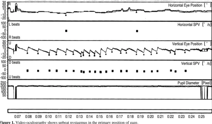
Computerized infrared video-oculography showed upbeat nystagmus with a constant veloci- ty decay of the slow phases in the primary posi- tion (Fig. 1). Brain MRI revealed high signal intensity in the paramedian area of the caudal medulla on axial and sagittal T2-weighted images (Fig. 2).
Intravenous methylprednisolone was given with
단독 상방 안진으로 재발한 다발성 경화증
전북대학교 의과대학 신경과학교실, 전북대학교병원 임상연구소
임의성∙신병수
Relapse of Multiple Sclerosis with Isolated Upbeat Nystagmus
Eui-Seong Lim, M.D., Byoung-Soo Shin, M.D.
Department of Neurology, Chonbuk National University Medical School, Chonbuk National University Hospital Research Institute of Clinical Medicine
Multiple sclerosis is an autoimmune demyelinating disorder of the nervous system. The ocular manifestation includes optic neuritis, internuclear opthalmoplegia and nystagmus. Upbeat nystagmus is a rare manifestation of multiple sclero- sis. We report a patient with relapsing multiple sclerosis who presented with upbeat nystagmus from a circumscribed lesion in the caudal medulla.
Key Words: Upbeat nystagmus, Multiple sclerosis, Relapse
Address for correspondence Byoung-Soo Shin, M.D.
Department of Neurology,
Chonbuk National University Hospital and Medical School
634-18, Keumamdong, Jeonju, Chonbuk, 561-712, Republic of Korea Tel: +82-63-250-1896 FAX: +82-63-251-9363
E-mail : [email protected]
a dose of 1g per day for 3 days, which was fol- lowed by oral prednisolone 60 mg daily in a tapering schedule. The patient left the hospital 5 days after her admission. We tried gabapentin (1200 mg/day) for control the upbeat nystagmus, and there was partial response.
Discussion
UBN is characterized by a downward drift of the eyes when the patient attempts to maintain the primary position of gaze. The drift is interrupted by saccadic movements which correct eye position back towards the center and give rise to the
임의성∙신병수
180 J Korean Society for Clinical Neurophysiology / Volume 8 / December, 2006 Figure 1. Video-oculography shows upbeat nystagmus in the primary position of gaze.
Figure 2. Axial and sagittal T2-weighted MRIs show a high signal intensity lesion in the paramedian area of the caudal medulla.
upward beating appearance of the nystagmus.2 Few cases of upbeat nystagmus were reported with low-grade astrocytoma, cystic tumor, exten- sive demyelination, pontine hemorrhage associat- ed with lesion of the anterior cerebellar vermis, perihypoglossal and inferior olivary nuclei of the medulla, pontine tegmentum, brachium conjunc- tivum, midbrain, and brainstem diffusely.4-6
Medullary lesions invariably involve the perihy- poglossal nucleus and adjacent medial vestibular nucleus, nucleus intercalates (NI), and ventral tegmentum, which contain projections from vestibular nuclei that receive inputs from the anterior semicircular canals.7-10The perihypoglos- sal nucleus consists of three small subnuclei, the nucleus prepositus hypoglossi (NPH), the nucleus of Roller (NR), and nucleus intercalates.8 The NI, the most caudal of the perihypoglossal nuclei, has strong reciprocal connections with the NPH. It receives afferents from the medial and inferior vestibular nuclei and has numerous projections including those to the cerebellum and the ocular motor nuclei.10
The pathophysiology of UBN is not well known.
Since most lesions were located inferiorly to the NPH in the posterior paramedian part of the medulla, it has at times been suggested that the NI, lying just caudally to the NPH, could be involved.8,10,11 Pierrot-Deseilligny et al. suggested that no obvious link with UBN can be found if the NPH or NI circuitry is considered.12 By contrast, the NR appears to be a better candidate to play a role in upward vestibular eye movements. This small nucleus is located at the same caudal medullary levels as the NI, lying slightly anteri- orly and medially to the superior part of this nucleus. Therefore, the NR was probably also damaged in most, if not all, of the caudal medullary lesions resulting in UBN.12
The caudal medulla may receive a collateral branch from the superior vestibular nucleus and project to the flocculus via a probably inhibitory pathway. UBN in the caudal medullary lesion may be ascribed to an impairment of this inhibitory pathway. The result would be disinhibition of the
inhibitory flocculovestibular neurons and overin- hibition of the ventral tegmental tract with a slow downward deviation of the eye.12
In our case, symmetrical demyelinating lesion was found in the lower medulla around the IN and NR. We propose that UBN can be a manifes- tation of relapsing multiple sclerosis.
REFERENCES
01. Chen L, Gordon LK. Ocular manifestations of multiple sclerosis. Curr Opin Ophthalmol 2005;16:315-20.
02. Fisher A, Gresty M, Chambers B, Rudge P. Primary posi- tion upbeating nystagmus. A variety of central positional nystagmus. Brain 1983;106 (Pt 4):949-64.
03. Ohkoshi N, Komatsu Y, Mizusawa H, Kanazawa I.
Primary position upbeat nystagmus increased on down- ward gaze: clinicopathologic study of a patient with multi- ple sclerosis. Neurology 1998;50:551-3.
04. Brazis P, Masdeu J, Biller J. Localization in clinical neu- rology. 4th ed. Philadelphia: Lippincott Williams &
Wilkins, 2001;249.
05. Tokumasu K, Fujino A, Yoshio S, Nitta K, Goto K, Yoneda S. Upbeat nystagmus in primary eye position.
Acta Otolaryngol Suppl 1991;481:366-8.
06. Kanaya T, Nonaka S, Kamito M, Unno T, Sako K, Takei H. Primary position upbeat nystagmus localizing value.
ORL J Otorhinolaryngol Relat Spec 1994;56:236-8.
07. Kato I, Nakamura T, Watanabe J, Harada K, Aoyagi M, Katagiri T. Primary position upbeat nystagmus. Localizing value. Arch Neurol 1985;42:819-21.
08. Hirose G, Ogasawara T, Shirakawa T, et al. Primary posi- tion upbeat nystagmus due to unilateral medial medullary infarction. Ann Neurol 1998;43:403-6.
09. Keane JR, Itabashi HH. Upbeat nystagmus: clinicopatho- logic study of two patients. Neurology 1987;37:491-4.
10. Munro NA, Gaymard B, Rivaud S, Majdalani A, Pierrot- Deseilligny C. Upbeat nystagmus in a patient with a small medullary infarct. J Neurol Neurosurg Psychiatry 1993;56:1126-8.
11. Janssen JC, Larner AJ, Morris H, Bronstein AM, Farmer SF. Upbeat nystagmus: clinicoanatomical correlation. J Neurol Neurosurg Psychiatry 1998;65:380-1.
12. Pierrot-Deseilligny C, Milea D, Sirmai J, Papeix C, Rivaud-Pechoux S. Upbeat nystagmus due to a small pon- tine lesion: evidence for the existence of a crossing ventral tegmental tract. Eur Neurol 2005;54:186-90.
단독 상방 안진으로 재발한 다발성 경화증
J Korean Society for Clinical Neurophysiology / Volume 8 / December, 2006 181