Copyright © 2018 Korean Neurological Association 251
JCN
Open AccessSelf-Injurious Behavior Revealing Advanced Primary Progressive Multiple Sclerosis
with a Massive Right Temporal Lesion
Dear Editor,
A 40-year-old left-handed carpenter working in an art gallery was admitted for investiga- tion of disabling walking difficulties due to imbalance and shaky legs that had insidiously evolved over the previous year. Physical examination found extended hyperkeratotic skin le- sions consistent with lichenification (lichen simplex chronicus) on both upper extremities with radial distribution (Fig. 1). Clinical observation uncovered compulsive self-biting of both arms during unobserved moments. Further medical history-taking revealed that the self-injurious behavior (SIB) had started around 7 years previously. Not only did the patient bite his hands and arms, but he also poked his eyes, banged his head, and shouted loudly when he was alone, to such an extent that he was expelled from his apartment due to nocturnal noise. He ex- plicitly denied itching in his arms, and related his behavior to intermittent feelings of loneli- ness, but denied being depressed.
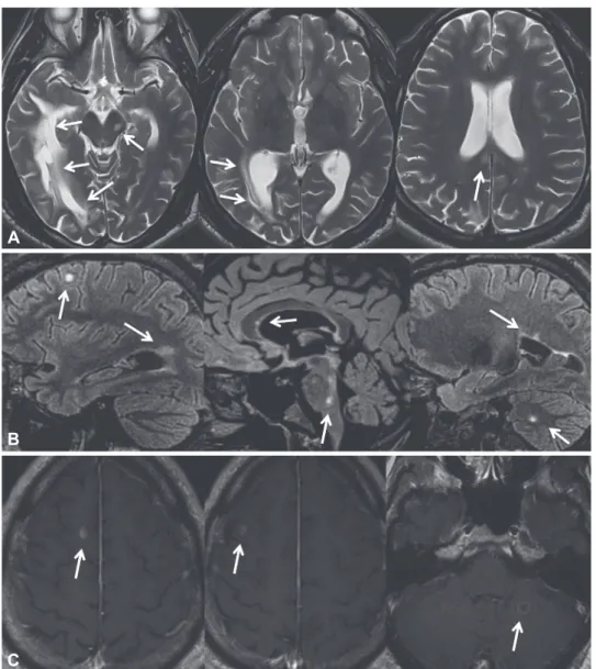
Neurological examination revealed an attentive and friendly man with unremarkable be- havior showing cerebellar dysarthria and ataxia and tetrapyramidal signs that caused his unsteady ataxospastic gait, but with no sensory difficulties in the arms, legs, or trunk. Brain and spine MRI showed extensive periventricular white-matter damage in the right tempo- ral lobe (Fig. 2A), as well as multiple demyelinating lesions juxtacortical in the corpus callo- sum, pons, midbrain, and cerebellum (Fig. 2B), some of which showed also Gd+ contrast en- hancement (Fig. 2C). However, there were virtually no lesions in the frontal lobe.
An extended infectiological and immunological workup did not find any evidence of acute or chronic infection (including PCRs of the CSF), nor of autoimmune connective-tis- sue disorder or paraneoplastic disease. The only deficit in vitamin levels was in vitamins B12
and D, for which supplements were administered. Flow cytometry findings were unremark- able. CSF analysis showed a white blood cell count of 15,000/µL (lymphocytes), elevated proteins (0.61 g/L) with a highly elevated albumin quotient at 8.36×10⁻³, positivity for oligo- clonal bands (IgG index 1.3), and normal interleukin-6. A neuropsychological evaluation re- vealed memory problems (visuospatial, verbal, short-term, and working memory), and marked executive difficulties (mental inflexibility and psychomotor slowing). Based on the pattern of the brain and spine lesions fulfilling McDonald’s revised criteria, we diagnosed primary progressive multiple sclerosis (PPMS) with radiological progression based on the neurological symptoms (notably walking difficulties) for longer than 1 year combined with progressive accumulation of handicap without recovery. An offline treatment with rituximab was introduced.
SIB is an intriguing human behavior that is seen in a range of neurological and psychiat- ric disorders but remains poorly understood. It occurs most frequently in developmental disorders and in psychiatric or neurological conditions involving striatofrontal dysfunction, Markus Gschwinda
Agustina Maria Lascanoa Gürkan Kayab
Frederic Assala
a Departments of Neurology and
b Dermatology, Geneva University Hospital, Geneva, Switzerland
pISSN 1738-6586 / eISSN 2005-5013 / J Clin Neurol 2018;14(2):251-253 / https://doi.org/10.3988/jcn.2018.14.2.251
Received October 19, 2017 Revised January 3, 2018 Accepted January 3, 2018 Correspondence Markus Gschwind, MD Department of Neurology, Geneva University Hospital, Rue Gabrielle-Perret-Gentille 4, Geneva 1211, Switzerland Tel +41-79-5533793 Fax +41-22-3728340
E-mail [email protected]
cc This is an Open Access article distributed under the terms of the Creative Commons Attribution Non-Com- mercial License (http://creativecommons.org/licenses/by-nc/4.0) which permits unrestricted non-commercial use, distribution, and reproduction in any medium, provided the original work is properly cited.
LETTER TO THE EDITOR
252 J Clin Neurol 2018;14(2):251-253
Self-Injurious Behavior from Temporal Lobe Lesion in PPMS
JCN
such as in Tourette’s, Lesch-Nyhan, Rett’s, Prader-Willi, or frag- ile-X syndromes.1 However, anterior temporal dysfunction has also been reported as an SIB-causing condition in patients with frontotemporal dementia2 or temporal lobe epilepsy.3 Very little is known about SIB in multiple sclerosis. One neu- roimaging study described damage to the right inferior and middle temporal gyri and the inferior frontal cortex as being associated with compulsive behavior in 16 patients with mul- tiple sclerosis.4 Another study suspected that right temporo- parietal injury can result in actions related to SIB by discon- necting the networks of agency and body ownership, often in the presence of an existing focal neurological deficit (e.g., limb paresthesia).5
In our case, SIB over a period of years resulted in extended chronic lichenification on the patient’s arms, hands, and fin-
Fig. 2. Magnetic resonance imaging of the patient’s brain. A: Transverse T2-weighted images show an extensive periventricular white-matter damage in the right temporal lobe (arrows). B: Sagittal FLAIR images show multiple demyelinating lesions juxtacortical in the corpus callosum, pons, midbrain, and cerebellum (arrows). C: T1-weighted images with Gd+ show lesions with contrast enhancement (arrows). Images displayed in accordance with the usual radiological convention. FLAIR: fluid attenuated inversion recovery.
Fig. 1. The patient’s arms, hands, and fingers were covered with hy- perkeratotic skin lesions consistent with lichen simplex chronicus.
A
B
C
www.thejcn.com 253
Gschwind M et al.
JCN
gers. He only sought medical advice after walking difficulties became disabling. The first brain MRI led to the discovery of advanced PPMS with an unusually massive white-matter le- sion in the right temporal lobe. Evidence can be found in the literature that establish a causal link of this lesion to the pa- tient’s SIB.
Our case highlights the utility of early brain MRI in cases of newly appearing SIB, since there is increasing recognition that focal brain lesions—due to potentially treatable condi- tions—can be implicated in SIB.
Conflicts of Interest
The authors have no financial conflicts of interest.
REFERENCES
1. Stein DJ, Zohar J, Simeon D. Compulsive and impulsive aspects of self-
injourious behavior. In: Davis KL, Charney D, Coyle JT, Nemeroff C, editors. Neuropsychopharmacology: The Fifth Generation of Progress- An Official Publication of the American College of Neuropsychopharma- colog. Philadelphia, PA: Lippincott Williams & Wilkins, 2002:1743-1758.
2. Mendez MF, Bagert BA, Edwards-Lee T. Self-injurious behavior in frontotemporal dementia. Neurocase 1997;3:231-236.
3. Critchley HD, Simmons A, Daly EM, Russell A, van Amelsvoort T, Robertson DM, et al. Prefrontal and medial temporal correlates of re- petitive violence to self and others. Biol Psychiatry 2000;47:928-934.
4. Tinelli E, Francia A, Quartuccio EM, Morreale M, Contessa GM, Pas- cucci S, et al. Structural brain MR imaging changes associated with obsessive-compulsive disorder in patients with multiple sclerosis.
AJNR Am J Neuroradiol 2013;34:305-309.
5. Borah S, McConnell B, Hughes R, Kluger B. Potential relationship of self-injurious behavior to right temporo-parietal lesions. Neurocase 2016;22:269-272.