Kor. J. Fertil. Steril., Vol. 31, No. 2, 2004, 6
GnRH-agonist에 의한 인간 과립-황체화 세포의 세포사멸과 PBR 단백질의 발현
연세대학교 의과대학 산부인과학교실1, 한양대학교 자연과학대학 생명과학과2, 을지의과대학교 생명과학연구소3
김세광1・염윤희2・윤정미1・배상욱1・양현원3・조동제1・윤용달2・송찬호1
Apoptosis and Peripheral Benzodiazepin Receptor (PBR) Expression in Human Granulosa-Luteal Cells by GnRH-agonist
Sei Kwang Kim
1, Yun-Hee Youm
2, Jeong-Mi Yoon
1, Sang Wook Bai
1, Hyunwon Yang
3, Dong Jae Cho
1, Yong-Dal Yoon
2, Chan Ho Song
11
Department of Obstetrics and Gynecology, College of Medicine, Yonsei University,
2
Department of Life Sciences, College of Natural Sciences, Hanyang University Life Science Institute,
3Eulji University School of Medicine
Objective: To investigate whether GnRH-agonist (GnRH-Ag) using in IVF-ET affects apoptosis of human granulosa-luteal cells and expression of peripheral benzodiazepine receptor (PBR) protein involved in the apoptosis of the cells.
Methods: Granulosa-luteal cells obtained during oocyte retrieval were cultured and treated with 10
-5M GnRH-Ag. Apoptosis of the cells by the treatment was confirmed using DNA fragmentation analysis 24 h after culture. The presence of PBR protein within the cells was examined by immunofluorescence staining and the expression of the protein was analyzed by Western blotting. In addition, it was measured for progesterone and nitric oxide (NO) produced by granulosa-luteal cells after GnRH-Ag treatment. To evaluate the relationship between NO production and PBR expression, sodium nitroprusside (SNP) as a NO donor was added in media and investigated the expression of PBR protein by Western blotting.
Results: Apoptosis increased in the granulosa-luteal cells 24 h after GnRH-Ag treatment, whereas the expression of PBR protein significantly decreased. Furthermore, the production of progesterone and nitric oxide (NO) by the cells significantly fell from 12 h after the treatment. In the results of Western blotting after SNP treatment, the expression of PBR protein increased in the treatment with SNP alone to the granulosa-luteal cells, but was suppressed in the treatment with GnRH-Ag and SNP. Additionally, the staining result of PBR protein in the cells showed the even distribution of it through the cell.
Conclusion: These results demonstrate that GnRH-Ag treatment induces apoptosis, decreasing expression of PBR protein and NO production in human granulosa-luteal cells. The present study suggests that one of the apoptosis mechanism of human granulosa-luteal cells by GnRH-Ag might be a
주관책임자: 김세광, 우) 120-752 서울특별시 서대문구 신촌동 134, 연세대학교 의과대학 산부인과학교실 Tel: (02) 361-5499, Fax: (02) 313-8357, e-mail: [email protected]
*
본 논문은 2002학년도 연세대학교 의과대학 일반교수 연구비 지원에 의하여 연구되었음.
signal transduction pathway via NO and PBR.
Key Words: Apoptosis, GnRH-agonist, Granulosa-luteal cells, Nitric Oxide (NO), Peripheral benzodiazepine receptor (PBR)
성선자극호르몬 분비호르몬 (GnRH)은 시상하부 에서 만들어지는 10개의 아미노산으로 구성된 단백 질 호르몬으로 뇌하수체에 존재하는 수용체와 결합 하여 성선자극호르몬인 FSH와 LH의 분비를 자극 하고, 이러한 성선자극호르몬들이 생식소 및 그 부 속기관에 영향을 미친다.1 또한 GnRH는 뇌하수체 를 통하지 않고 난소에 직접적으로 작용하여 난소 내 과립 세포의 생리적 변화와 난자의 성장과 배란 에 영향을 미치는 것으로 보고되고 있다.2 그러나 시상하부에서 분비되는 GnRH는 반감기가 매우 짧 아 혈청 내에서 거의 측정되지 않으므로 난소 내에 서 GnRH의 작용은 난소 자체에서 발현되는 GnRH 에 의해 일어나는 것으로 알려져 있다.3 아울러 GnRH 수용체가 인간의 난소 내에서 발견되면서 난 소에 대한 GnRH의 직접적인 작용에 대한 많은 연 구가 이루어지고 있다.4
GnRH가 난소에 직접적으로 미치는 작용으로 난 소에서 생성되는 스테로이드 호르몬 합성 억제를 들 수 있다. 즉, GnRH는 과립 세포의 cyclic nucleotide 의 축적을 억제시키고 LH와 prolactin 수용체의 형 성을 억제시킬 뿐만 아니라 FSH, LH, IGF-1 수용체 의 농도를 감소시켜 progesterone, androgen 및 estro- gen의 합성을 억제시키는 것으로 보고되고 있다.5 또한 다량으로 투여된 GnRH는 생체 내에서 난포의 발생과 배란을 억제시키며,6 배양된 과립 세포에 처 리하면 inhibin의 생성을 억제하는 것으로 알려지고 있다.7
한편 난소에 미치는 GnRH의 작용 중 난소 내 세포사멸을 직접적으로 유도한다는 것이 밝혀지면 서 GnRH에 의해 일어나는 세포사멸 기전 연구가 활발히 진행되고 있다.8,9 GnRH는 난소 내 IGFBP-4 의 생성을 유도하여 난포의 폐쇄 (atresia)를 유도하 고, 난소 내 과립 세포 및 황체 세포의 세포사멸을 직접적으로 유발시키는 것으로 알려지고 있다.10 또 한 GnRH는 황체 세포에서 세포사멸과 관련된 Bax 유전자의 발현을 증가시키는 반면 생존과 관련된 Bcl-xL 유전자는 감소시키는 것으로 보고하고 있
다.11 최근에 GnRH-Ag가 황체 세포의 세포사멸과 함께 세포 내 스테로이드 이동과 관련된 peripheral benzodiazepine receptor (PBR) 및 steroidogenic acute regulatory (StAR) 단백질의 합성을 억제시키는 것이 밝혀졌다.12 특히 PBR은 미토콘드리아 외막에 존재 하면서 StAR에 의해 미토콘드리아 외막까지 이동 한 콜레스테롤을 미토콘드리아 내막까지 이동시키 는 단백질로 알려져 있다.13 이러한 PBR은 또한 미 토콘드리아의 막 투과성을 조절하면서 세포사멸에 관여하는 것으로 알려지고 있으며, 미토콘드리아의 기능 이상으로 인하여 PBR 단백질이 작용을 하지 못하면 스테로이드 합성이 억제되고 세포사멸이 유 발되는 것으로 보고되고 있다.14
Nitric oxide (NO)는 여러 조직에서 다양한 생리적 역할을 수행하는 세포 내 신호 전달 물질로 알려져 있다.15 최근 NO 합성 효소인 nitric oxide synthase (NOS)가 토끼,16 생쥐,17 및 인간18의 황체와 난포 에서 발견되었고, 난포의 발달,19 배란,20 난자의 성 숙21 등 다양한 여성 생식 기능에 관여하는 것으로 보고되고 있다. 특히 NO는 난포 및 황체에 의한 estradiol과 progesterone의 합성을 억제시키면서 난 소의 기능을 조절하는 것으로 밝혀졌다.19,22 또한 NO는 과립 세포의 세포사멸을 억제하는 효과를 보 이면서 난포 내에 존재하는 생존 인자로 알려지고 있다.23 더욱이 소 난포 내 과립 세포에서 높은 농 도의 NO는 세포사멸을 억제시키는 반면, 낮은 농 도의 NO는 세포사멸을 촉진시키는 것으로 보고하 고 있다.24
위에서 기술한 바와 같이 난소에서 일어나는 스 테로이드 호르몬 합성과 세포사멸에 대한 NO의 작 용이 GnRH에 의한 작용과 유사한 것으로 보아, GnRH-Ag가 과립-황체화 세포의 스테로이드 호르 몬 합성 억제하고 세포사멸을 유도하는데 필요한 신호 전달 물질로 작용할 수 있다고 추측된다. 따라 서 본 연구는 인간 과립-황체화 세포를 배양하면서 GnRH-Ag와 NO 합성제를 처리한 후 생성되는 NO 양을 측정하고, 또한 PBR 단백질의 발현과 세포사
멸 양상을 비교함으로써 GnRH에 의한 인간 과립- 황체화 세포의 세포사멸 기전을 밝혀보고자 하였다.
연구 대상 및 방법
1. 과립-황체화 세포의 획득
인간 과립-황체화 세포는 체외수정 및 배아이식 시술 (IVF-ET)를 시행하는 환자로부터 난자를 채취 하는 과정에서 얻어 사용하였다. 과배란을 유도하기 위하여 사용된 성선자극호르몬으로는 hMG (Meri- onal; IBSA, Swiss)과 hFSH (Metrodin; Serono, Swiss) 이 사용되었다. 외인성 성선자극호르몬 투여 방법은 기본적으로 단계적 용량 감소법 (step-down fashion) 을 채택하였으며, hMG나 hFSH 투여 시작 후 4일 후부터 질식 초음파 (Medison 128; Medison Co., Korea)를 이용하여 난포의 성장을 감시하였고, 난포 의 성장 정도에 따라 hMG의 용량을 조절하였다.
최대 난포의 평균 직경이 18 mm에 도달하였거나 평균 직경이 17 mm 이상인 난포가 2개 이상 있는 경우에 hCG (Profasi; Serono, Swiss) 10,000 IU를 근 육 주사하였다. hCG 투여 후 35~36시간째에 질식 초음파 유도 하에서 난자 흡인을 시행하였다.
각각의 난포에서 추출된 난포액은 배양 접시에 옮기고 해부 현미경 하에서 난자를 수집한 다음 난 자의 상태를 판정한 후 배양액에 옮겨 배양하였다.
이후 난포액 내에 존재하는 과립-황체화 세포들을 채취하였으며 배양액에 옮긴 후 혈구 세포들을 분 리하기 위해 과립-황체화 세포가 들어 있는 배양액 1 ml를 40% percoll 3 ml 위에 조심스럽게 올려놓고 300 xg에서 20분간 원심분리하였다. 원심분리 후 과 립-황체화 세포들은 중간에 층을 형성하게 되며 혈 구 세포들은 바닥에 가라앉는다. 과립-황체화 세포 들을 채취하여 배양액으로 3번 세척한 다음 0.1%
collagenase (Sigma, St. Louis, MO)가 들어 있는 배양 액으로 옮겨 주었다. 37℃ 배양기에서 30분 경과 후 과립-황체화 세포들을 26 G 주사 바늘을 이용하여 여러 번 흡입과 배출을 해줌으로써 뭉쳐져 있던 세 포 덩어리를 단일 세포로 분리시킨 다음 heamocy- tometer를 이용하여 세포 수를 계산하였다. 생존율 을 조사하기 위하여 trypan blue을 사용하여 염색한 후 염색되지 않은 세포의 수를 계산하여 생존율을
분석하였다. 배양에 사용한 세포들은 생존율 70%
이상을 보이는 것으로 배양액 1 ml당 100,000개의 세포를 넣어 배양하였다.
2. 과립-황체화 세포의 배양
준비된 과립세포 덩어리황체화 세포들은 24-well culture plate (Nunc, Denmark)의 1 well (배양액 1 ml) 당 100,000개의 세포를 넣어, 37℃에서 95% 공기와 5% CO2가 공급되고 100% 습도가 유지되는 배양기 내에서 배양하였다. 배양액은 Dulbecco's Modified Eagle Medium (dMEM; GIBCO BRL)에 10% fetal bo- vine serum (FBS; GIBCO BRL)과 2 mM L-glutamine (GIBCO BRL), 100 U/ml penicillin (GIBCO BRL), 100 µg/ml streptomycin (GIBCO BRL)을 첨가하여 사용하였다. 준비된 과립-황체화 세포를 24-well culture plate에 옮겨서 24시간 배양한 후 plate 바닥 에 세포들이 충분히 부착되어 있는 것을 확인하고 배양액을 갈아주었다.
3. GnRH-Ag 및 SNP 처리
배양된 과립-황체화 세포에 10-6 M GnRH-Ag (Sigma, St. Louis, MO) 또는 대조군으로 생리식염수 를 처리하였다. 한편 NO가 PBR 단백질 발현에 미 치는 영향을 알아보고자 NO 합성제인 SNP (Sigma, St. Louis, MO)를 GnRH-Ag 또는 Hemonglobin (Sigma, St. Louis, MO)와 함께 처리하였다. 처리 후 24시간 에 세포들을 수거하여 세포사멸을 조사하였고 또한 PBR 단백질 발현과 NO 농도를 분석하였다.
4. 세포사멸 확인을 위한 DNA 분절화 분석 획득된 과립-황체화 세포에 0.2 ml 분쇄 완충액을 첨가하고 조직분쇄기를 이용하여 분쇄하였다. 시료 에 12.5 µl의 10% SDS를 넣고 65℃에서 30분간 방 치하였다. 여기에 35 µl의 8 M potassium acetate를 넣고 시료를 잘 섞은 후 60분간 단백질이 가라앉도 록 얼음에 방치한 후 시료를 4℃, 5000 xg에서 10분 간 원심분리하였다. 상층액은 1.5 ml 미량원심분리 시험관으로 옮기고 동량의 phenol: chloroform: iso- amyl alcohol (25:24:1, V:V:V)를 첨가하여 DNA를 추출하였으며, 다시 동량의 chloroform: isoamyl alco- hol (24:1, V:V)로 재추출하였다. 상층액을 1.5 ml 미
량원심분리 시험관에 옮기고, 0℃에서 보관한 2.5배 부피의 100% ethanol을 첨가, -70℃ 초저온 냉동고 에서 1시간 이상 침전시켰다. 이들 시료를 4℃에서 14,000 xg로 30분간 원심분리하여 DNA를 추출하고 침전물은 50 µl의 1X TE buffer (10 mM Tris-HCl, 1 mM EDTA, pH 8.0)에 용해시킨 후 1 µl의 DNase- free RNase (500 µg/ml; Boehringer-Mannheim, IN)을 첨가하고 60분 동안 37℃에서 방치하였다. 시료의 DNA를 동량의 phenol : chloroform : isoamyl alcohol 로 추출 후, 동량의 chloroform: isoamyl alcohol로 재추출하였다. 상층액을 모아서 0.1배 부피의 3 M sodium acetate와 0℃에 보관한 2.5배의 100% ethanol 로 DNA를 침전시키고 -70℃ 초저온 냉동고에서 적 어도 60분 이상 방치시켰다. 이것을 4℃에서 14,000 xg로 30분간 원심분리하고, 0℃에 보관된 0.2 ml의 80% ethanol로 세척하고 건조시켰다. 압축 결정물을 25 µl의 증류수에 녹이고, 260 nm의 흡광도에서 DNA 양을 측정한 다음 -20℃에 보관하였다. 이렇게 추출된 DNA를 lane당 5 µg의 농도로 1.5% agarose gel에 loading하고, running buffer로는 TBE 용액을 사용하였으며, 50 V에서 3시간 동안 전기영동을 시 행한 후 ethidium bromide로 염색하여 자외선 tran- silluminator로 확인하였다.
5. 배양액에서 NO 농도 측정
배양액 내 과립-황체화 세포에 의해 분비된 NO 의 양은 nitrate/nitrite colorimetric assay kit (Alexis Biochemicals, San Diego, CA)를 이용하여 측정하였 다. 먼저 배양액은 10 kDa molecular mass cut-off filter (Amicon, Millipore Co., Bedford, MA)을 이용하 여 여과하였으며, 여과된 배양액은 nitrate reductase 와 enzyme cofactor가 포함된 용액과 함께 상온에서 3시간 동안 반응시켰다. 반응이 끝난 후 sulfanila- mide와 N-[1-naphthyl] ethylenediamine을 첨가하고 10분간 방치한 다음 microplate reader (Spectra Max 250, Molecular Devices, Sunnyvale, CA)을 이용하여 540 nm에서 측정하였다.
6. PBR의 Western blot 분석
획득된 과립-황체화 세포들은 50 mM Tris-base (pH 7.4), 150 mM NaCl, 10 mM EDTA, 0.1% Tween-
20, protease inhibitors (0.1 mM phenyl methyl-sulfonyl- fluoride, 5 g/ml aprotinin, and 5 g/ml leupeptin)가 포함 된 homogenization buffer에서 분쇄한 후 12,000 xg 에서 30분간 원심분리하였다. 단백질의 농도는 DC protein assay kit (Bio-Rad Laboratories, Inc., Hercules, CA, U.S.A.)를 이용하여 측정한 후 동일한 양을 10%
SDS-PAGE에서 전기영동을 시행하였다.
전기영동에 의해 분리된 단백질들은 blotting 방 법으로 nitro-cellulose membrane으로 옮기고, 5%
non-fat dry milk가 포함된 Tris-buffered saline (TTBS;
10 mM Tris (pH 7.6), 150 mM NaCl, 0.1% Tween-20) 용액에 1시간 동안 담가 비특이적 결합을 방지하기 위한 blocking을 수행하였다. Blocking된 membrane 은 rabbit polyclonal anti-human PBR (Trevigen, Gai- thersbug, MD) 일차 항체들이 1:1,000으로 희석된 TTBS에서 1시간 동안 반응시켰다. TTBS로 1분씩 3회 세척 후 이차 항체인 anti-rabbit horseradish peroxidase-conjugated antibody (Santa Cruz Biotechno- logy, Santa Cruz, CA)를 TTBS에 1:1,000으로 희석 시킨 후 40분 동안 상온에서 반응시켰다. 반응이 끝 난 membrane은 chemiluminescence 용액 (ECL kit;
Amersham Life Science, Buckinghamshire, U.K.)으로 발색시킨 다음 X-ray film (Hyperfilm, Amersham Life Science, Buckinghamshire, U.K.)으로 감광하였다.
7. PBR의 면역세포화학적 염색
배양된 과립-황체화 세포에서 PBR에 대한 면역 세포화학적 염색을 위하여, 먼저 배양된 세포를 1%
paraformaldehyde 용액에서 고정한 후 PBS로 세척 하였다. 비특이적 염색을 방지하기 위하여 goat nor- mal serum을 가지고 30분간 상온에서 반응시켰다.
본 실험에서 사용한 일차 항체들은 rabbit polyclonal anti-human PBR 일차 항체로서, 1:100으로 희석시 켜 1시간 동안 상온에서 반응시켰다. PBS로 세척 후 fluorescein isothiocyanate (FITC)가 결합되어 있는 anti-rabbit IgG (Jackson Immuno Research laboratories, West Grove, PA)을 가지고 30분 동안 상온에서 반응 시켰다. 증류수로 세척한 후 Propidium Iodide를 가 지고 핵을 이차 염색한 다음 봉입하였다. 배양된 황 체 세포에 염색된 PBR의 발현은 형광 현미경 (Ni- kon, Tokyo, Japan)을 이용하여 관찰하였다.
8. 통계학적 분석
통계학적 유의성 검정은 one-way ANOVA과 stu-
dent's t-test 방법을 사용하였으며 p 값이 0.05보다 작은 경우를 유의하다고 판정하였다.
결 과
1. GnRH-Ag에 의한 세포사멸 확인
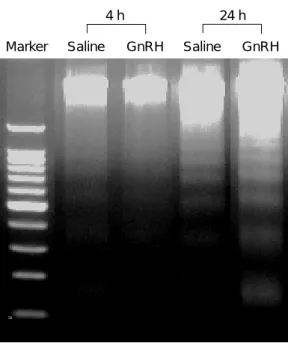
GnRH-Ag에 의해 인간 과립-황체화 세포의 세포 사멸을 확인하기 위하여 배양된 세포에서 DNA를 추출한 후 분절화 현상을 확인하였다. 배양 후 4시 간에서는 대조군과 GnRH-Ag을 처리한 군에서 모 두 DNA 분절화 현상은 관찰되지 않았다. 그러나 배양 후 24시간에 DNA 분절화 현상은 GnRH-Ag를 처리한 군에서 대조군에 비해 증가하였다 (Figure 1).
Marker Saline GnRH Saline GnRH
4 h 24 h
PBR
β-actin
Saline GnRH Saline GnRH Saline GnRH 4 h 12 h 24 h A
Figure 1. Effects of GnRH-Ag treatment on DNA
fragmentation in the cultured human granulosa-luteal cells. The cells cultured for 24 h showed an increase in DNA fragmentation in GnRH-Ag treatment compared with those in the saline.Figure 2. Nitric oxide levels in the medium (mean ±
EM; n = 5) at points timed after initiation of the treat- ment. *p<0.05 compared with corresponding saline con- trols.Figure 3. Western blot analysis (mean ± EM; n = 5)
of PBR proteins in the granulosa-luteal cells at points timed after the commencement of treatment. *p<0.05 compared with corresponding saline controls.B
2. GnRH-Ag에 의한 NO 합성 억제
Figure 2는 GnRH-Ag 처리 후 배양액 내 NO 농 도를 측정한 결과를 보여 주고 있다. 배양액 내 NO
농도는 배양 후 12시간에 GnRH-Ag 처리군에서 3.33±0.66 µM/ml로 대조군 6.21±1.52 µM/ml에 비 해 유의하게 감소하였으며 (p<0.05), 24시간에서도 각각 2.35±0.91 µg/ml과 4.04±1.23 µg/ml로 유의하
Figure 4. Western blot analysis (mean ± EM; n = 5)
of PBR protein in the granulosa-luteal cells (A & B) and nitric oxide levels (mean ± EM; n = 5) in the medium 24 h after sodium nitroprusside (SNP) treatment in a dose-dependent manner. *p<0.05 compared with corre- sponding saline controls.Figure 5. Western blot analysis (mean ± EM; n = 5)
of PBR protein in the granulosa-luteal cells (A & B) and nitric oxide levels (mean ± EM; n = 5) in the medium 24 h after treatment with GnRH-Ag, sodium nitroprus- side (SNP), or hemoglobin (Hb). *p<0.05 compared with corresponding saline controls.B
C PBR
β-actin
SNP (mM) Saline 0.01 0.1 1 A
PBR
β-actin
Saline SNP SNP+ SNP Hb GnRH Hb +GnRH
A
B
C
게 감소하였다 (p<0.05).
3. GnRH-Ag에 의한 PBR 단백질 발현 억제 배양된 과립-황체화 세포에서 발현되는 PBR 단 백질 양을 분석한 결과, 4시간에는 차이를 보이지 않다가, 12시간에 GnRH-Ag를 처리한 군 (71.34±
10.21)에서 대조군 (48.67±6.98)에 비해 유의하게 감 소를 보였다 (p<0.05). 또한 24시간에서도 GnRH- Ag를 처리한 군 (72.78±12.32)은 대조군 (43.34±
5.22)에 비해 유의하게 감소하였다 (p<0.05) (Fi- gure 3).
4. SNP에 의한 PBR 단백질 발현 촉진 NO 합성제인 SNP를 배양된 과립-황체화 세포 에 처리한 후 PBR 단백질의 발현을 조사한 결과, 먼저 PBR 단백질의 발현은 0.01 mM SNP (62.48±
9.20)과 0.1 mM SNP (121.09±11.11)에서 증가하다 가 1 mM SNP (48.76±4.23)에서 급격히 감소하였다 (Figure 4B). 반면 배양액 내 NO 농도는 처리된 SNP 농도 의존적으로 증가하는 것을 알 수 있었다 (Figure 4C). Figure 5은 0.1 mM SNP, Hb 및 GnRH- Ag를 함께 처리 후 PBR 단백질의 발현 양상과 배 양액 내 NO 농도를 분석한 결과를 보여 주고 있다.
먼저 PBR 단백질의 발현은 SNP만을 처리한 군 (239.11±23.12)에서 급격히 증가하였으나 Hb을 함 께 처리한 군 (178.56±13.32)에서는 유의하게 감소 하는 것을 알 수 있었다. 더욱이 GnRH-Ag를 함께 처리한 경우 (123.98±21.87) SNP에 의한 PBR 단백 질의 발현이 완전히 억제되었다. 또한 Hb (72.13±
17.09)과 GnRH-Ag (81.98±19.09)를 단독으로 처 리한 경우에서도 PBR 단백질의 발현이 대조군 (113.60±13.54)에 비해 감소함을 보여주었다 (Figure 5B). 배양액 내 NO 농도의 변화는 SNP만을 처리한 군 (23.21±2.12 µg/ml)에서 급격히 증가하였으나, Hb을 함께 처리한 군 (8.95±1.93 µg/ml)에서는 급격 히 감소하는 것을 알 수 있었다. 반면 GnRH-Ag를 함께 처리한 경우 NO 농도가 SNP만을 처리한 군 과 비슷한 수준인 19.78±2.67 µg/ml을 보였다. 또한 Hb (2.10±1.09 µg/ml)과 GnRH-Ag (1.97±0.91 µg/ml) 를 단독으로 처리한 경우에서도 PBR 단백질의 발 현이 대조군 (5.66± 1.24 µg/ml)에 비해 감소함을 보여주었다 (Figure 5C).
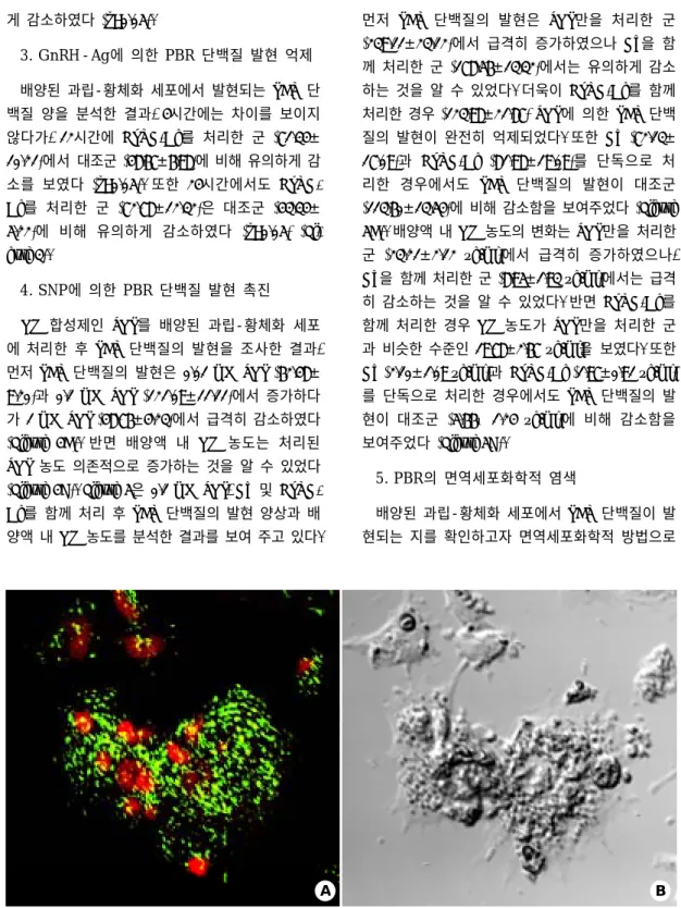
5. PBR의 면역세포화학적 염색
배양된 과립-황체화 세포에서 PBR 단백질이 발 현되는 지를 확인하고자 면역세포화학적 방법으로
Figure 6. Localization of PBR protein in the cultured human granulosa-luteal cells. Green spots displaying PBR
expression localize on mitochondria in even throughout the cell. A; Fluorescent and B; Light microphotographs. Original magnification X400.B
A
염색한 결과, 녹색 형광을 보이는 점들이 세포 전 체에 균일하게 분포하고 있음을 관찰할 수 있었다 (Figure 6).
고 찰
GnRH는 인간 난소 내 세포들에 직접적으로 작 용하여 세포사멸을 유도하며, 이러한 GnRH에 의한 세포사멸으로 인하여 과배란 유도 과정에서 다량으 로 사용되고 있는 GnRH-Ag 또한 난소에 직접적으 로 작용하여 난소의 기능을 억제할 수 있는 것으로 알려지고 있다.25 그러나 과배란 유도 과정 중 함께 사용하고 있는 FSH는 이러한 GnRH-Ag의 세포사멸 기전을 억제시킬 수 있는 것으로 밝혀졌으며, 오히 려 GnRH-Ag를 과배란 유도 과정에 사용한 경우 일정한 성숙도를 가진 다량의 난자를 획득할 수 있 는 것으로 보고하고 있다.26 그러나 현재 난소에 대 한 GnRH의 직접적인 기전을 설명하기 위한 많은 논문들이 보고되고 있으나, 아직 GnRH-Ag에 의한 난소 세포의 세포사멸 기전은 잘 알려져 있지 않다.
따라서 본 연구에서는 난소 세포에서 GnRH-Ag에 의한 세포사멸 기전을 밝히고자 인간 과립-황체화 세포를 이용하여 GnRH-Ag 처리 후 세포 내 NO 합성과 PBR 단백질의 발현 변화를 조사함으로써 GnRH-Ag에 의한 세포사멸 신호 전달 기전을 알아 내고자 하였다.
먼저 GnRH-Ag에 의해 인간 과립-황체화 세포의 세포사멸이 일어나는 지를 확인하기 위하여 배양된 과립-황체화 세포에 GnRH-Ag를 처리한 후 DNA 를 추출하여 분절화 현상을 조사하였다. 본 실험 결 과 배양 후 24시간에 DNA 분절화가 GnRH-Ag를 처리한 군에서 대조군에 비해 증가함을 알 수 있었 다. GnRH-Ag에 의한 DNA 분절화 현상은 인간 난 소 세포에서 뿐만아니라 흰쥐, 소, 돼지의 난소에서 도 보고되고 있으며, 이러한 결과들은 GnRH-Ag가 인간에서 뿐만아니라 다른 포유류의 난소에서도 세 포사멸을 유도할 수 있다는 것을 보여주고 있다. 흰 쥐를 이용한 실험에서 다량으로 투여된 GnRH-Ag 는 투여 시간과 투여 량에 비례하여 과립 세포의 세포사멸을 증가시키는 것으로 보고하였으며,27 또 한 GnRH-Ag는 황체 세포에서 세포사멸과 관련된
Bax 유전자의 발현을 증가시키는 반면 생존과 관련 된 Bcl-xL 유전자는 감소시키는 것으로 보고하고
있다.11,14 이러한 결과들은 과립 세포 또는 황체 세
포에 존재하는 특정한 GnRH 수용체에 GnRH-Ag 가 결합하여 직접적으로 유도된 것으로 생각하고 있다.28
현재 GnRH-Ag에 의한 난소 내 세포사멸 기전 연구는 기존에 알려진 신호 전달 물질에 대한 연구 결과를 기초로 활발히 진행되고 있다. 특히 대표적 인 신호 전달 물질인 NO는 난소 내 스테로이드 호 르몬 합성 조절자로서 뿐만아니라 세포의 죽음을 억제하는 생존 조절자로서 중요한 역할을 하는 것 으로 알려져 있다.29 그러나 난소 내 세포사멸과 연 관된 NO의 역할에 대해서는 스테로이드 호르몬 합 성에서와 같이 많은 논쟁이 있어 왔다.30 높은 농도 의 NO는 난포 내 과립 세포의 세포사멸을 억제하 는 반면, 낮은 농도에서는 세포사멸을 유도하는 것 으로 보고하였다.31 이러한 결과들은 세포 내 NO가 스테로이드 호르몬 합성에 미치는 영향과 마찬가지 로 세포의 종류나 농도에 따라 세포사멸에도 다른 효과를 나타낼 수 있다는 것을 시사하고 있다. 본 실험 결과에서도 GnRH-Ag에 의해 유도된 NO 합 성의 감소와 함께 과립-황체화 세포의 세포사멸이 증가됨을 관찰할 수 있었다. 이러한 결과는 GnRH- Ag 처리에 의한 NO 합성의 감소가 과립-황체화 세포의 세포사멸을 유발시키는 하나의 요인으로 작 용할 수 있다는 것을 보여주고 있다. 최근 흰쥐를 이용한 실험에서 체내로 GnRH-Ag를 투여한 경우 황체 내 NO와 NOS의 합성은 감소하면서 세포사멸 은 증가하였고, 또한 체외에서 배양된 황체 세포에 GnRH-Ag를 처리한 경우에서도 마찬가지로 NO와 NOS의 합성이 감소하면서 세포사멸은 증가한다는 것을 보고하였다.32 반면 배양된 과립 세포에 NO 합성제인 S-nitroso-N-acetyl-DL-penicillamine을 처리 한 경우 직접적으로 세포사멸이 억제된다는 보고가 있었으며,33 또한 흰쥐 난포에 SNP를 처리한 경우 FSH를 처리했을 때 만큼이나 세포사멸을 억제시키 는 것을 보고하였다.34
본 실험 결과 GnRH-Ag를 처리한 과립-황체화 세포에서 NO 농도의 감소와 함께 PBR 단백질 발 현이 감소함을 관찰할 수 있었다. NO 농도와 PBR
단백질 발현과의 관계를 알아보고자 먼저 NO 합성 제인 SNP를 농도별로 처리하고 PBR 단백질의 발현 변화를 조사한 결과, NO 농도의 증가와 함께 PBR 단백질의 발현도 증가함을 알 수 있었다. 그러나 1 mM SNP를 처리한 경우 NO의 농도는 급격히 증가 하였으나 PBR 단백질의 발현은 반대로 감소하는 것을 관찰할 수 있었다. 이는 고 농도의 NO가 가지 고 있는 세포 독성으로 인해 배양된 세포들이 죽음 으로써 일어난 것으로 사료된다. NO 합성과 PBR 단백질 발현과의 관계를 좀 더 정확히 분석하기 위 하여 NO 합성제인 SNP와 반대로 합성된 NO를 제 거하는 것으로 알려진 Hb를 함께 처리하면서 PBR 단백질의 발현을 조사하였다. SNP만을 처리한 군에 서 PBR 단백질의 발현은 급격히 증가하였으나, Hb 을 함께 처리함으로서 유의하게 감소함을 알 수 있 었다. 더욱이 GnRH-Ag를 함께 처리한 경우 SNP에 의한 PBR 단백질 발현이 완전히 억제되었다. 이는 SNP로 인해 증가된 NO이 Hb에 의해 제거됨으로 서 PBR 단백질의 발현이 감소한 것으로 사료된다.
그러나 GnRH-Ag에 의한 PBR 단백질 발현의 감소 는 세포 내 NO 농도가 SNP에 의해 충분히 증가되 어 있는 것으로 보아 NO가 아닌 또 다른 신호 전 달 기전이 관여하고 있을 것으로 추측된다.
GnRH-Ag에 의한 과립-황체화 세포의 세포사멸에 관여할 것으로 판단되는 PBR은 분자량이 18 kDa 인 단백질로써 난소를 포함한 여러 조직에 존재하 는 것이 밝혀졌다.35 미토콘드리아 외막에 위치하고 있는 PBR은 Ro5-4864 또는 isoquinoline carboxa- mide와 같은 benzodiazepine들과 결합하여 voltage- dependent anion channels을 조절하는 것을 알려져 있다.36 더욱이 이러한 조절을 통하여 oxidative pho- sphorylation에 관여하고, 특히 콜레스테롤을 미토콘 드리아 외막에서 내막 쪽으로 이동시키는 중요한 역할을 하는 것으로 보고되었다.13 최근에는 이러한 PBR이 세포의 세포사멸에 직접적으로 관여한다는 논문들이 보고되고 있다.32 미토콘드리아는 세포사 멸 과정에서 중심적인 역할을 하는 세포 내 소기관 이다.38 이는 본 실험에서도 PBR에 대한 면역화학 적염색을 통해 세포 내 위치가 미토콘드리아임을 재확인하였다. 미토콘드리아의 투과성을 조절하는 통로 (mitochondrial permeability transition pore; MPT)
의 개방은 미토콘드리아 내막의 potential 변화를 유 도하여 미토콘드리아 내부로부터 향세포사멸 (pro- apoptotic) 단백질의 방출을 촉진시킨다. PBR은 미토 콘드리아 막에 존재하면서 MPT 통로와 연관이 있 는 것으로 알려져 있으며, 이로 인하여 미토콘드리 아 내막의 potential이 조절되고 세포사멸이 유도될 수 있다. 최근 PBR ligand에 의해 세포사멸이 조절 될 수 있다는 논문들이 보고되고 있다. 즉, leukae- mic cells (HL 60)에 PBR ligand로 알려진 PK 11195, Ro5-4864, pyrrolo-1,5-benzoxazepines 등을 처리한 경우 세포사멸이 유도되는 것을 관찰할 수 있었 다.39 한편 혈액 또는 상피 근원 세포주에서는 Ro5- 4864와 PK 11195가 세포사멸을 유도하지 못하는 것으로 보고하고 있다.40 이러한 결과들은 PBR 또 한 NO와 마찬가지로 세포의 종류에 따라 세포사멸 조절 기전에 다른 영향을 미칠 수 있다는 것을 보 여주고 있다.
본 연구에서 GnRH-Ag에 의한 인간 과립-황체화 세포의 세포사멸은 세포 내 NO 합성의 감소로 인 한 PBR 단백질 발현의 감소와 연관이 있는 것으로 보여주었다. 이러한 결과는 NO가 GnRH에 의한 세 포사멸 기전에 중요한 신호 전달 물질로 작용하고 있음을 시사하고 있으며, 또한 NO에 의한 PBR 단 백질 발현 조절을 통해서 미토콘드리아의 투과성에 변화를 유도함으로써 인간 과립-황체화 세포의 세 포사멸을 유도하는 것으로 사료된다.
참 고 문 헌
1. Barbieri RL, Hornstein MD. Assisted reproduction-in vitro fertilization success is improved by ovarian stimulation with exogenous gonadotropins and pi- tuitary suppression with gonadotropin-releasing hor- mone analogues. Endocr Rev 1999; 20: 249-52.
2. Beckers NG, Macklon NS, Eijkemans MJ, Ludwig M, Felberbaum RE, Diedrich K, et al. Nonsupple- mented luteal phase characteristics after the admi- nistration of recombinant human chorionic gonado- tropin, recombinant luteinizing hormone, or gona- dotropin-releasing hormone (GnRH) agonist to in- duce final oocyte maturation in in vitro fertilization
patients after ovarian stimulation with recombinant follicle-stimulating hormone and GnRH antagonist cotreatment. J Clin Endocrinol Metab 2003; 88:
4186-92.
3. Peng C, Fan NC, Ligier M, Vaananen J, Leung PC.
Expression and regulation of gonadotropin-releasing hormone (GnRH) and GnRH receptor messenger ribonucleic acids in human granulosa-luteal cells.
Endocrinology 1994; 135: 1740-6.
4. Minaretzis D, Jakubowski M, Mortola JF, Pavlou SN. Gonadotropin-releasing hormone receptor gene expression in human ovary and granulosa-lutein cells. J Clin Endocrinol Metab 1995; 80: 430-4.
5. Erickson GF, Magoffin DA, Dyer CA, Hofeditz C.
The ovarian androgen producing cells: a review of structure function relationships. Endocr Rev 1985;
6: 371-99.
6. Rippel RH, Johnson ES. Inhibition of hCG-induced ovarian and uterine weight augmentation in the im- mature rat by analogs of GnRH. Proc Soc Exp Biol Med 1976; 152: 432-6.
7. Bicsak TA, Tucker EM, Cappel S, Vaugham V, Ri- vier J, Vale W, et al. Hormonal regulation of granu- losa cell inhibin biosyuthesis. Endocrinology 1986;
114: 2711-9.
8. Parborell F, Dain L, Tesone M. Gonadotropin-relea- sing hormone agonist affects rat ovarian follicle development by interfering with FSH and growth factors on the prevention of apoptosis. Mol Reprod Dev 2001; 60: 241-7.
9. Takekida S, Matsuo H, Maruo T. GnRH agonist action on granulosa cells at varying follicular stages.
Mol Cell Endocrinol 2003; 202: 155-64.
10. Erickson GF, Li D, Sadrkhanloo R, Liu XJ, Shima- saki S, Ling N. Extrapituitary actions of gonado- tropin-releasing hormone: stimulation of insulin-like growth factor-binding protein-4 and aterisa. Endo- crinology 1994; 134: 1365-72.
11. Sridaran R, Hisheh S, Dharmarajan AM. Induction of apoptosis by a gonadotropin-releasing hormone agonist during early pregnancy in the rat. Apoptosis
1998; 3: 51-7.
12. Sridaran R, Lee MA, Haynes L, Srivastava RK, Ghose M, Sridaran G, et al. GnRH action on luteal steroidogenesis during pregnancy. Steroids 1999;
64: 618-23.
13. Papadopoulos V, Amri H, Boujrad N, Cascio C, Culty M, Garnier M, et al. Peripheral benzodiaze- pine receptor in cholesterol transport and steroido- genesis. Steroids 1997; 62: 21-8.
14. Papadopoulos V, Dharmarajan AM, Li H, Culty M, Lemay M, Sridaran R. Mitochondrial peripheral-type benzodiazepine receptor expression: correlation with gonadotropin-releasing hormone (GnRH) agonist- induced apoptosis in the corpus luteum. Biochem Pharmacol 1999; 58: 1389-93.
15. Moncada S, Palmer RM, Higgs EA. Nitric oxide:
physiology, pathophysiology, and pharmacology.
Pharmacol Rev 1991; 43: 109-42.
16. Hesla JS, Preutthipan S, Maguire MP, Chang TS, Wallach EE, Dharmarajan AM. Nitric oxide modu- lates human chorionic gonadotropin-induced ovu- lation in the rabbit. Fertil Steril 1997; 67: 548-52.
17. Jablonka-Shariff A, Olson LM. The role of nitric oxide in oocyte meiotic maturation and ovulation:
meiotic abnormalities of endothelial nitric oxide synthase knock-out mouse oocytes. Endocrinology 1998; 139: 2944-54.
18. Van Voorhis BJ, Dunn MS, Snyder GD, Weiner CP.
Nitric oxide: an autocrine regulator of human granulosa-luteal cell steroidogenesis. Endocrinology 1994; 135: 1799-806.
19. Dixit VD, Parvizi N. Nitiric oxide and the control of reproduction. Anim Reprod Sci 2001; 65: 1-16.
20. Shukovski L, Tsafriri A. The involvement of nitric oxide in the ovulatory process in the rat. Endo- crinology 1994; 135: 2287-90.
21. Jablonka A, Olson LM. The role of nitric oxide in oocyte meiotic maturation and ovulation: meiotic abnormalities of endothelial nitric oxide synthase knock-out mouse oocytes. Endocrinology 1998; 139:
2944-54.
22. Olson LM, Jones-Burton CM, Jablonka-Shariff A.
Nitric oxide decreases estradiol synthesis in rat luteal cells in vitro: possible role for nitric oxide in functional luteal regression. Endocrinology 1996;
137: 3531-39.
23. Smith CJ, Richards JS, Yasin K, Sangster JN, Sri- daran R. Changes in rat luteal ultrastructure and P450scc mRNA and protein content after in vivo treatment with a gonadotropin-releasing hormone agonist. Biol Reprod 1991; 44: 382-91.
24. Nelson SE, McLean MP, Jayatilak PG, Gibori G.
Isolation, characterization, and culture of cell sub- populations forming the pregnant rat corpus luteum.
Endocrinology 1992; 130: 954-66.
25. Pellicer A, Tarin JJ, Miro F, Sampaio M, De los Santos MJ, Remohi J. The use of gonadotrophin releasing-hormone analogues (GnRHa), in in-vitro fertilization: some clinical and experimental inve- stigations of a direct effect on the human ovary.
Hum Reprod 1992; 7 Suppl 1: 39-47.32.
26. Herman A, Ron-El R, Golan A, Raziel A, Soffer Y, Caspi E. Pregnancy rate and ovarian hyperstimu- lation after luteal human chorionic gonadotropin in in vitro fertilization stimulated with gonadotropin- releasing hormone analog and menotropins. Fertil Steril 1990; 53: 92-6.
27. Billig H, Furuta I, Hsueh AJW. Gonadotropin-relea- sing hormone (GnRH) directly induces apoptotic cell death in the rat ovary: biochemical and in situ detection of deoxyribonucleic acid fragmentation in granulosa cells. Endocrinology 1994; 134: 245-52.
28. Peng C, Fan NC, Ligier M, Vaananen J, Leung PCK Expression and regulation of gonadotropin-releasing hormone (GnRH) and GnRH receptor messenger ribonucleic acids in human granulosa luteal cells.
Endocrinology 1994; 135: 1740-6.
29. Chun SY, Eisenhauer KM, Kubo M, Hsueh AJW.
Interleukin-1β suppresses apoptosis in rat ovarian follicles by increasing nitric oxide production. Endo- crinology 1995; 136: 3120-7.
30. Kim YM, Bombeck CA, Billiar TR. Nitric oxide as
a bifunctional regulator of apoptosis. Circ Res 1999;
84: 253-6.
31. Basini G, Baratta M, Ponderato N, Bussolati S, Ta- manini C. Is nitric oxide an autocrine modulator of bovine granulosa cell function? Reprod Fertil Dev 1998; 10: 471-8.
32. Yang H, Bhat GK, Wadley R, Wright KL, Chung BM, Whittaker JA, et al. Gonadotropin-releasing hormone-agonist inhibits synthesis of nitric oxide and steroidogenesis by luteal cells in the pregnant rat. Biol Reprod 2003; 68: 2222-31.
33. Matsumi H, Koji T, Yano T, Yano N, Tsutsumi O, Momoeda M, et al. Evidence for an inverse relati- onship between apoptosis and inducible nitric oxide synthesis expression in rat granulosa cells: a possi- ble role of nitric oxide in ovarian follicle atresia.
Endocr J 1998; 45: 745-51.
34. Chun SY, Eisenhauer KM, Minami S, Billig H, Per- las E, Hsueh AJW. Hormonal regulation of apo- ptosis in early antral follicle: follicle-stimulating hormone as a major survival factor. Endocrinology 1996; 137: 1447-56.
35. Gavish M, Bachmann I, Shoukrun R, Katz Y, Veen- man L, Weisinger G, et al. Enigma of the peripheral benzodiazepine receptor. Pharmacol Rev 1999; 51:
629-50.
36. McEnery MW, Snowman AM, Trifiletti RR, Snyder SH. Isolation of the mitochondrial benzodiazepine receptor: association with the voltage-dependent anion channel and the adenine dinucleotide carrier.
Proc Natl Acad Sci USA 1992; 89: 3170-4.
37. Bono F, Lamarche I, Prabonnaud V, Le Fur G, Her- bert JM, Peripheral benzodiazepine receptor agonists exhibit potent anti-apoptotic activities. Biochem Biophys Res Commun 1999; 265: 457-61.
38. Susin SA, Zamzami N, Kroemer G. Mitochondria as regulators of apoptosis: doubt no more. Biochim Biophys Acta 1998; 1366: 151-65.
39. Fennell DA, Corbo M, Pallaska A, Cotter FE. Bcl-2 resistant mitochondrial toxicity mediated by the isoquinoline carboxamide PK 11195 involves de
novo generation of reactive oxygen species. Br J Cancer 2001; 84: 1397-404.
40. Xia W, Spector S, Hardy L, Zhao S, Saluk A, Alemane L, et al. Tumor selective G2/M cell cycle
arrest and apoptosis of epithelial and hematological malignancies by BBL22 a benzazepine. Proc Natl Acad Sci USA 2000; 97: 7494-9.