ISSN: 2233-601X (Print) ISSN: 2093-6516 (Online)
Received: February 19, 2016, Revised: April 24, 2016, Accepted: April 26, 2016, Published online: February 5, 2017
Corresponding author: In Kyu Park, Department of Thoracic and Cardiovascular Surgery, Seoul National University Hospital, Seoul National University College of Medicine, 101 Daehak-ro, Jongno-gu, Seoul 03080, Korea
(Tel) 82-2-2072-2342 (Fax) 82-2-764-3664 (E-mail) [email protected]
© The Korean Society for Thoracic and Cardiovascular Surgery. 2017. All right reserved.
This is an open access article distributed under the terms of the Creative Commons Attribution Non-Commercial License (http://creativecommons.org/
licenses/by-nc/4.0) which permits unrestricted non-commercial use, distribution, and reproduction in any medium, provided the original work is properly
cited.
Primary Intrapulmonary Thymoma Presenting as a Solitary Pulmonary Nodule
Woohyun Jung, M.D., Chang Hyun Kang, M.D., Young Tae Kim, M.D., In Kyu Park, M.D.
Department of Thoracic and Cardiovascular Surgery, Seoul National University Hospital, Seoul National University College of Medicine
Primary intrapulmonary thymoma (PIT) is a very rare lesion of uncertain pathogenesis. PIT should be con- sidered when the histopathological appearance of a lung tumor shows features that are uncommon but sim- ilar to those of a thymoma. In this case report, we discuss the case of a 5 9-year-old female with a solitary pulmonary nodule that was confirmed to be PIT on the basis of pathological tests. Treatment with complete resection showed good results.
Key words: 1. Thymoma
2. Solitary pulmonary nodule 3. Diagnosis
4. Histology
Case report
Primary intrapulmonary thymoma (PIT) was first reported by McBurney in 1951. PIT displays the characteristic histological features of a thymoma, with the additional characteristic of being surrounded by lung or visceral pleura without evidence of a thymic lesion in the anterosuperior mediastinum [1]. Due to its rarity, variety, unclear natural course, and non- specific features on diagnostic imaging, a formal diag- nosis can be made only based on a pathological examination. Therefore, surgical resection should be considered for an exact diagnosis, unlike is the case for mediastinal thymomas. Herein, we report the case of a patient who was initially suspected to have lung cancer on the basis of a bronchoscopic biopsy but was later diagnosed with PIT after surgery followed by a pathological examination. Written informed con- sent was obtained from the patient for the pub-
lication of this case report and all accompanying images.
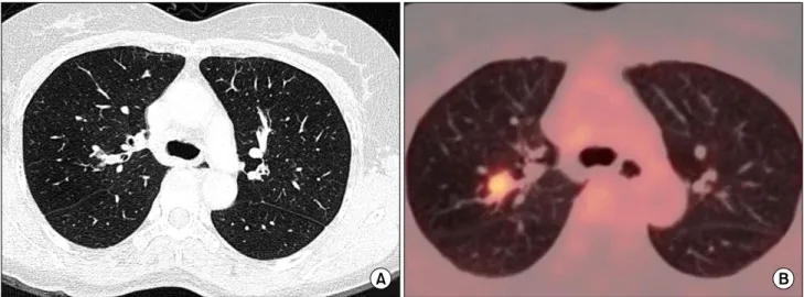
A 59-year-old asymptomatic female with a history of partial thyroidectomy for papillary thyroid cancer visited for the evaluation of pulmonary nodules ob- served in a low-dose chest computed tomography (CT) scan. There was a 1.1-cm nodule in the right upper lobe (RUL) and a 5-mm small ground glass nodule in the left upper lobe. The 1.1-cm RUL nodule was suspected to be primary lung cancer, and a con- trast CT scan was performed. In the contrast CT scan, the RUL nodule was observed as a 1.5-cm fin- ger-shaped lesion with a smooth margin in the sub- segmental bronchus of the posterior segment. It had an expansile appearance and showed mild homoge- neous enhancement (Fig. 1A). In positron emission tomography (PET) with 18-F fluorodeoxyglucose (FDG), the maximum standardized uptake value (SUV
max
) of the RUL endobronchial nodule was 4.1 (Fig. 1B).
https://doi.org/10.5090/kjtcs.2017.50.1.54