J Korean Soc Radiol 2016;74(3):204-209 http://dx.doi.org/10.3348/jksr.2016.74.3.204
INTRODUCTION
Malignant mixed tumors of the salivary glands are classified into three distinct histologic types: carcinoma ex pleomorphic adenoma, carcinosarcoma (malignant mixed tumor), and me- tastasizing pleomorphic adenoma. Metastasizing pleomorphic adenoma, a subset of pleomorphic adenoma that spreads to distant sites, is rare; very few cases have been recorded. So far, there has been no case report of metastasizing pleomorphic ad- enoma focusing on radiologic features of the tumor using mul- tiple imaging tools. Previous reported cases of metastasizing pleomorphic adenoma presenting as pulmonary metastasis in- volved multiple pulmonary metastases. Also, most patients
with metastasizing pleomorphic adenoma have a history of at least one local recurrence of pleomorphic adenoma prior to the detection of distant metastasis (1-7). Therefore, metastasizing pleomorphic adenoma presenting as a solitary pulmonary nod- ule without a history of local tumor recurrence is a rare mani- festation of the disease. This article presents a case of this very rare condition with a discussion of the clinical presentation and a review of the literature; we focus on the radiologic features of the adenoma visualized with multiple imaging modalities.
CASE REPORT
A 46-year-old female patient visited our hospital for evalua-
Multimodality Imaging of Metastasizing Pleomorphic Adenoma Presenting as a Solitary Pulmonary Nodule without Local Tumor Recurrence: A Case Report
국소 재발 병력 없이 고립성 폐 결절로 보이는 전이성 다형 선종의 다양한 영상 소견: 증례 보고
Seo Young Choi, MD
1, Woocheol Kwon, MD
1*, In Soo Hong, MD
1, Soon Hee Jung, MD
2Departments of 1Radiology, 2Pathology, Wonju Severance Christian Hospital, Yonsei University Wonju College of Medicine, Wonju, Korea
Pleomorphic adenoma is the most common neoplasm of the salivary gland. It is usually a well-circumscribed and slow-growing benign tumor. In rare instances, be- nign pleomorphic adenomas may metastasize and spread to distant sites in which case they are described as metastasizing pleomorphic adenomas. So far, there has been no case report of metastasizing pleomorphic adenoma focusing on radiologic features of the tumor using several different imaging tools. Furthermore, only a few cases of pleomorphic adenoma with metastasis to pulmonary sites have been re- ported, which usually present as multiple lung nodules. We report a rare case of metastasizing pleomorphic adenoma presenting as a solitary pulmonary nodule without prior history of local tumor recurrence with a particular focus on multimo- dality imaging of the tumor.
Index terms
Adenoma, Pleomorphic Neoplasm Metastasis Solitary Pulmonary Nodule
Received May 12, 2015 Revised September 15, 2015 Accepted September 19, 2015
*Corresponding author: Woocheol Kwon, MD Department of Radiology, Wonju Severance Christian Hospital, Yonsei University Wonju College of Medicine, 20 Ilsan-ro, Wonju 26426, Korea.
Tel. 82-33-741-1467 Fax. 82-33-732-8281 E-mail: [email protected]
This is an Open Access article distributed under the terms of the Creative Commons Attribution Non-Commercial License (http://creativecommons.org/licenses/by-nc/3.0) which permits unrestricted non-commercial use, distri- bution, and reproduction in any medium, provided the original work is properly cited.
tion and treatment of an incidentally discovered solitary pul- monary nodule. She had a medical history of asthma and surgi- cally excised pleomorphic adenoma of the left submandibular gland 9 years prior. An earlier computed tomography (CT)
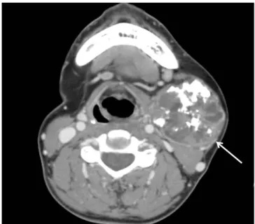
scan of the neck showed a 4.1 × 5.1 cm well-defined non-ho- mogenous enhancing mass in the left submandibular gland with internal calcifications (Fig. 1). She was asymptomatic at the time of the initial visit. All initial laboratory values were within nor- mal ranges.
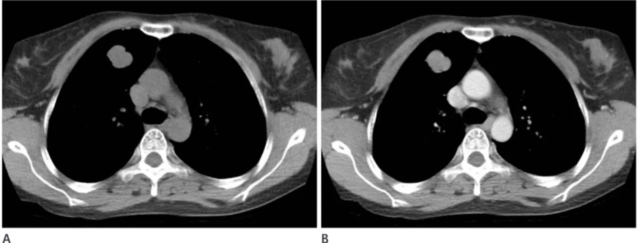
A chest radiograph showed a 2.4 cm, oval, well-defined soli- tary pulmonary nodule in the right upper lobe (Fig. 2). We re- viewed the chest CT scan and found a 2.2 × 2.0 × 2.2 cm well- defined nodule with lobulated margin and relatively homoge- nous enhancement (12 to 44 Hounsfield units) in the anterior segment of the right upper lobe. The pulmonary nodule showed no sign of cavitation, necrosis, or calcification (Fig. 3).
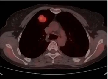
The initial list of differential diagnoses included sclerosing hemangioma, carcinoid, and, less likely, lung cancer. Magnetic resonance imaging (MRI) was performed in order to further characterize the structure of the affected tissues. MRI revealed intermediate to high signal intensity on T1-weighted images and heterogenous high signal intensity on T2-weighted images with strong enhancement on gadolinum-enhanced T1-weight- ed images (Fig. 4). A positron emission tomography-computed tomography (PET-CT) scan revealed increased fluorodeoxy- glucose (FDG) uptake in the right upper lobe nodule with a
Fig. 2. Chest radiograph of a 46-year-old female with metastasizing pleomorphic adenoma.
A, B. Chest radiograph of posterior-anterior projection (A) and right lateral projection (B) show a 2.4 cm, oval, well-defined solitary pulmonary nodule in the right upper lobe (arrows).
A B
Fig. 1. CT scan of the neck of a 46-year-old female with a history of left submandibular pleomorphic adenoma 9 years prior. Axial post- contrast image reveals a 4.1 × 5.1 cm well-defined non-homogenous enhancing mass in the left submandibular gland with internal calcifi- cations (arrow).
maximum standardized uptake value of 4.98 (Fig. 5). There was no other uptake in the maximum intensity projection image of the torso. Because of the notable FDG uptake, which is not usu- ally found in sclerosing hemangioma or carcinoid, we considered lung cancer to be a likely diagnosis after performing PET-CT.
CT-guided core-needle biopsy was performed for pathologic diagnosis. Given the presence of a previous submandibular gland tumor, the pathologic findings were consistent with me- tastasizing pleomorphic adenoma with the standard features of predominant proliferation of epithelial and mucoepithelial cells, immersed in a chondromyxoid stroma. Immunohistochemistry of the biopsy specimen showed diffuse strong positive reaction
for S-100 protein, multifocal weakly positive reaction for smooth muscle actin, and negative reaction for thyroid transcription fac- tor-1 (Fig. 6). Because there was a single metastasizing pulmo- nary pleomorphic adenoma, a decision to surgically remove the mass was made. Thus, the patient underwent right upper lobe lobectomy with video assisted thoracoscopic surgery. She has exhibited no remarkable postoperative complications during the 15 months since the procedure.
DISCUSSION
Pleomorphic adenoma is the most common neoplasm of the
Fig. 4. MR images of metastasizing plemorphic adenoma.
A-C. Axial T1-weighted (A), T2-weighted (B), and post-contrast enhanced T1-weighted image (C) of the lung show a solitary pulmonary nodule in the anterior segment of the right upper lobe with intermediate to high signal intensity on T1-weighted images, heterogenous high signal in- tensity on T2-weighted images, and with strong enhancement on gadolinium-enhanced T1-weighted images.
MR = magnetic resonance
A B C
Fig. 3. CT images of metastasizing plemorphic adenoma.
A, B. Axial pre-contrast image (A) and post-contrast image (B) show a 2.2 × 2.0 × 2.2 cm well-defined nodule with lobulated margin and rela- tively homogenous enhancement (12 to 44 Hounsfield units) in the anterior segment of the right upper lobe without cavitation, necrosis, or cal- cification.
CT = computed tomography
A B
salivary gland. It is usually a benign, slow-growing, and well- circumscribed tumor. Rarely, pleomorphic adenoma metasta- sizes to distant sites (1-7). The World Health Organization de- fines metastasizing pleomorphic adenoma as a “histologically benign pleomorphic adenoma that inexplicably manifests local or distant metastasis” (5-7). The mechanism underlying the metastatic behavior of salivary pleomorphic adenoma is not clear. The seeming paradox that benign entities occasionally ex- hibit aggressive behavior and also manifest as multiple metasta- ses is also true for other neoplasms of different organs such as metastatic giant cell tumor of bone, metastasizing leiomyoma-
tosis of the uterus, benign pheochromocytomas of the adrenal gland, lymphangiomatosis of the lymphatic system, and intra- cranial benign meningioma.
Most patients (72.8%) with metastasizing pleomorphic ade- noma have a history of at least a single incidence of local recur- rence of pleomorphic adenoma prior to the detection of distant metastasis. A meta-analysis of 81 case reports published from 1942 through 2014 uncovered no reports of metastasizing pleo- morphic adenoma as a solitary lung lesion without local tumor recurrence (5). There were two case reports of metastasizing pulmonary pleomorphic adenoma with no history local tumor recurrence that did not cite the number of lung lesions (8, 9).
We found incorrect classification in one of the two case reports:
it was in fact a case of hepatic metastasizing pleomorphic ade- noma. The other case was metastasizing pulmonary pleomor- phic adenoma with two lung lesions. Therefore, our case is the first report of solitary pulmonary metastasizing pleomophic adenoma without a history of local tumor recurrence.
CT findings of metastasizing pulmonary pleomorphic ade- noma have been reported as multiple, well-defined, variable- sized, ovular or round lung nodules (1-4). To our knowledge, MRI findings of metastasizing pulmonary pleomorphic adeno- ma have not been reported. Our case showed heterogenous high signal intensity on T2-weighted MRI images (Fig. 4). These findings are corroborated by histological findings of a mixture of epithelial tissue intermingled with chondromyxoid stroma.
The chondroid area with myxoid interstitium showed high sig- Fig. 5. FDG PET-CT image of metastasizing pleomorphic adenoma.
Axial fusion image shows increased FDG uptake (maximum standard- ized uptake value of 4.98) in the right upper lobe.
FDG = fluorodeoxyglucose, PET-CT = positron emission tomography- computed tomography
Fig. 6. Microscopic findings of pathologically confirmed metastasizing pleomorphic adenoma.
A, B. Low power micrograph (A) of the metastasizing pleomorphic adenoma shows an intrapulmonary well-demarcated cellular nodule (hema- toxylin and eosin, × 20). High power micrograph (B) of another field of the metastasizing pleomorphic adenoma shows proliferation of epithelial and myoepithelial cells immersed in a chondromyxoid stroma (arrow) (hematoxylin and eosin, × 100).
A B
nal intensity, whereas epithelial tissue showed relatively low sig- nal intensity, on T2-weighted images (10). Our case also points out that MRI is better than CT in tissue characterization as it more clearly shows heterogenous components of the lesion. In- deed, CT findings in our case suggested a relatively homoge- nous appearance. PET-CT scan findings of metastasizing pleo- morphic adenoma have been described as FDG-accumulated masses; our case is similar (7).
The treatment of choice for metastasizing pleomorphic ade- noma is total surgical resection. The effectiveness of adjuvant primary radiotherapy is a controversial issue. Recurrence after complete surgical removal is rare, and the prognosis is excel- lent. However, multiple metastases are deemed invariably fatal (1, 2, 5-7).
In conclusion, we described multimodality imaging features of a rare case of metastasizing pleomorphic adenoma present- ing as a solitary pulmonary nodule without local tumor recur- rence based on the findings of CT, MRI, and PET-CT. Hence, radiologists should be aware that metastasizing pleomorphic adenoma may be a differential diagnosis of solitary pulmonary nodule in cases with a prior history of pleomorphic adenoma.
REFERENCES
1. Rodríguez-Fernández J, Mateos-Micas M, Martínez-Tello FJ, Berjón J, Montalvo JJ, Forteza-González G, et al. Meta- static benign pleomorphic adenoma. Report of a case and review of the literature. Med Oral Patol Oral Cir Bucal 2008;
13:E193-E196
2. Sit KY, Chui WH, Wang E, Chiu SW. Multiple pulmonary metastases from benign pleomorphic adenoma. Asian Car- diovasc Thorac Ann 2008;16:62-64
3. Zhang Y, Gomez-Fernandez CR, Jorda M, Ganjei-Azar P.
Fine-needle aspiration (FNA) and pleural fluid cytology di- agnosis of benign metastasizing pleomorphic adenoma of the parotid gland in the lung: a case report and review of literature. Diagn Cytopathol 2009;37:828-831
4. Raja V, China C, Masaki KH, Nakano G. Unusual presenta- tions of uncommon tumors: case 1. Benign metastasizing pleomorphic adenoma. J Clin Oncol 2002;20:2400-2403 5. Knight J, Ratnasingham K. Metastasising pleomorphic ad-
enoma: systematic review. Int J Surg 2015;19:137-145 6. Santaliz-Ruiz LE, Morales G, Santini H, Sánchez-Santiago
M, Arroyo A. Metastasizing pleomorphic adenoma: a fas- cinating enigma. Case Rep Med 2012;2012:148103 7. Nouraei SA, Ferguson MS, Clarke PM, Sandison A, Sandhu
GS, Michaels L, et al. Metastasizing pleomorphic salivary adenoma. Arch Otolaryngol Head Neck Surg 2006;132:
788-793
8. Youngs GR, Scheuer PJ. Histologically benign mixed pa- rotid tumour with hepatic metastasis. J Pathol 1973;109:
171-172
9. Landolt U, Zöbeli L, Pedio G. Pleomorphic adenoma of the salivary glands metastatic to the lung: diagnosis by fine needle aspiration cytology. Acta Cytol 1990;34:101-102 10. Kinoshita T, Ishii K, Naganuma H, Okitsu T. MR imaging
findings of parotid tumors with pathologic diagnostic clues: a pictorial essay. Clin Imaging 2004;28:93-101
국소 재발 병력 없이 고립성 폐 결절로 보이는 전이성 다형 선종의 다양한 영상 소견: 증례 보고
최서영
1· 권우철
1* · 홍인수
1· 정순희
2다형성 선종은 가장 흔한 타액선 종양이다. 이는 흔히 경계가 좋고 천천히 자라는 양성 종양이다. 드물게, 조직학적 양성 의 다형성 선종은 원위부로 전이를 할 수 있는데 이를 전이성 다형 선종이라고 한다. 현재까지 전이성 다형 선종의 다면 영 상 소견에 초점을 둔 보고는 없다. 더욱이, 폐 전이를 동반한 전이성 다형 선종은 일부 드물게 보고되나, 주로 다발성 폐 전 이의 형태이다. 저자들은 원발부의 재발 병력 없이, 고립성 폐 결절로 보이는 드문 형태의 전이성 다형 선종 증례의 영상장 비별 다양한 영상 소견에 초점을 두어 보고하고자 한다.
연세대학교 원주의과대학 원주세브란스기독병원 1영상의학과, 2병리과