Clinical characteristics of second primary
pancreatic cancer
Jung Hyun Jo, In Rae Cho, Jang Han Jung, Hee Seung Lee, Moon Jae Chung,
Seungmin Bang, Seung Woo Park, Jae Bock Chung, Si Young Song, Jeong Youp Park*
Division of Gastroenterology, Department of Internal Medicine, Severance Hospital, Yonsei University College of Medicine, Seoul, Korea
*SENSASS@yuhs.ac
Abstract
Purpose
Several studies reported the increased risk of second primary pancreatic ductal adenocarci-noma (2nd PDAC) in cancer survivors. However, data on the characteristics of 2nd PDAC are insufficient.
Methods
This retrospective cohort study included 1759 patients with PDAC. They were classified as having 2nd PDAC or first primary PDAC (1st PDAC) according to a prior diagnosed cancer of different origin, at least 6 months before PDAC diagnosis.
Results
There were 110 patients (6.4%) with 2nd PDAC and 1606 (93.6%) patients with 1st PDAC. Patients with 2nd PDAC presented with older age (66.5 vs. 62.2 years, p<0.001) and higher rate of resectability (26.4% vs. 15.9%, p = 0.004) at diagnosis than those with 1st PDAC. Multivariate analysis without considering resectable status showed that 2nd PDAC (hazard ratio [HR] 0.73, 95% confidence interval [CI] 0.56–0.94, p = 0.016) was associated with bet-ter overall survival. Afbet-ter adjusting for resectable status, however, 2nd PDAC (HR 0.85, 95% CI 0.66–1.09, p = 0.198) was no longer associated with overall survival. When sub-groups were separately analyzed according to initial treatment modality, the effectiveness of surgery and chemotherapy were similar between 2nd and 1st PDAC (33.1 vs. 28.5 months, p = 0.860 and 10.8 vs. 10.7 months, p = 0.952).
Conclusions
The proportion of resectable cases was significantly higher in 2nd PDAC. When surgery with curative aim was possible, the overall survival was increased even in patients with 2nd PDAC. These results suggest the importance of screening for second primary cancer in can-cer survivors. a1111111111 a1111111111 a1111111111 a1111111111 a1111111111 OPEN ACCESS
Citation: Jo JH, Cho IR, Jung JH, Lee HS, Chung MJ, Bang S, et al. (2017) Clinical characteristics of second primary pancreatic cancer. PLoS ONE 12 (6): e0179784.https://doi.org/10.1371/journal. pone.0179784
Editor: Masaru Katoh, National Cancer Center, JAPAN
Received: February 3, 2017 Accepted: June 4, 2017 Published: June 26, 2017
Copyright:© 2017 Jo et al. This is an open access article distributed under the terms of theCreative Commons Attribution License, which permits unrestricted use, distribution, and reproduction in any medium, provided the original author and source are credited.
Data Availability Statement: All relevant data are within the paper.
Funding: The authors received no specific funding for this work.
Competing interests: The authors have declared that no competing interests exist.
Introduction
Second primary cancer is a new neoplasm that is biologically independent of a prior cancer.[1] The second primary cancer may share genetic factors or environmental background with the prior cancer, such as alcohol consumption, smoking, genetic syndromes/predisposition, and underlying diseases, which may be precancerous factors. Moreover, exposure to chemotherapy and radiation for prior cancer and receiving surgery can be predisposing factors to a second primary cancer. There were previous reports about an increased risk of secondary malignan-cies in cancer survivors.[2–4] The development of diagnostic and therapeutic techniques for malignancy has increased both the life span and the risk of second malignancies in cancer sur-vivors. In the United States, the proportion of cancer survivors among the total population is estimated to be 3.5%, and about 10% of newly diagnosed cancers develop in cancer survivors. [5,6] These data imply that understanding the characteristics of second primary cancers has become more important in developing screening and management programs for cancer survivors.
Pancreatic ductal adenocarcinoma (PDAC) is the fourth most common cause of cancer-related deaths and known to have an extremely poor prognosis, with a 5-year survival rate of
<6%.[5] The median overall survival (OS) is 9 months for locally advanced pancreatic cancer and 3–6 months for metastatic disease.[7] The only potentially curative therapy for pancreatic cancer is surgical resection, and only 20% of pancreatic cancers are resectable at the time of diagnosis. In addition to this dismal prognosis, to date, there is no effective screening tool for high-risk patients and early screening for PDAC is not recommended for the general popula-tion in terms of cost-effectiveness.[8]
Similar to patients with other second primary malignancies, there have been patients with PDAC who developed another primary tumor in another organ or with a different histology. To date, there are only a few studies about second primary PDAC (2nd PDAC). A study about 2nd PDAC with pooled analysis of international multicenter cancer registries reported that the prevalence of 2nd PDAC among all patients with PDAC was about 6.3%.[9] There have been a few studies which reported pancreatic cancer as a second primary malignancy, and observed that the risk of second primary pancreatic cancer was increased after the development of vari-ous malignancies.[10] Recently, a report about second primary pancreatic cancer in colorectal cancer survivors showed that these patients have an increased risk of developing second pri-mary pancreatic cancer.[11] However, although several studies have reported the increased risk of 2nd PDAC in cancer survivors, data on the characteristics of 2nd PDAC are insuffi-cient. Thus, we conducted a study on the characteristics of 2nd PDAC, with the premise that our results might help in developing follow-up and screening strategies for cancer survivors, and in elucidating the treatment for these patients with fatal PDAC.
Materials and methods
Study population and definitions
A retrospective patient cohort from Severance Hospital, Seoul, Korea, was examined. A total of 1759 patients with PDAC, diagnosed as having ductal adenocarcinoma of the pancreas according to histopathological findings between 2006 and 2014, were included in this study. The age of the participants ranged from 44 to 91 years old.
The patients in the cohort were classified as having 2nd PDAC or first primary PDAC (1st PDAC) according to the history of a prior cancer. Patients who met the following criteria were considered as having 2nd PDAC: 1) the histological type of the prior cancer was not adenocarcinoma; 2) if the histological type of the previous tumor was adenocarcinoma, the
possibility of primary pancreatic cancer should be higher than that of metastasis or recur-rence of the previous tumor based on specific immunohistochemical staining or a reliable review by an experienced pathologist; and 3) PDAC was diagnosed at least 6 months after the diagnosis of the prior cancer to exclude synchronous PDAC. If the patient had a history of more than one prior cancer, the time of diagnosis of the first cancer was applied into the criteria.
The following clinical characteristics of patients were collected: sex, age, smoking status, alcohol consumption, history of diabetes and hypertension, CA19-9 level at diagnosis, primary location of pancreatic cancer, resectability at diagnosis, initial treatment modality, origin of another primary tumor, period between the time of diagnosis of another primary tumor and that of the 2nd PDAC, and OS.
Statistical analysis
Data were analyzed by using theχ2, Fisher’s exact, and Student’s t-tests to compare variables between the groups, and a p-value of <0.05 was considered statistically significant. The OS and cumulative incidence rate of the 2nd PDAC after the diagnosis of other tumors were cal-culated and analyzed by using Kaplan-Meier analysis with a log-rank test. Hazard ratios (HRs), 95% confidence intervals (95% CIs), and p-values were calculated with a univariate Cox proportional hazards model for OS, by using the variables that were statistically cant. A multivariate Cox proportional hazards model was built to evaluate possible signifi-cant factors, taking into account the possible influence of confounding clinical variables. Only those variables that were significant in univariate analysis were entered into the multi-variate analysis. Statistical analyses were carried out with Predictive Analytics Software statis-tics version 23.
Ethical review
All procedures performed in studies involving human participants were in accordance with the ethical standards of the institutional research committee and with the 1964 Helsinki decla-ration and its later amendments or comparable ethical standards. As a retrospective analysis, Yonsei University Health System, Severance Hospital, Institutional Review Board approval was obtained and the need for informed consent was waived.
Results
Baseline characteristics
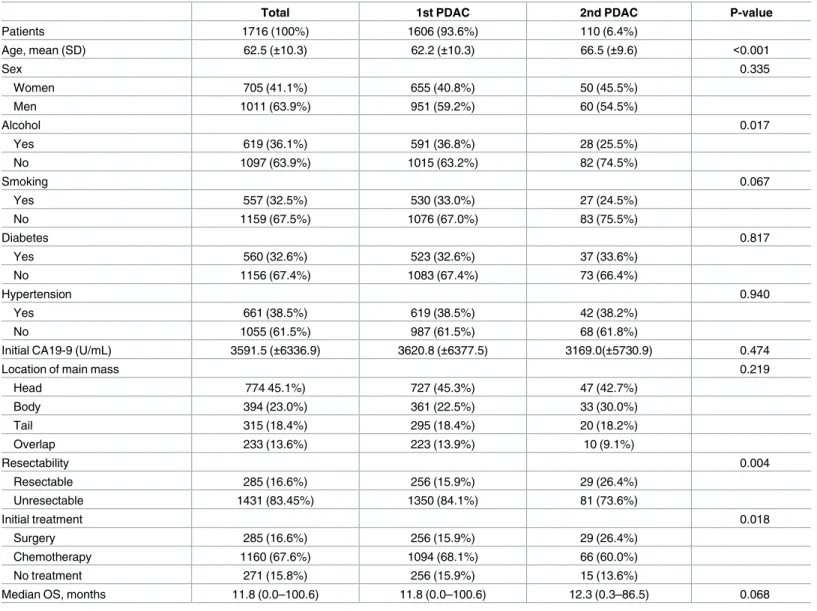
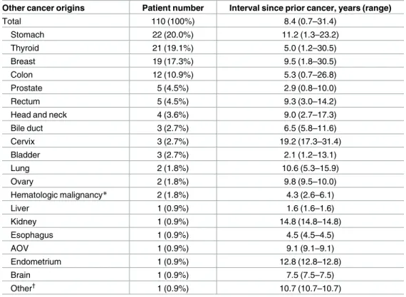
A total of 1759 patients with PDAC were included in the cohort. Forty-three patients were classified as having synchronous 2nd PDAC and excluded from the analysis. There were 1606 patients (93.6%) with 1st PDAC and 110 patients (6.4%) with 2nd PDAC in the study popula-tion (Table 1). Six patients (0.4%) had more than one prior cancer. In the comparison of base-line characteristics between the 1st PDAC and 2nd PDAC groups (Table 1), patients with 2nd PDAC presented significantly older age at diagnosis (66.5 vs. 62.2 years, p < 0.001), lower rate of alcohol consumption (25.5 vs. 36.8%, p = 0.017), higher rate of resectability of PDAC (26.4 vs. 15.9%, p = 0.004), and higher rate of receiving surgery as the initial treatment (26.4 vs. 15.9%, p = 0.018) than patients with 1st PDAC. The origins of prior cancers accompanied with 2nd PDAC are summarized inTable 2. The most common origin of prior cancers was the stomach (22 of 110, 20.0%), followed by the thyroid (21 of 110, 19.1%), breast (19 of 110, 17.3%), colon (12 of 110, 10.9%), and others.
Survival analysis
The OS was slightly longer in patients with 2nd PDAC; however, the difference was not signifi-cant (11.8 vs. 12.3 months, p = 0.068). Multivariate analysis without resectable status showed that 2nd PDAC (HR 0.73, 95% CI 0.56–0.94, p = 0.016), age at diagnosis (HR 1.02, 95% CI 1.01–1.02, p < 0.001), and alcohol consumption (HR 1.28, 95% CI 1.13–1.47, p < 0.001) were significantly related to OS (Table 3). When resectable status was included in multivariate anal-ysis, age at diagnosis (HR 1.02, 95% CI 1.01–1.02, p < 0.001), alcohol consumption (HR 1.25, 95% CI 1.11–1.42, p = 0.001), and resectable status at diagnosis (HR 0.30, 95% CI 0.25–0.36, p < 0.001) were significantly associated with OS. However, 2nd PDAC (HR 0.85, 95% CI 0.66–1.09, p = 0.198) was no longer significantly associated with OS after adjusting for resect-able status. This analysis suggested that the association between 2nd PDAC and survival was owing to the higher resectability rate.
Table 1. Baseline characteristics of subgroups.
Total 1st PDAC 2nd PDAC P-value
Patients 1716 (100%) 1606 (93.6%) 110 (6.4%) Age, mean (SD) 62.5 (±10.3) 62.2 (±10.3) 66.5 (±9.6) <0.001 Sex 0.335 Women 705 (41.1%) 655 (40.8%) 50 (45.5%) Men 1011 (63.9%) 951 (59.2%) 60 (54.5%) Alcohol 0.017 Yes 619 (36.1%) 591 (36.8%) 28 (25.5%) No 1097 (63.9%) 1015 (63.2%) 82 (74.5%) Smoking 0.067 Yes 557 (32.5%) 530 (33.0%) 27 (24.5%) No 1159 (67.5%) 1076 (67.0%) 83 (75.5%) Diabetes 0.817 Yes 560 (32.6%) 523 (32.6%) 37 (33.6%) No 1156 (67.4%) 1083 (67.4%) 73 (66.4%) Hypertension 0.940 Yes 661 (38.5%) 619 (38.5%) 42 (38.2%) No 1055 (61.5%) 987 (61.5%) 68 (61.8%)
Initial CA19-9 (U/mL) 3591.5 (±6336.9) 3620.8 (±6377.5) 3169.0(±5730.9) 0.474
Location of main mass 0.219
Head 774 45.1%) 727 (45.3%) 47 (42.7%) Body 394 (23.0%) 361 (22.5%) 33 (30.0%) Tail 315 (18.4%) 295 (18.4%) 20 (18.2%) Overlap 233 (13.6%) 223 (13.9%) 10 (9.1%) Resectability 0.004 Resectable 285 (16.6%) 256 (15.9%) 29 (26.4%) Unresectable 1431 (83.45%) 1350 (84.1%) 81 (73.6%) Initial treatment 0.018 Surgery 285 (16.6%) 256 (15.9%) 29 (26.4%) Chemotherapy 1160 (67.6%) 1094 (68.1%) 66 (60.0%) No treatment 271 (15.8%) 256 (15.9%) 15 (13.6%)
Median OS, months 11.8 (0.0–100.6) 11.8 (0.0–100.6) 12.3 (0.3–86.5) 0.068
Abbreviations: PDAC, pancreatic ductal adenocarcinoma; SD, standard deviation; OS, overall survival.
To exclude the confounding effect of the resectability of PDAC on OS, we divided the whole cohort according to the initial treatment modality for PDAC: 1) patients who received curative surgery and 2) patients who received chemotherapy. Then, the OS of patients with 1st PDAC and those with 2nd PDAC was compared with Kaplan-Meier analysis according to treatment modality (Fig 1). In the subgroup of patients who received curative surgery (Fig 1B), the median OS was 28.5 months (95% CI, 23.0–34.1) in the 1st PDAC group compared with 33.1 months (95% CI, 9.0–27.2) in the 2nd PDAC group (n: 259 vs. 29, p = 0.860). In the sub-group of patients who received chemotherapy (Fig 1C), the median OS was 10.7 months Table 2. Origins of prior cancers accompanied with second primary PDAC.
Other cancer origins Patient number Interval since prior cancer, years (range)
Total 110 (100%) 8.4 (0.7–31.4) Stomach 22 (20.0%) 11.2 (1.3–23.2) Thyroid 21 (19.1%) 5.0 (1.2–30.5) Breast 19 (17.3%) 9.5 (1.8–30.5) Colon 12 (10.9%) 5.3 (0.7–26.8) Prostate 5 (4.5%) 2.9 (0.8–10.0) Rectum 5 (4.5%) 9.3 (3.0–14.2)
Head and neck 4 (3.6%) 9.0 (2.7–17.3)
Bile duct 3 (2.7%) 6.5 (5.8–11.6) Cervix 3 (2.7%) 19.2 (17.3–31.4) Bladder 3 (2.7%) 2.1 (1.2–13.1) Lung 2 (1.8%) 10.6 (5.3–15.9) Ovary 2 (1.8%) 9.8 (9.5–10.0) Hematologic malignancy* 2 (1.8%) 4.3 (2.6–6.1) Liver 1 (0.9%) 1.6 (1.6–1.6) Kidney 1 (0.9%) 14.8 (14.8–14.8) Esophagus 1 (0.9%) 4.5 (4.5–4.5) AOV 1 (0.9%) 9.1 (9.1–9.1) Endometrium 1 (0.9%) 12.8 (12.8–12.8) Brain 1 (0.9%) 7.5 (7.5–7.5) Other† 1 (0.9%) 10.7 (10.7–10.7)
*Hematologic malignancy: one multiple myeloma and one aplastic large-cell lymphoma. †Other: a paratesticular tumor (soft tissue cancer).
Abbreviations: PDAC, pancreatic ductal adenocarcinoma; AOV, ampulla of Vater.
https://doi.org/10.1371/journal.pone.0179784.t002
Table 3. Cox proportional analysis for the contribution of clinical factors to overall survival.
Univariate Multivariate Multivariate*
P-value HR (95% CI) P-value HR (95% CI) P-value
Second PDAC 0.093 0.73 (0.56–0.94) 0.016 0.85 (0.66–1.09) 0.198
Older age <0.001 1.02 (1.01–1.02) <0.001 1.02(1.01–1.02) <0.001
Male sex 0.056 1.04 (0.90–1.19) 0.627 1.03(0.90–1.19) 0.645
Alcohol use (vs. non-alcohol use) 0.001 1.28 (1.13–1.47) <0.001 1.25 (1.11–1.42) <0.001 Resectable disease (vs. unresectable) <0.001 Not included 0.30 (0.25–0.36) <0.001
Abbreviations: PDAC, pancreatic ductal adenocarcinoma; SD, standard deviation; HR, hazard ratio; CI, confidence interval.
*Multivariate analysis including resectable disease and multivariate analysis without resectable disease are separately presented.
(95% CI, 10.0–11.4) in 1st PDAC compared with 10.8 months (95% CI, 9.2–12.3) in 2nd PDAC (n: 1094 vs. 66, p = 0.952). The effectiveness of either surgery or chemotherapy was sim-ilar between the 2nd and 1st PDAC groups.
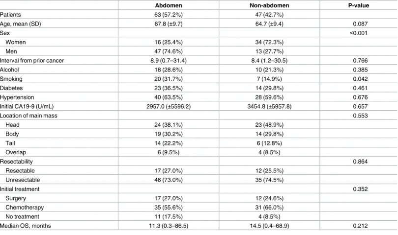
To compare differences according to the follow-up modality that might be different depending on the location of the prior cancer, we divided patients with 2nd PDAC into those with a prior cancer originating from the abdomen and those with prior cancer of non-abdomi-nal origin (Table 4). The origin of abdominon-abdomi-nal prior cancers included the gastrointestinon-abdomi-nal tract (stomach, colon, rectum), hepatobiliary tract (liver, bile duct), genitourinary tract (prostate, bladder, kidney), gynecologic system (cervix, endometrium, and ovary), and other sites in the abdominal and pelvic cavities. Other cancers originating from the thyroid, breast, head and neck, lung, and brain were included in the non-abdominal prior cancer group. There were 63 patients (57.2%) with abdominal prior cancer and 47 patients (42.7%) with non-abdominal prior cancer. In the comparison analysis, there were no significant differences in resectability (27.0 vs. 25.5%, p = 0.864) or OS (11.3 vs. 14.5 months, p = 0.212) between groups.
In addition, we analyzed ratios of incidental findings on abdominal follow-up diagnostics in comparison to symptomatic 2nd primary PDAC, which led to scan-diagnostics in the first place. There were 47 patients who were diagnosed with 2nd PDAC on follow-up abdominal CT/MRI scans and 63 patients who were diagnosed with 2nd PDAC by symptoms. Comparing the two groups, the rate of resectable disease was significantly higher in the patients who were diagnosed by follow-up abdominal CT/MRI scans than those diagnosed by symptoms presen-tation (36.2% vs. 19.0%, p = 0.044).
Cumulative incidence rate of 2nd PDAC after diagnosis of another
primary tumor
The median interval between the diagnosis of the 2nd PDAC and the diagnosis of the prior cancer was 8.4 years (range 0.7–31.4 years) in the 2nd PDAC group (Table 2). Among prior cancers with a ratio of >10%, the median interval of stomach cancer was the longest at 11.2 years (range 1.3–23.2 years). Thyroid cancer presented the shortest interval to 2nd PDAC at 5.0 years (range 1.2–30.5 years).Fig 2shows the cumulative incidence rate of 2nd PDAC after the diagnosis of prior cancer in the 2nd PDAC group. The trend of the first 5 years was 7 cases per year (35 cases every 5 years). Thereafter, every 5 years, the value decreased continuously to Fig 1. Kaplan-Meier analysis of overall survival (OS) according to subgroup. (A) First primary
pancreatic ductal adenocarcinoma (1st PDAC) vs. second primary PDAC (2nd PDAC) in the whole cohort (n: 1606 vs. 110): median OS 11.8 vs. 12.3 months, p = 0.068 by log-rank test. (B) 1st PDAC vs. 2nd PDAC in patients who received curative surgery as initial treatment (n: 256 vs. 29): median OS 28.5 vs. 33.1 months, p = 0.860 by log-rank test. (C) 1st PDAC vs. 2nd PDAC in patients who received chemotherapy as initial treatment (n: 1094 vs. 66): median OS 10.7 vs. 10.8 months, p = 0.952 by log-rank test.
6.4, 3.8, 2.8, 1.2, and 0.6 cases per year until 30 years. The cumulative incidence rate was 60.9% at 10 years, 90.9% at 20 years, and 99.1% at 30 years after the diagnosis of prior cancer.
Discussion
Previously, there were a few studies about 2nd PDAC with a population-based analysis. These studies reported that cancer survivors have an increased risk of developing 2nd PDAC.[10,12, 13] Although there have been reports about the increased risk of 2nd PDAC in cancer survi-vors, there have been few studies about how the 2nd PDAC was different from the 1st PDAC. Hackertet al reported features of patients with PDAC and history of extrapancreatic
malig-nancy compared to patients with PDAC as only tumor, however, the results of the study were limited to patients who underwent surgery for PDAC.[14] Considering the fatality of pancre-atic cancer, an increased risk of pancrepancre-atic cancer in cancer survivors necessitates more atten-tion on 2nd PDAC. In that context, we aimed to analyze the characteristics of 2nd PDAC in this study.
In this study, there were 110 patients with 2nd PDAC among a total of 1716 patients with PDAC. The proportion of patients with 2nd PDAC was about 6.4%, and this percentage was compatible with that reported in a previous study.[9] The considerable proportion of patients with 2nd PDAC in the entire pancreatic cohort again suggests that more attention should be paid to this specific subset of patients with pancreatic cancer.
Table 4. Comparison of subgroups according to the location of the prior cancer in second PDAC.
Abdomen Non-abdomen P-value
Patients 63 (57.2%) 47 (42.7%)
Age, mean (SD) 67.8 (±9.7) 64.7 (±9.4) 0.087
Sex <0.001
Women 16 (25.4%) 34 (72.3%)
Men 47 (74.6%) 13 (27.7%)
Interval from prior cancer 8.9 (0.7–31.4) 8.4 (1.2–30.5) 0.766
Alcohol 18 (28.6%) 10 (21.3%) 0.385
Smoking 20 (31.7%) 7 (14.9%) 0.042
Diabetes 23 (36.5%) 14 (29.8%) 0.461
Hypertension 40 (63.5%) 28 (59.6%) 0.676
Initial CA19-9 (U/mL) 2957.0 (±5596.2) 3454.8 (±5957.8) 0.657
Location of main mass 0.553
Head 24 (38.1%) 23 (48.9%) Body 19 (30.2%) 14 (29.8%) Tail 14 (22.2%) 6 (12.8%) Overlap 6 (9.5%) 4 (8.5%) Resectability 0.864 Resectable 17 (27.0%) 12 (25.5%) Unresectable 46 (73.0%) 35 (74.5%) Initial treatment 0.352 Surgery 17 (27.0%) 12 (24.6%) Chemotherapy 35 (55.6%) 31 (66.0%) No treatment 11 (17.5%) 4 (8.5%)
Median OS, months 11.3 (0.3–86.5) 14.5 (0.4–68.9) 0.212
Abbreviations: SD, standard deviation; OS, overall survival.
In our results, the patient age at diagnosis of 2nd PDAC was older than that of 1st PDAC (66.5 vs. 62.2 years, p < 0.001). Similarly, in a previous report, there were more patients >75 years old (46% vs. 32%) and fewer patients <65 years old (22% vs. 37%) in the 2nd PDAC group than in the 1st PDAC group.[9] This result concurs with another study which reported that patients with PDAC and history of extrapancreatic neoplasm presented at older age than patients with PDAC as only tumor in patients who underwent surgery (68 vs. 64 years, p < 0.0001).[14] There seemed to be a tendency that patients with second primary cancer are older than patients with first primary cancer at the time of diagnosis. A previous study about second primary non-small cell lung cancer (NSCLC) reported that the group of patients with NSCLC with a history of another tumor presented older age at diagnosis (71 vs. 66 years) than did patients with NSCLC as the only tumor, similar to our result.[15] The age at diagnosis might be different because old age is one of the risk factors of cancer and patients with second primary cancer had lived long enough to survive the prior cancer.
The rate of alcohol consumption (25.5 vs. 36.8%, p = 0.017) was lower in the 2nd PDAC group in our results. This might be because of the tendency of patients to stop drinking alcohol upon the diagnosis of cancer. Like our study, a previous study reported that the onset of cancer was significantly associated with cessation of alcohol consumption, based on cohort data from 9001 Korean patients.[16]
In the multivariate analysis after adjusting for confounding factors such as older age at diag-nosis of 2nd PDAC, 2nd PDAC was significantly associated with a longer OS compared with 1st PDAC (HR 0.73, p = 0.016). However, after adjusting for resectability at diagnosis, 2nd Fig 2. Cumulative curve of diagnosis of second pancreatic ductal adenocarcinoma (PDAC).
Cumulative incidence rate of second primary PDAC (2nd PDAC) after the diagnosis of prior cancer in the 2nd PDAC group. The median interval between the diagnosis of the 2nd PDAC and the diagnosis of the prior cancer was 8.4 years. The cumulative rates (cumulative cases for every 5 years after the diagnosis of prior cancer) are presented as a table.
PDAC was no longer significantly associated with OS. In fact, the rate of resectability of 2nd PDAC was significantly higher than that of 1st PDAC (26.4 vs. 15.9%, p = 0.004). Furthermore, more patients with 2nd PDAC could receive curative surgery, which could potentially increase their OS. When the whole cohort was divided into subgroups according to initial treatment modality (curative surgery or chemotherapy), there was no difference in OS between 2nd PDAC and 1st PDAC in both subgroups. These results suggest that the OS of the 2nd PDAC group seemed to be longer because there were more resectable cases at the time of diagnosis. Concerning the treatment, it can be presumed that that the nature of 2nd PDAC and 1st PDAC was not much different. When surgery of curative aim was possible, the OS was increased even in patients with 2nd PDAC. This result suggests the need to develop screening programs for detecting resectable pancreatic cancer in cancer survivors.
To identify the effect of follow-up methods that might be different depending on the ana-tomical distribution of the prior cancer, we compared the subgroups divided according to the location of prior cancer in the 2nd PDAC group. When comparing cancers of abdominal origin, which were closer to the pancreas, and those of non-abdominal origin, there were no significant differences between subgroups in terms of resectability (p = 0.864) or OS (p = 0.212). It seems that the origin of prior cancer was not related to the tendency of early detection of 2nd PDAC. Interestingly, the rate of resectable disease was significantly higher in the patients who were diagnosed by follow-up abdominal CT/MRI scans than those diag-nosed by presentation of symptomatic 2nd PDAC, which led to scan-diagnostics in the first place. It seemed that there was a correlation between early detection and follow-up methods, which suggested the necessity of developing screening programs for 2nd PDAC. However, there were only 110 patients with 2nd PDAC in this study, and follow-up intervals and methods were heterogeneous. A larger study with subgroup analysis according to the cell types or common etiologies shared by prior cancers should be conducted in the future. Most importantly, an optimal follow-up program for 2nd PDAC should be developed and evaluated.
The cumulative curve of diagnosis of 2nd PDAC is presented inFig 2. According to our analysis, the median interval from prior cancer to 2nd PDAC was 8.4 years, and the cumulative rate of diagnosis of 2nd PDAC continuously increased for 10 years with inclination similar to the first 5 years (6.4 and 5.8 cases per year). The graph was steadily increasing for 20 years with a 90.9% cumulative rate and then reached a plateau. This result implies that cancer screening in cancer survivors and regular follow-up need to continue up to 20 years after the diagnosis of prior cancer, which is longer than the usual current follow-up period for most cancers. In our study, the most common prior cancer of 2nd primary PDAC was stomach cancer (20.0%), fol-lowed by thyroid cancer (19.1%), breast cancer (17.3%), colon cancer (10.9%), and others. This result was compatible with the prevalence and OS of these cancers in Korea. According to Cancer Statistics 2013 in Korea,[17] the most common origin of cancer was the stomach (17.8%) and colon (14.6%) in men. In women, thyroid cancer (30.5%) was the most common and breast cancer (15.4%) was the second common malignancy. In our study, there were only a few patients with lung cancer or liver cancer as the prior cancer, although these cancers are highly prevalent in Korea. This result was presumed to be because of the poor survival of patients with liver and lung cancers.
This study has several limitations. The number of patients with 2nd PDAC was small com-pared with those with 1st PDAC, although the proportion of 2nd PDAC in the total cases of PDAC was compatible with a previous study. In addition, we did not consider the possibility of multicancer syndromes accompanied with genetic disorders and the presence of premalig-nant lesions of PDAC such as intraductal papillary mucinous tumor. To overcome such limita-tions, larger prospective studies are needed in the future.
Conclusions
Second primary pancreatic cancer had a higher rate of resectability, and there was no differ-ence in the effectiveness of curative surgery and chemotherapy between 2nd and 1st PDAC. Therefore, when curative surgery for 2nd PDAC is possible, it should be conducted similarly to curative surgery for 1st PDAC. Considering the increased risk of 2nd PDAC in cancer survi-vors and the fact that surgery is the only curative treatment for this fatal cancer, more efforts are needed to develop screening programs for second primary pancreatic cancer in cancer survivors.
Author Contributions
Conceptualization: JH Jo IRC JH Jung HSL MJC SB SWP SYS JBC JYP. Data curation: JH Jo JYP.
Formal analysis: JH Jo JYP. Investigation: JH Jo JYP.
Methodology: JH Jo IRC JH Jung HSL MJC SB SWP SYS JBC JYP.
Project administration: JH Jo IRC JH Jung HSL MJC SB SWP SYS JBC JYP. Resources: JH Jo JYP.
Supervision: JYP.
Validation: IRC JH Jung HSL MJC SB SWP SYS JBC. Visualization: JH Jo JYP.
Writing – original draft: JH Jo JYP.
Writing – review & editing: JH Jo IRC JH Jung HSL MJC SB SWP SYS JBC JYP.
References
1. Boice JD Jr., Storm HH, Curtis RE, Jensen OM, Kleinerman RA, Jensen HS, et al. Introduction to the study of multiple primary cancers. Natl Cancer Inst Monogr. 1985; 68:3–9. PMID:4088304
2. Soliman H, Agresta SV. Current issues in adolescent and young adult cancer survivorship. Cancer Con-trol. 2008; 15(1):55–62. PMID:18094661
3. Kebudi R, Ozdemir GN. Second malignant neoplasms in childhood cancer survivors. Curr Pediatr Rev. 2016.
4. Donin N, Filson C, Drakaki A, Tan HJ, Castillo A, Kwan L, et al. Risk of second primary malignancies among cancer survivors in the United States, 1992 through 2008. Cancer. 2016; 122(19):3075–86. https://doi.org/10.1002/cncr.30164PMID:27377470
5. Howlader N NA, Krapcho M, Miller D, Bishop K, Altekruse SF, Kosary CL, Yu M, Ruhl J, Tatalovich Z, Mariotto A, Lewis DR, Chen HS, Feuer EJ, Cronin KA (eds). SEER Cancer Statistics Review, 1975– 2013, National Cancer Institute. Bethesda, MD,http://seer.cancer.gov/csr/1975_2013/, based on November 2015 SEER data submission, posted to the SEER web site, April 2016. [updated April 2016. 6. Ryerson AB, Eheman CR, Altekruse SF, Ward JW, Jemal A, Sherman RL, et al. Annual Report to the
Nation on the Status of Cancer, 1975–2012, featuring the increasing incidence of liver cancer. Cancer. 2016; 122(9):1312–37.https://doi.org/10.1002/cncr.29936PMID:26959385
7. Chong I, Cunningham D. Pancreatic Cancer. 2012 [cited October 11, 2012.]. In: Harrison’s Principles of Internal Medicine [Internet]. New York: McGraw-Hill. 18th [cited October 11, 2012.].http://www. accessmedicine.com.ymlib.yonsei.ac.kr/content.aspx?aID=9116312.
8. Canto MI, Harinck F, Hruban RH, Offerhaus GJ, Poley JW, Kamel I, et al. International Cancer of the Pancreas Screening (CAPS) Consortium summit on the management of patients with increased risk for
familial pancreatic cancer. Gut. 2013; 62(3):339–47.https://doi.org/10.1136/gutjnl-2012-303108PMID: 23135763
9. Shen M, Boffetta P, Olsen JH, Andersen A, Hemminki K, Pukkala E, et al. A pooled analysis of second primary pancreatic cancer. Am J Epidemiol. 2006; 163(6):502–11.https://doi.org/10.1093/aje/kwj073 PMID:16421239
10. Neugut AI, Ahsan H, Robinson E. Pancreas cancer as a second primary malignancy. A population-based study. Cancer. 1995; 76(4):589–92. PMID:8625151
11. Rahimi E, Batra S, Thosani N, Singh H, Guha S. Increased Incidence of Second Primary Pancreatic Cancer in Patients with Prior Colorectal Cancer: A Population-Based US Study. Dig Dis Sci. 2016; 61 (6):1652–60.https://doi.org/10.1007/s10620-016-4170-xPMID:27107866
12. Amin S, McBride RB, Kline JK, Mitchel EB, Lucas AL, Neugut AI, et al. Incidence of subsequent pancre-atic adenocarcinoma in patients with a history of nonpancrepancre-atic primary cancers. Cancer. 2012; 118 (5):1244–51.https://doi.org/10.1002/cncr.26414PMID:21887676
13. Hemminki K, Li X. Familial and second primary pancreatic cancers: a nationwide epidemiologic study from Sweden. Int J Cancer. 2003; 103(4):525–30.https://doi.org/10.1002/ijc.10863PMID:12478670 14. Hackert T, Tjaden C, Muller S, Hinz U, Hartwig W, Strobel O, et al. Extrapancreatic malignancies in
patients with pancreatic cancer: epidemiology and clinical consequences. Pancreas. 2012; 41(2):212– 7.https://doi.org/10.1097/MPA.0b013e3182240602PMID:21934549
15. Duchateau CS, Stokkel MP. Second primary tumors involving non-small cell lung cancer: prevalence and its influence on survival. Chest. 2005; 127(4):1152–8.https://doi.org/10.1378/chest.127.4.1152 PMID:15821189
16. Park JE, Ryu Y, Cho SI. The Association Between Health Changes and Cessation of Alcohol Consump-tion. Alcohol Alcohol. 2016.
17. Oh CM, Won YJ, Jung KW, Kong HJ, Cho H, Lee JK, et al. Cancer Statistics in Korea: Incidence, Mor-tality, Survival, and Prevalence in 2013. Cancer Res Treat. 2016; 48(2):436–50.https://doi.org/10. 4143/crt.2016.089PMID:26987395