J Korean Soc Radiol 2016;74(4):267-272 http://dx.doi.org/10.3348/jksr.2016.74.4.267
INTRODUCTION
Thyroid cancer is usually considered a slow growing malig- nancy with good prognosis. Common manifestation of thyroid carcinoma is a palpable neck mass due to primary thyroid nod- ule or less frequently, metastases in regional lymph nodes. How- ever, distant metastasis from thyroid carcinoma is often report- ed in the advanced stage of disseminated disease and lung and bone are the major sites (1, 2). Metastasis to the breast from thy- roid carcinoma is quite rare, with approximately 16 cases report- ed in the English literature, to our best knowledge. Neverthe-
less, recognizing metastases from thyroid cancer has a significant impact on the clinical decision and patient’s prognosis. We re- ported an uncommon case of 2 metastatic breast tumors and a small neck nodule from poorly differentiated thyroid carcinoma in a 72-year-old woman, 6 years after total thyroidectomy for pap- illary thyroid carcinoma. These lesions were detected on the posi- tron emission tomography/computed tomography (PET/CT) and confirmed by ultrasonography (USG)-guided biopsy after lesion delineation with the fusion images of computed tomography (CT) navigation and USG. This case report was approved by the ethics committee at our institution and informed consent was waived.
Two Breast Metastases from Thyroid Carcinoma Presented 6 Years Later after Total Thyroidectomy: A Case Report
갑상선 전절제술 6년 후 두 개의 유방 전이로 재발한 갑상선암: 증례 보고
Gene Hyuk Kwon, MD
1, Bong Joo Kang, MD
1*, Na Young Jung, MD
2, Sung Hun Kim, MD
1, Ahwon Lee, MD
3Departments of 1Radiology, 3Hospital Pathology, Seoul St. Mary’s Hospital, College of Medicine, The Catholic University of Korea, Seoul, Korea
2Department of Radiology, Bucheon St. Mary’s Hospital, College of Medicine, The Catholic University of Korea, Bucheon, Korea
Thyroid carcinoma is usually indolent with good prognosis, as compared to other ma- lignancy. Distant metastases from thyroid cancer are rare and usually manifest as multiple lesions especially in lungs, bones and lymph nodes, in advanced stages of the disease. Metastasis to the breast from thyroid carcinoma is extremely rare, with about 16 cases reported in the English literature. Herein, we reported a case of met- astatic poorly differentiated thyroid carcinoma, which presented as 2 breast masses in a 72-year-old woman, 6 years after total thyroidectomy for papillary thyroid car- cinoma. Although the computed tomography (CT) and ultrasonography (USG) image findings are nonspecific oval mass with circumscribed or partially indistinct margin, metastases from thyroid cancer should be included in the differential diagnosis when recurrence of thyroid carcinoma is suspected. Also, fusion images of CT and USG are helpful to the radiologists in localizing the targeted lesion and conducting accurate USG-guided biopsy.
Index terms Thyroid Carcinoma Breast
Metastasis
Computed Tomography Ultrasonography
Positron-Emission Tomography
Received August 18, 2015 Revised October 1, 2015 Accepted November 25, 2015
*Corresponding author: Bong Joo Kang, MD Department of Radiology, Seoul St. Mary’s Hospital, College of Medicine, The Catholic University of Korea, 222 Banpo-daero, Seocho-gu, Seoul 06591, Korea.
Tel. 82-2-2258-6253 Fax. 82-2-2258-6771 E-mail: [email protected]
This is an Open Access article distributed under the terms of the Creative Commons Attribution Non-Commercial License (http://creativecommons.org/licenses/by-nc/3.0) which permits unrestricted non-commercial use, distri- bution, and reproduction in any medium, provided the original work is properly cited.
CASE REPORT
A 72-year-old woman with a palpable posterior neck mass for several months visited our hospital and underwent mass excision, which was confirmed as poorly differentiated thyroid carcinoma.
She had a history of total thyroidectomy 6 years prior for papillary thyroid carcinoma, followed by neck lymph nodes excision and radioisotope therapy due to recurrent neck lymphadenopathy 5 years prior. Her thyroglobulin (Tg) antigen level after the posteri- or neck mass excision was elevated to 1.87 ng/mL. Also, no ab- normality was noted on a mammography taken 6 years prior.
The patient underwent the PET/CT (Biograph 40 True Point;
Siemens Medical Solutions, Knoxville, TN, USA) and chest CT scan for the systemic evaluation of recurrent thyroid carcinoma at the posterior neck. Homogeneous 18F-fluorodeoxyglucose (FDG) uptake was detected in the 2 nodular lesions, measuring from 8.2 to 10.9 maximum standardized uptake value (SUVmax).
The non-enhanced axial images of PET/CT and enhanced axial images of chest CT (SOMATOM Definition, Siemens, Erlar- gen, Germany) showed 2 soft tissue attenuated nodules in the right breast with moderate enhancement (Hounsfield unit in pre-contrast 30–35; after contrast enhancement 55–65): 1 in
periareolar region of inner portion and the other in peripheral region of the outer lower portion (Fig. 1A, B). Breast USG (AplioTM 500 TUS-A500, Toshiba Medical Systems, Tokyo, Ja- pan) revealed 2 slight hypoechoic masses in the right breast, 1 in the periareolar region of 3 o’clock direction and the other in peripheral region of 8 o’clock direction (Fig. 1C). The periareolar mass was an oval, circumscribed, non-parallel oriented lesion, measuring about 1.2 × 0.7 × 0.9 cm. On the Doppler image, peri-tumoral increased vascularity was present and intermedi- ate elastogram score was detected. On the other hand, the 8 o’clock peripheral breast mass was an about 1.0 × 0.7 × 0.5 cm sized oval lesion with microlobulated or partially indistinct margin, surrounded by fat. Both lesions were co-registered with CT nav- igated fusion USG, and FDG-avid 2 nodules on PET/CT were matched to the 2 hypoechoic masses on CT-USG fusion images.
The patient underwent USG-guided core needle biopsy. On microscopic examination, the mass consisted of small nests of atypical or malignant cells with increased mitosis. The small nests of cells were predominantly solid in nature, but focally con- tained eosinophilic colloidal materials (Fig. 2A). Immunohisto- chemical staining was positive for Tg and thyroid transcription factor-1 (TTF1) (Fig. 2B, C). The above histological and immu-
A B C
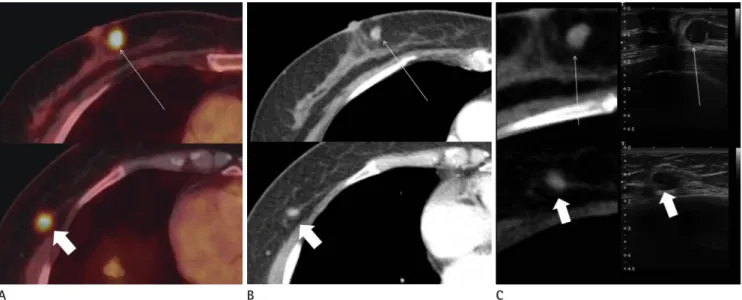
Fig. 1. Imaging findings of metastatic tumors in the right breast from thyroid carcinoma in a 72-year-old woman, 6 years after initial thyroidectomy.
A-C. Periareolar lesion in the 3 o’clock direction (long thin white arrows; upper column) and another peripheral lesion in the 8 o’clock direction (short thick white arrows; lower column) of the right breast are shown in the positron emission tomography/computed tomography (PET/CT), chest CT, ultrasonography (USG), and CT navigated USG fusion images. 18F-fluorodeoxyglucose (FDG) uptake in 2 nodular lesions on axial image of PET/CT (A) is noted in the inner portion and outer lower portion of the right breast. The lesions are soft tissue attenuated and homogenously enhanced after contrast injection on axial image of chest CT scan (B). On CT navigated USG fusion images (C), the periareolar lesion is oval, cir- cumscribed, non-parallel oriented mass with hypo- to isoechogenicity, surrounded by premammary fat. The other lesion is visualized as oval, par- tially indistinct marginated and hypoechoic mass with parallel orientation. CT navigated USG fusion image reaffirms that prior FDG-avid nodules are matched to the hypoechoic masses on sonogram, surrounded by fat.
nohistochemical findings were consistent with poorly differen- tiate thyroid carcinoma, which was the same pathology found in the excised palpable posterior neck mass.
DISCUSSION
Papillary carcinomas of the thyroid are characterized by indo- lent and slow clinical course. It usually remains localized to the thyroid gland. Distant metastases are seen in a minority of pa- tients and presence of distant metastases is the most significant poor prognostic factor for survival. The lung is the most common site of distant metastases (2). Other distant metastases are rela- tively rare and involve the bones, brain, liver, kidney, muscle, skin and breast, as in the presented case.
Breast is an uncommon site of metastasis and it is usually as- sociated with other disseminated systemic metastases. The most common metastases in the breast are from carcinomas arising in the contralateral breast, lung cancer, malignant melanoma, lymphoma, ovarian carcinoma, and gastrointestinal or genitouri- nary carcinomas (3). Metastasis to the breast from thyroid can-
cer is quite rare and few cases are reported in the English litera- ture (1, 4-10). Clinico-radiological features of reported cases in available references and our cases were summarized on Table 1.
Most of the cases were in females and histologic types of prima- ry thyroid carcinoma were heterogeneous; however, most com- mon type was from the medullary carcinoma followed by pap- illary carcinoma. Metastatic thyroid carcinoma to the breast tends to be superficially located, especially fat-glandular inter- face, as presented our case (2, 3). Contrary to expectation, of the 10 cases with imaging featuring metastatic breast tumors from thyroid carcinoma, single mass involving unilateral breast was noted as the most common manifestation (6 out of 10, 60%), fol- lowed by multiple masses involving both breasts (2 out of 10, 20%). Multiple masses involving single breast is least likely to oc- cur among all cases (1 out of 10, 10%), which is the currently pre- sented case. The echogenicity of metastatic tumors in the breast from thyroid carcinoma are variable. According to published research, 7 cases presented as a breast mass after thyroidectomy, of which, 5 occurred > 5 years after initial treatment for differ- entiated thyroid carcinoma; hence, the necessity for careful life-
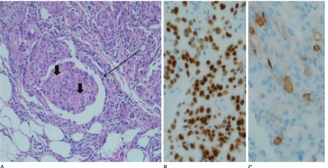
Fig. 2. Pathologic findings of metastatic tumors in the right breast from thyroid carcinoma in a 72-year-old woman, 6 years after initial thyroid- ectomy.
A. Microscopic photomicrograph illustrates that small nests of tumor cells are predominantly solid and focally contain eosinophilic colloidal mate- rials (short thick black arrows). The neoplastic cell nests show increased mitosis and locate within cells of a mammary duct and interstitium (long thin black arrow) (original magnification, × 200; H&E stain).
B, C. Immunohistochemical staining shows the tumor cells are positive for TTF1 (B) and thyroglobulin (C), which appear brown suggestive of thyroid tissue origin (original magnification, × 400). Scanty expression of thyroglobulin and increased Ki-67 labeling index confirms thyroid carcinoma with poor differentiation.
H&E = hematoxylin and eosin, TTF1 = thyroid transcription factor-1
A B C
Table 1. Literature Review of Breast Metastasis from Thyroid Cancer and Its Characteristics Reference YearPrimary PathologySexAgeOther MetastasisInterval between Initial Thyroidectomy and Breast Metastasis (Year)
Involving Breast (Right or Left or Both)
Metastatic Breast Tumor Features† (Mass Number, Size, Location, Imaging Features on Mammography or Ultrasound) Chisholm et al. (6)1980Follicular carcinomaFemale75Lymph nodes, skeletal muscle, skin, lungs
9 yearsRightSingle, 4 × 5 cm, upper inner quadrant, firm mass partially fixed to the chest Ordóñez et al. (4)1988Medullary carcinomaFemale72Lymph nodes22 yearsRightSingle, 5 cm, upper, firm mass Cristallini et al. (5)1994Follicular carcinomaFemale57Lymph nodesSynchronously detectedLeftSingle, 2 cm, superficial, upper outer quadrant Ill-defined margin without microcalcification on mammography Loureiro et al. (1)1997Papillary carcinomaFemale64Subcutaneous nodules, skin
7 yearsRightSingle, 1.5 cm Al-Abed et al. (8)2008Hurthle cell carcinomaFemale77Lymph nodes10 months*LeftSingle, 1.1 cm, upper inner quadrant Hyperdense, oval, lobulated mass on mammography Marcy et al. (10)2009Medullary carcinomaFemale43Lymph nodes, lungs, bones15 yearsBothMultiple Angeles-Angeles et al. (7)2009Papillary carcinomaFemale58Lymph nodes17 yearsLeftSingle, 8.4 cm Mandanas et al. (9)2015Medullary carcinomaMale67Lymph nodes, lungs, bones, liver
N/ABothMultiple Hyperdense, oval, lobulated masses on mammography Hypoechoic, oval to irregular, circumscribed masses on ultrasound The presented case2015Papillary carcinomaFemale72Posterior neck subcutaneous
6 yearsRightTwo, less than 1.2 cm Hypoechoic, oval, parallel or non-parallel, lobulated or indistinct marginated on ultrasound N/A: not applicable or no comment about the information on the paper or described as only ‘during the periods of follow-up’. *Less than 1 year between thyroidectomy and breast metastatic tumor confirmation. †Described in the order of features mentioned above. Features are described only when not described on previous reference.
long follow-up in the case of thyroid carcinoma.
Although USG and CT image findings were nonspecific in ur case, the newly noted metastatic breast tumors showed oval, hy- poechoic, circumscribed or partially indistinct marginated mass- es with peri-tumoral vascularity and intermediate elastogram that should be categorized as 4B by BIRADS, indicative as patho- logic confirmation. Moreover, avid FDG uptake in both newly noted breast nodules on PET/CT at the fat-glandular interface supported the possibility of metastatic breast tumors. During USG-guided procedure, the navigated fusion images of CT and USG helped radiologists match FDG-avid tumors on PET/CT to the hypoechoic masses on USG, facilitating easier localization of breast mass from other cysts or similar hypoechoic lesions in the same breast.
Several immunohistochemical tumor markers are used in the diagnosis of cancers and combination of these markers is benefi- cial to identify the origins of malignancy. Tg, produced by thy- roid tissues alone, is a valuable marker for distinguishing cancers of thyroid origin from other organs (7). In our case, poorly dif- ferentiated thyroid carcinoma within the breast metastasis oc- curred 6 years after initial thyroidectomy for papillary carcino- ma. Compared with the initial thyroidectomy specimen, tissue sampled under USG-guidance showed decreased but mildly ex- pressed Tg in the metastatic breast tumors, suggesting its origin.
In conclusion, the imaging findings of breast metastasis from thyroid carcinoma are nonspecific for metastatic or primary breast malignancy, but differential diagnosis should be included in the clinical setting of thyroid cancer recurrence. Incidentally found benign or malignant breast tumors on CT or PET/CT scans can be easily localized on USG/CT navigated fusion imag- es, facilitating more accurate USG-guided biopsy.
REFERENCES
1. Loureiro MM, Leite VH, Boavida JM, Raposo JF, Henriques MM, Limbert ES, et al. An unusual case of papillary carci- noma of the thyroid with cutaneous and breast metasta- ses only. Eur J Endocrinol 1997;137:267-269
2. Song HJ, Xue YL, Xu YH, Qiu ZL, Luo QY. Rare metastases of differentiated thyroid carcinoma: pictorial review. En- docr Relat Cancer 2011;18:R165-R174
3. Akçay MN. Metastatic disease in the breast. Breast 2002;
11:526-528
4. Ordóñez NG, Katz RL, Luna MA, Samaan NA. Medullary thy- roid carcinoma metastatic to breast diagnosed by fine-nee- dle aspiration biopsy. Diagn Cytopathol 1988;4:254-257 5. Cristallini EG, Ascani S, Nati S, Liberati F, Farabi R. Breast
metastasis of thyroid follicular carcinoma. Acta Oncol 1994;
33:71-73
6. Chisholm RC, Chung EB, Tuckson W, Khan T, White JE. Fol- licular carcinoma of the thyroid with metastasis to the breast. J Natl Med Assoc 1980;72:1101-1104
7. Angeles-Angeles A, Chable-Montero F, Martinez-Benitez B, Albores-Saavedra J. Unusual metastases of papillary thyroid carcinoma: report of 2 cases. Ann Diagn Pathol 2009;13:
189-196
8. Al-Abed Y, Gray E, Wolfe K, Watters GW, Philpott JM.
Metastatic Hurthle Cell Carcinoma of the thyroid present- ing as a breast lump: a case report. Int Semin Surg Oncol 2008;5:14
9. Mandanas S, Margaritidou E, Christoforidou V, Karoglou E, Geranou C, Chrisoulidou A, et al. Breast metastasis from medullary thyroid carcinoma in a male patient: case report and review of the literature. Rare Tumors 2015;7:5765 10. Marcy PY, Thariat J, Peyrottes I, Dassonville O. Bilateral
breast involvement in medullary thyroid carcinoma. Thy- roid 2009;19:197-199
갑상선 전절제술 6년 후 두 개의 유방 전이로 재발한 갑상선암:
증례 보고
권진혁
1· 강봉주
1* · 정나영
2· 김성헌
1· 이아원
3갑상선암종은 다른 악성 종양과 비교하여 진행이 더디고, 좋은 예후를 보인다. 갑상선암에서 전이는 드문 편이며, 주로 폐, 뼈, 림프절 등의 다발성 병변으로 병의 말기에 나타난다. 갑상선암에서 유방으로의 전이는 매우 드물며, 16개 증례의 영문 문헌보고가 있었다. 이에 저자들은 72세 여성에서 갑상선 유두암으로 전절제술 시행 6년 후, 두 개의 유방 종괴로 재발한 전이성 미분화성 갑상선암종의 증례를 보고하고자 한다. 전산화단층촬영과 초음파 영상소견은 타원모양으로, 경계가 좋 거나 또는 일부 불명확한 종괴의 비특이적 소견이나, 갑상선암의 재발이 의심되는 상황에서 유방의 전이성 병변을 감별진 단으로 반드시 고려해야 한다. 또한 전산화단층촬영과 초음파의 융합영상은 영상의학과 의사가 목표하는 병변의 위치를 파악하여 정확한 초음파 유도하 조직검사를 시행하는 데 도움을 준다.
가톨릭대학교 의과대학 서울성모병원 1영상의학과, 3병리과, 2가톨릭대학교 의과대학 부천성모병원 영상의학과