INTRODUCTION
Although distant metastases from renal cell carcinoma (RCC) are not uncommon, the appearance of metastatic lesions in the extracranial region of head and neck is unusual. There have been a limited number of such cases published in the literature, mostly cancers located in the parotid gland, thyroid gland, pa- ranasal sinuses and the orbit. Intramasseteric neoplasms are re- ported with a low incidence, and there is no specific imaging finding. Therefore, it is important that radiologists have knowl- edge about the possible primary and secondary diseases of masseter muscle, and can suggest appropriate further workup or prompt management. We present a rare case of intermasse- teric metastasis of RCC.
CASE REPORT
A 66-year-old male presented at our hospital with a 15 day history of a nontender palpable mass measuring approximately
1.5 cm in his left cheek. He refused further evaluation at that time and we visited him again after three months. The lesion became progressively larger in size but did not, as ever, cause pain or other symptoms. There was no evidence of ongoing in- flammatory process such as pain or erythema. He had no tris- mus and no history of trauma. He denied medical histories ex- cept pulmonary tuberculosis about 40 years ago. Physical examination revealed an approximately 3-cm firm, immobile and non-tender mass in the left buccal space. There was no other abnormal palpable finding in the neck region. Facial nerve func- tion was normal on both sides. Computed tomography (CT) examination demonstrated a well-circumscribed, lobulated, soft tissue mass hypodense to adjacent muscle, and measuring 3 × 2.5 × 2.5 cm, in the left masticator space (Fig. 1). Necrosis, hemorrhage, and focal calcification were absent. After intrave- nous administration of contrast agent, the mass showed strong and heterogeneous enhancement, and prominent serpentine vascular structures were noted near the lesion which were thought to be feeding vessels. The mass was confined to the masseter
J Korean Soc Radiol 2012;66(2):123-127
Received September 22, 2011;
Accepted October 27, 2011
Corresponding author: Han Bee Lee, MD Department of Radiology, Sanggye Paik Hospital, Inje University College of Medicine, 1342 Dongil-ro, Nowon-gu, Seoul 139-707, Korea.
Tel. 82-2-950-1296 Fax. 82-2-950-1220 E-mail: [email protected]
Copyrights © 2012 The Korean Society of Radiology
Renal cell carcinoma (RCC) is often concomitant with distant metastasis, and these metastases are the first sign of an otherwise occult primary. Whereas metastasis of RCC to the head and neck has been reported, metastasis to the masseter muscle, which is composed of skeletal muscle, is quite rare. We now report the case of a 66-year-old man who had a past history of pulmonary tuberculosis, with RCC metas- tasis of a well-defined intensely enhancing hypervascular mass in the masseter mus- cle as the initial presentation. We present the imaging findings of this case and a liter- ature review about radiologic differential diagnosis of intramasseteric masses.
Index terms
Metastatic Renal Cell Carcinoma Masseter Muscle
Masticator Space
Initial Presentation of Renal Cell Carcinoma as a Metastatic Mass within the Masseter Muscle: A Case Report and Literature Review
1 저작근을 침범한 전이종괴로 처음 발견된 신세포암: 증례 보고와 문헌조사1Kyung Eun Bae, MD
1, Han Bee Lee, MD
1, Woo Ho Cho, MD
1, Jae Hyung Kim, MD
1, Jihae Lee, MD
1, Mi-Jin Kang, MD
1, Hyun-Jung Kim, MD
2Departments of 1Radiology, 2Pathology, Sanggye Paik Hospital, Inje University College of Medicine, Seoul, Korea
yellowish gray to brown without hemorrhage and necrosis. The final pathological diagnosis was metastatic carcinoma, morpho- logically and immunohistochemically compatible with a clear cell type of RCC with positive staining at CD10, pancytokeratin and vimentin with frequent lympho-vascular invasion (Fig. 2).
With this information, an abdominopelvic CT was performed, which showed a lobulated contour bulging mass (6.1 × 4.7 cm) in the upper pole of the left kidney with a heterogeneously well- enhancing appearance in the corticomedullary phase (Fig. 3).
The patient underwent left radical nephrectomy and the mass was found to be clear cell RCC (Fig. 4). Lung metastasis was found in both lungs on chest CT. The patient received chemo- therapy and immunotherapy. However, follow-up chest CT af- muscle without evidence of either fascial invasion or bone de-
struction. There was no significant cervical lymphadenopathy.
With this information, our initial impression of the mass was hemangiopericytoma because of the hypervascular mass with prominent adjacent engorged vascular structures, despite its rarity in head and neck regions. Other possible diagnoses were soft tissue sarcomas and metastasis. Because the mass grew progressively over three months, the physicians decided resec- tion of the mass considering the possibility of malignancy.
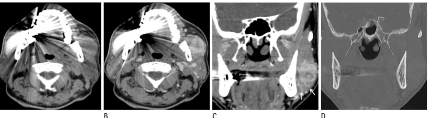
Excision of the mass was performed through a submandibu- lar incision. Intraoperatively, a localized round mass (3 × 2.8 × 2.8 cm) was found in the left masseter muscle. The lesion was excised with a clear margin. The cut surface of the mass was Fig. 1. CT images in a 66-year-old man with left facial swelling.
A. Axial pre-contrast image shows a soft tissue mass in the left masseter muscle, which is iso-dense with the adjacent muscle.
B. An axial post-contrast image obtained at the same level shows that the mass is lobulated and well-circumscribed with heterogeneous well en- hancement. And the mass is confined within the left masseter muscle.
C. Coronal post-contrast image shows prominent engorged serpentine vessels (arrows) peripheral to the mass.
D. On the bone window setting of the coronal image, there is no destruction or erosion of the adjacent mandible.
A B C D
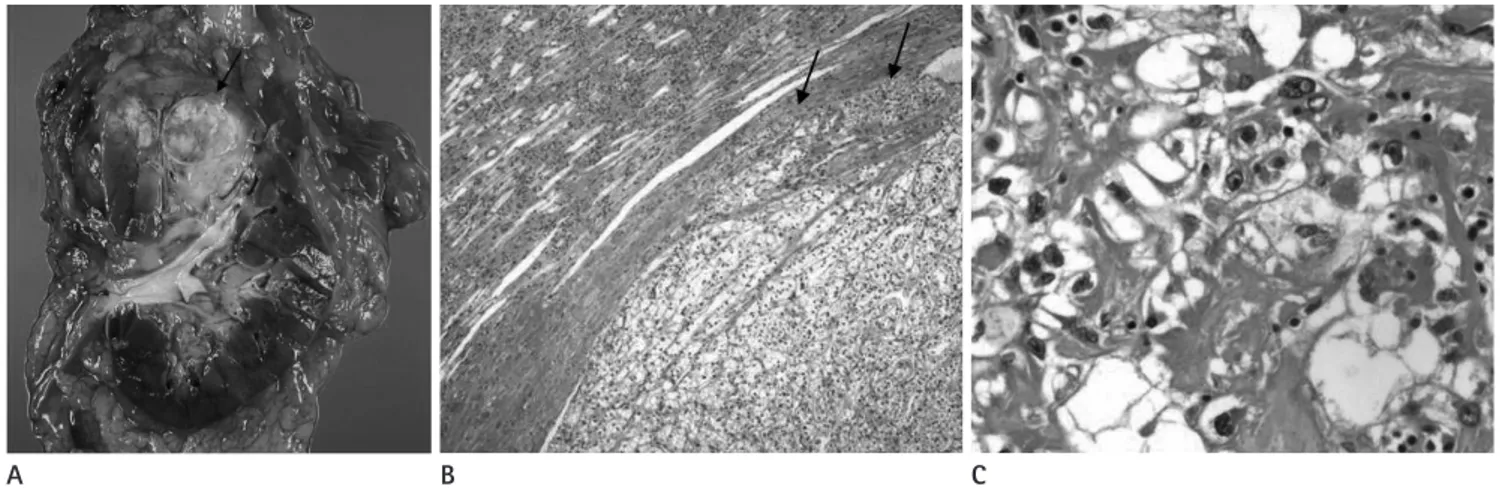
Fig. 2. Pathologic findings of the mass in left masseter muscle.
A. Gross specimen of left masseter muscle shows a 3 cm well-defined, lobulated firm mass. The cut surface looks whitish-gray with petechial hemorrhages without necrosis.
B. On the microscopy (H&E staining, × 40), the intramuscular mass is well-delineated from skeletal muscle fibers. The tumor cells are surrounded by sinusoidal vessels and composed of round epithelial cells with clear cytoplasms.
A B
the thoracic duct (1).
Skeletal muscle is one of the most unusual sites of reported metastasis of RCC. Masseter muscle is striated muscle that con- tributes to mastication. In general, striated muscle is a rare site either for primary or metastatic tumors. According to the liter- ature, the reasons for the rarity of metastatic tumors in skeletal muscles could be related to its dominant lactic acid metabo- lism, high tissue pressure and possession of diffusible protease and other inhibitors in its connective tissue. And variable vas- cular flow in mastication may contribute to the mechanical ar- rest of metastatic emboli (2). To our knowledge, there are only 3 cases of metastatic lesions to the masseter muscle in the litera- ture (2, 4, 5). In two cases, patients already had undergone uni- ter 3 months showed progression of lung metastasis and after 8
months, bone metastasis was detected at the L2 vertebra. He died 1 year after the initial diagnosis.
DISCUSSION
RCC frequently presents with distant metastases. Because RCC is slow-growing and encapsulated, the symptoms related to the primary site such as hematuria or flank pain tend to be not obvious in early stages. Therefore, initial presentation of RCC is often associated with a metastatic lesion (1). Almost 25% of patients with newly diagnosed RCC have evidence of metastases at presentation (2). The most frequent sites are lung (76%), regional lymph nodes (66%), bone (42%), and liver (41%) (3). Extracranial regions of head and neck are unusual meta- static sites, only accounting for approximately 15% of patients with RCC, involving nose, tongue, paranasal sinuses, larynx, mandible, temporal bone, thyroid glands and parotid glands.
Most lesions are in the sinonasal tract. RCC metastasis to the masseter muscle is extremely rare. Possible routes of metastasis to the head and neck include arterial, venous and lymphatic circulations, which could similarly explain the intramasseteric metastasis. Through a local vascular network invasion and Bat- son’s venous plexus extending from skull to sacrum, RCC can be hematogeneously spread to anywhere in the body including head and neck. And lymphatic spread is also possible through
Fig. 3. Cortical phase CT images demonstrate the left renal lesion (ar- row) with characteristic hypervascularity expected from clear cell renal cell carcinoma.
Fig. 4. Pathologic findings of the renal cell carcinoma in left kidney.
A. Gross finding of left radical nephrectomy. A relatively circumscribed, 6.5 cm-sized mass is found at the upper pole. The cut surface is yellow- ish-gray with lobulating contours. It is confined to the renal parenchyma (arrow).
B. Microscopic findings of the left renal tumor (H&E staining, × 40). The tumor is sharply demarcated from intact renal cortex. The tumor cells are arranged in a sinusoidal pattern and composed of clear cells, similar to a masseteric mass. There are a few intra-vascular tumor emboli (arrows).
C. Microscopic findings of the left renal tumor (H&E staining, × 400). Most parts of the tumor cells reveal Fuhrman’s nuclear grade 2/4, however focally 4/4 (pleomorphic).
A B C
the masseter muscle. CT may reveal the characteristic phlebo- liths and intense enhancement (6, 7). Hemangiopericytoma is a rare vascular tumor, representing 1% of all vasoformative neo- plasms. Hemangiopericytoma in the head and neck have been described on CT examinations as expansile, bone-remodeling lesions with variable patterns of enhancement. Some authors have mentioned the presence of prominent serpentine vessels in the periphery of hemangiopericytomas, reflecting the vascu- lar supply (7). On MR images, the tumor may be heteroge- neously isointense on T1-weighted images and isointense or hyperintense on T2-weighted images (7).
Benign or malignant schwannoma can also appear in the mas- ticator space. This tumor is related to mandibular division of trigeminal nerve as it passes superficial to the masseter muscle.
Therefore, location of the lesion tends to be not confined within the masseter muscle. The tumor may extend through the fora- men ovale and adjacent middle cranial fossa. CT and MR im- ages demonstrate a heterogeneously enhancing mass (8).
The most common primary malignant tumor of the mastica- tor space is sarcoma. A CT finding of a primary soft tissue sar- coma is not specific, which is a heterogeneously enhanced soft tissue tumor with or without destruction of adjacent bone. It is indistinguishable from other hypervascular tumors (8).
Secondary malignant involvement of the masseter muscle have been reported and may also show well-enhancing circum- scribed masses on CT scans. Reported primary sites are lung, breast, and kidney (1, 5, 9).
The prognosis of patients with RCC is poor. The 5-year sur- vival of patients after treatment for primary RCC ranges from 60% to 75% without metastasis, but the presence of distant me- tastasis at the time of diagnosis is a strong predictor of poor out- come with a 5-year survival rate of 10-20%. The head and neck metastasis is even worse; most patients die within 1 year after detection of such lesions (5, 10).
In RCC, the primary site can remain occult for long periods of time, sometimes presenting years after the appearance of metastatic disease. Therefore, even though the image finding of a metastatic tumor from an RCC in the head and neck region is not characteristic, it is important that radiologists should include it in the differential diagnosis of a hypervascular mass in the head and neck, including the masseter muscle, in older adults.
It also should be remembered that high-grade malignancies lateral nephrectomy for RCC. After 3 months and 4 years in
each of these two cases, a hypervascular mass developed in the masseter muscle. While one case was a solitary metastasis to the masseter muscle, the other case showed brain metastasis in the left temporal lobe as a hypervascular mass (2, 4). In the third case, a metastatic mass in the masseter muscle was the initial symptom of RCC, one that presented with right facial swelling.
After the mass excision and confirmation of the pathological diagnosis, an abdominal CT was performed, which showed the primary malignancy at the left kidney (5).
All of the reported metastatic RCC masses in masseter mus- cles including our case have hypervascularity as homogenous or heterogeneous well-enhancing masses on contrast-enhanced CT images with well-circumscribed margins, which resemble primary renal lesions. Angiography can also show the meta- static lesion of RCC as a hypervascular mass and may show a tumor feeder, which is useful for preoperative embolization (4).
In a reported case with MR images, an intramasseteric RCC le- sion was a lobulated well-delineated soft tissue mass with a slightly hyperintense signal compared with the masseter mus- cle on T1 weighted images. This is a nonspecific finding and there is no pathognomonic imaging finding of RCC metastasis in masseter muscle (5). However, according to a previous study for RCC metastasis in head and neck, intense enhancement af- ter contrast injection, destruction of adjacent bones, and lack of tumor calcification should suggest metastatic RCC as a part of the differential diagnosis (1). Although imaging has a limited role in the differential diagnosis of masseter muscle tumors, all imaging modalities are helpful in accurate delineation of the extent of the lesion, which is pivotal for directing surgical inter- vention or radiotherapy planning. Biopsy is the only way to confirm the diagnosis. In fact, as in our case, since high-grade malignancies can arise within the masticator space without evi- dence of either bone destruction or fascial violation despite their aggressive nature, biopsy should be performed without delay when such a mass is seen within this space (6).
The differential diagnosis of a hypervascular mass in the mas- seter muscle includes hemangioma, hemangiopericytoma, be- nign or malignant schwannoma, sarcoma and other secondary malignant tumors. The most common tumor of the masseter muscle is intramuscular hemangioma. 15% of these tumors are found in the head and neck, one third of which are localized to
case. J Laryngol Otol 1996;110:172-174
5. Gal TJ, Ridley MB, Arrington JA, Muro-Cacho C. Renal cell carcinoma presenting as a masseteric space mass. Am J Otolaryngol 1997;18:280-282
6. Chong VF, Fan YF. Pictorial review: radiology of the masti- cator space. Clin Radiol 1996;51:457-465
7. Lim SM, Lee HK, Shin JH, Kim JK, Kim DH, Choi CG, et al.
CT, MR and angiographic findings of hemangiopericyto- mas. J Korean Radiol Soc 1999;41:9-16
8. Connor SE, Davitt SM. Masticator space masses and pseu- domasses. Clin Radiol 2004;59:237-245
9. Lin SP, Bierhals AJ, Lewis JS Jr. Best cases from the AFIP:
metastatic renal cell carcinoma. Radiographics 2007;27:
1801-1807
10. Marulli G, Sartori F, Bassi PF, dal Moro F, Gino Favaretto A, Rea F. Long-term results of surgical management of pul- monary metastases from renal cell carcinoma. Thorac Car- diovasc Surg 2006;54:544-547
can be present without fascial or adjacent bone invasion in this region. So radiologists should recommend histopathological confirmation without delay.
REFERENCES
1. Pritchyk KM, Schiff BA, Newkirk KA, Krowiak E, Deeb ZE.
Metastatic renal cell carcinoma to the head and neck. La- ryngoscope 2002;112:1598-1602
2. Yiotakis J, Hantzakos A, Kostakopoulos A, Adamopoulos G.
Intramasseteric metastasis of renal cell carcinoma. J Lar- yngol Otol 2001;115:65-67
3. Sgouras ND, Gamatsi IE, Porfyris EA, Lekka JA, Harkiolakis GC, Nikolopoulou SM, et al. An unusual presentation of a metastatic hypernephroma to the frontonasal region. Ann Plast Surg 1995;34:653-656
4. Nakagawa H, Mizukami Y, Kimura H, Watanabe Y, Kuwaya- ma N. Metastatic masseter muscle tumour: a report of a
저작근을 침범한 전이종괴로 처음 발견된 신세포암:
증례 보고와 문헌조사1
배경은
1· 이한비
1· 조우호
1· 김재형
1· 이지혜
1· 강미진
1· 김현정
2신세포암은 원격 전이를 잘 하는 악성 종양으로 알려져 있고, 신장의 이상보다 전이된 부분의 증상으로 첫 발현이 되는 경 우가 적지 않다. 두경부에 신세포암이 전이된 경우들 중 골격근 중의 하나인 저작근으로 전이된 경우는 매우 드물게 보고 된 바 있다. 저자들은 폐결핵의 과거력이 있던 66세 남자에서 좌측 저작근을 침범한 경계가 분명하고 강하게 조영증강되 는 과혈관성 종괴로 처음 발견된 신세포암 1예를 경험하였기에 영상의학적 감별진단에 관한 문헌고찰과 함께 보고하고 자 한다.
인제대학교 의과대학 상계백병원 1영상의학과학교실, 2진단병리과학교실