Angiolipoma is a rare and benign mesenchymal tumor that’s composed of mature adipocytes and abnormal vascular tissue (1). Their main pathologic characteristics are macroscopic mass formation with or without cap- sule, microscopic evidence of mature lipocytes as the major cell population and angiomatous cell proliferation inside the mass (2). Angiolipomas are further divided in- to the non-infiltrating (capsulated) and infiltrating (non- capsulated) types. The former is more common and the latter shows a tendency to recur after surgery (2, 3).
Angiolipomas are most commonly found in the subcuta- neous tissue of the trunk and extremities, but other sites have been reported as well. The craniospinal axis is an uncommon, but significant site. Spinal angiolipomas ac- count for 1.2% of all spinal axis tumor, 3% of the ex- tradural spinal tumors and 24% of the spinal lipomas.
Intracranial angiolipomas are extremely rare (4).
We describe here the clinical and imaging features of a
case of angiolipoma in the cerebrebellopontine angle.
Case Report
A 65-year-old female was admitted to our hospital be- cause of her prolonged headache that was intractable to medical treatment. No precipitating factor was identi- fied. The general physical and neurological examina- tions were negative. There was no special personal his- tory and no family history of any neurological disease such as seizure. On admission, a MR imaging demon- strated an extraaxial mass in the right cerebrebellopon- tine angle. The mass was shown as heterogeneous high signal intensity on both the T1-weighted image and T2- weighted image, and vascular signal voids were noted within the mass. A slightly enhanced mass was noted on the contrast enhanced image (Fig. 1). Cerebral angiogra- phy was subsequently performed to exclude any associ- ated vascular malformation. DSA revealed a vascular entangled mass that was fed by the right anterior inferi- or cerebellar artery in the posterior fossa. Neither arteri- ovenous shunting nor any early draining veins were seen (Fig. 2). The lesion was surgically removed by the suboccipital retromastoid approach. The pathologic di-
J Korean Radiol Soc 2006;55:535-537
─ 535 ─
Angiolipoma in the Cerebellopontine Angle:
A Case Report
1Seung Rim Kang, M.D., Hong Dae Kim, M.D., Han Myun Kim, M.D., Ik Yang, M.D.
1Department of Radiology, Hallym University College of Medicine Received April 15, 2006 ; Accepted June 2, 2006
Address reprint requests to : Hong Dae Kim, M.D., Department of Radiology, Hallym University, Kangnam Sacred Heart Hostipal, 948-1, Daerim 1-Dong, Yungdungpo-gu, Seoul 150-950, Korea.
Tel. 82-2-829-5241 Fax. 82-2-832-1845 E-mail: [email protected]
A 65-year-old female, who suffered with a longstanding headache, was admitted to our hospital. MR imaging and digital subtraction angiography (DSA) demonstrated a vascular fat-containing mass in the right cerebellopontine angle. The lesion was surgi- cally removed and the diagnosis of angiolipoma was established. The symptoms sub- sided after the operation.
Index words :Brain Angioma
Magnetic resonance (MR), image display Angiography
agnosis was angiolipoma (Fig. 3). After the operation, the patient’s headache completely subsided.
Discussion
Central nervous system angiolipomas are uncommon lesions that may arise from the abnormal development of primitive pluripotential mesenchymal cells from which adipose tissue, smooth muscle and vascular en- dothelium develop. Angiolipomas can be currently diag- nosed with a high degree of certainty based on the spe- cific MR imaging. Due to their rich fat content, angi-
olipomas usually show hyperintensity on the precon- trast T1-weighted image. Fat-suppression techniques are particularly helpful to confirm the lipomatous nature of this lesion and to demonstrate its enhancement (3, 5).
The contrast enhancement and hypervascularity on an- giograms differentiate angiolipomas from other lesions with fat content such as dermoids and lipomas. The lat- ter lesions do not show contrast enhancement on MR imaging and they do not show vascularity on angiogra- phy. The angiographic patterns of angiolipomas are vari- able, from an avascular pattern to a hypervascular pat- tern, by the relative amount of the vascular component.
Seung Rim Kang, et al : Angiolipoma in the Cerebellopontine Angle
─ 536 ─
A B
Fig. 2. Left vertebral angiogram. The vascular entangled mass fed by the right AICA is demonstrated.
Fig. 3. Photomicrograph (original magnification, ×40; H-E stain) shows mature adipose tissue and the irregular vascular structures within the pathologic specimen.
C
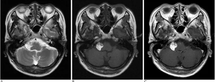
Fig. 1. A. The T2-weighted image shows an extraaxial mass in the right cerebrebellopontine angle. The mass contains multiple vas- cular signal voids.
B. The T1-weighted image demonstrates the bright high signal intensity of the mass, which suggests the fatty component of the mass.
C. The contrast enhanced image shows an enhanced mass in the right cerebrebellopontine angle.
On rare occasions intracranial angiolipomas may bleed and this represents a sporadic cause of subarachnoid he- morrhage. Although more evidence is needed in order to understand the natural history of intracranial angi- olipomas, they are usually considered to be benign and a conservative approach should be taken for their treat- ment (6).
Treatment of angiolipomas includes a surgical ap- proach with the goal of total removal. Most authors agree in the need of a wide excision for surgically re- moving infiltrating angiolipomas, and they have report- ed good outcomes after surgical excision of spinal angi- olipomas, and this is often in spite of the severe preoper- ative neurological deterioration. However, the outcome of intracranial angiolipoma patients are quite variable;
this is most probably related to the tumor location and the surgical complexity of removing these tumors (4).
In summary, intracranial angiolipoma can be specifi-
cally diagnosed by MR imaging. Preoperative angiogra- phy may be useful for the case that has a rich vascular component.
References
1. Howard WR, Helwig EB. Angiolipoma. Arch Dermatol 1960;82:
924-931
2. Lin JJ, Lin F. Two entities in angiolipoma: a study of 459 cases of lipoma with review of literature on infiltrating angiolipoma.
Cancer 1974;34:720-727
3. Turgut M. Spinal angiolipomas: report of a case and review of the cases published since the discovery of the tumour in 1890. Br J Neurosurg 1999;13:30-40
4. Andaluz N, Balko G, Bui H, Zuccarello M. Angiolipomas of the central nervous system. J Neurooncol 2000;49:219-230
5. Provenzale J, McLendon R. Spinal angiolipomas: MR features.
AJNR Am J Neuroradiol 1996;17:713-719
6. Vilela P, Saraiva P, Goulao A. Intracranial angiolipoma as cause of subarachnoid haemorrhage. Case report and review of the litera- ture. Neuroradiology 2005;47:91-96
J Korean Radiol Soc 2006;55:535-537
─ 537 ─
대한영상의학회지 2006;55:535-537
소뇌 연수각에서 생긴 지방 혈관종: 증례 보고1
1한림대학교 의과대학 방사선과학교실
강승림・김홍대・김한면・양 익
65세 여자 환자가 오랫동안 지속한 두통으로 병원을 방문하였다. 자기공명영상과 혈관 조영술을 시행하여 우측 소뇌 연수각에 혈관과 지방을 함유한 병소를 발견하였다. 그 병소는 수술로 제거되었고 혈관 지방종으로 확진되었 다. 수술 후 두통은 호전되었다.