복벽의 혈종은 흔하지는 않으나 잘 알려진 질환으로, 주로 외상에 의해 심장골회선 동맥, 하복벽 동맥, 요골 동맥, 늑간 동맥 등 복벽에 혈류를 공급하는 혈관이 손상되어 발생한다 (1-3). 드물게 혈종이 자발적으로 발생할 수 있으며, 이 경우 급성 복증을 일으키는 다른 질환과의 감별이 임상적으로 어려 워 진단이 지연될 수 있고, 불필요한 수술을 하는 경우도 발생 할 수 있다(4-8). 저자들은 CT와 혈관조영술로 진단하고, 코 일 색전술로 치료한, 심한 기침 후 자발적으로 발생한 심장골 회선 동맥 손상에 의한 전측복벽의 혈종 1예를 보고하고자 한 다.
증례 보고
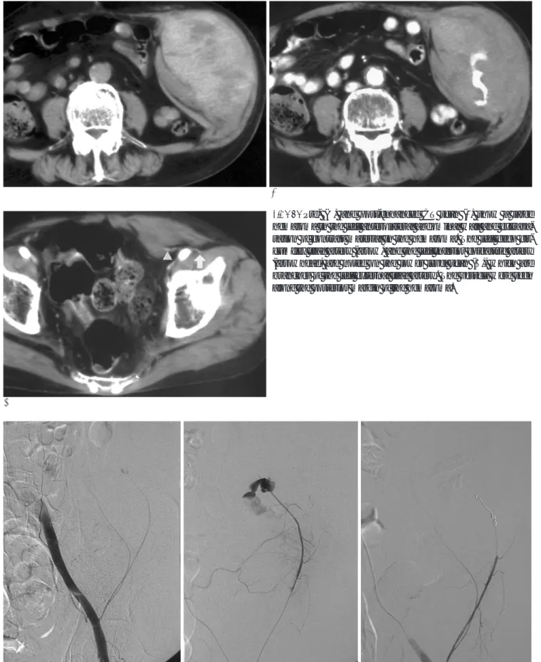
67세 남자 환자가 내원 당일 심한 기침 후에 발생한 좌하복 부의 동통성 종괴와 호흡곤란을 주소로 내원하였다. 환자는 천 식의 과거력이 있었으나, 출혈성 질환의 병력이나 항응고제 복 용의 과거력은 없었다. 내원 당시 약 10 cm 정도의 반상출혈 을 동반한 종괴가 좌복부에서 촉지되었으며, 혈압, 맥박, 혈액 및 생화학 검사 소견 등은 정상이었다. 내원 후 종괴의 크기는 점차 증가되었다. 조영 증강 전 CT에서 고음영의 혈종이 좌전 측복벽의 근육층에 관찰되었으며, 조영 증강 후 혈종 내부에 조영제의 누출이 있었다(Fig. 1A, B). 조영증강 후 CT에 혈종 의 하단부위 단면에서 확장된 두 개의 혈관이 외장골 동맥으 로부터 분지되어 혈종의 후면으로 주행하는 것이 확인되었다 (Fig. 1C, D). 외장골 동맥의 혈관조영검사로 심장골회선 동맥 의 손상에 의한 혈종을 진단하였으며, 심장골회선 동맥을 2.5- F 미세카테터로 초선택하여 혈관 손상부위를 확인하고, 2개의
코일 (MWCE-18-2.0-2-HILAL; Cook, Bloomington, U.S.A.)로 색전하였다 (Fig. 2). 시술 후 혈관조영 검사로 심 장골회선 동맥의 성공적인 색전을 확인하였고, 환자는 더 이상 의 치료 없이 혈종이 점차 감소하여 퇴원하였다.
고 찰
전측복벽에 혈액을 공급하는 주 혈관은 심장골회선동맥 (deep circumflex iliac artery), 하복벽 동맥(inferior epigas- tric artery), 상복벽동맥(superior epigastric artery), 요골동 맥(lumbar artery), 하부 늑간동맥(lower intercostal arter- ies) 등이다. 하복벽 동맥과 상복벽동맥이 주로 복직근에 혈액 을 공급하는 반면, 심장골회선 동맥은 복횡근, 내복사근, 외복 사근에 혈액을 공급한다. 심장골회선 동맥은 외장골 동맥의 분 지로 서혜 인대 부근에서 분지하여 복횡근막 내를 주행하다, 관통하여 위로 주행하며, 복횡근과 내복사근 사이에서 장요동 맥, 상둔동맥 등과 문합한다. 전상장골극 부위에서 상부로 주 행하는 동맥이 분지되며, 이는 복횡근과 내복사근 사이를 주행 하며, 두 근육에 혈액을 공급하고, 요동맥, 하복벽동맥과 문합 한다(9). 심장골회선동맥은 4가지 형태로 나눌 수 있는데, 첫 번째 형태는 하나의 주심장골회선 동맥이 하나의 상행혈관을 분지하는 경우이고, 두 번째 형태는 하나의 주심장골회선 동맥 이 여러 개의 작은 상행 혈관들로 분지되는 경우이다. 세 번째 형태는 두 개의 심장골회선 동맥과 하나의 상행 분지가 있는 경우, 네 번째 형태는 천장골회선 동맥과 심장골회선 동맥이 하나의 혈관에서 분지되며, 하나의 상행동맥이 심장골회선 동 맥에서 분지되는 경우이다(10).
하복벽 동맥이나 심장골회선 동맥의 근위부는 느슨하게 부 착되어 유동적인 반면, 근육 내를 주행하는 원위부는 근육에 대한영상의학회지 2004;50:423-426
─ 423 ─
자발적으로 발생한 심장골회선 동맥 손상에 의한 복벽 혈종의 코일 색전술 치료: 증례 보고1
백준현・박영하・전정수・황성수・인연권
복벽에 발생하는 혈종은 비교적 드문 질환이며, 외상 혹은 자발적으로 발생한다. 주로 복직 근초나 측복벽에서 하복벽 동맥이나 장골회선동맥의 손상에 의해 발생하며, 자발적으로 발생 한 복벽 혈종은 임상적으로 다른 급성 복증을 일으키는 질환과의 감별이 어려울 수 있으므로, 조기에 CT와 혈관조영술을 시행하여 진단하는 것이 중요하다. 저자들은 심한 기침 후 심장골 회선동맥 손상에 의해 발생한 전측복벽의 혈종을 코일을 이용한 혈관 색전술로 치료한 1예를 보고하고자 한다.
1가톨릭대학교 의과대학 방사선과학교실
이 논문은 2003년 11월 5일 접수하여 2004년 4월 2일에 채택되었음.
백준현 외: 자발적으로 발생한 심장골회선 동맥 손상에 의한 복벽 혈종의 코일 색전술 치료
─ 424 ─ A
C
B
Fig. 1. Pre- (A) and post-enhanced CT scan (B) show a large hematoma in the left anterolateral abdominal wall and extrava- sation of contrast material in the hematoma. The left deep cir- cumflex iliac artery (arrow) and the left inferior epigastric artery (arrowhead) are noted on the lower level scan (C), which are branches of the left external iliac artery. The vessels were seen along the posterior margin of the hematoma.
A B C
Fig. 2. Emergency angiography of the left common iliac artery (A) and selective angiography of the left deep circumflex iliac artery (B) demonstrate the extravastion from the ascending branch of the deep circumflex iliac artery. The ascending branch of the deep circumflex iliac artery was embolized with two microcoils. Postembolization angiography (C) shows complete occlusion of the artery.
단단하게 고정되어 있어, 기침, 재채기, 구토, 운동시 근육의 과 도한 수축으로 전단력이 발생할 경우 손상을 받을 수 있다. 이 외에도 동맥경화, 염증성 질환, 분만, 비만, 노화 등으로 혈관 벽이 약해지거나, 근육 저항이 감소하는 경우, 출혈성 질환 혹 은 항응고제 사용하는 경우 등에서도 자발적 출혈이 발생할 수 있다(5, 6).
본 증례는 천장골회선 동맥과 심장골회선 동맥이 하나의 혈 관에서 분지되며, 내복사근, 외복사근, 복횡근에 혈액을 공급하 는 하나의 상행동맥이 심장골회선 동맥에서 분지하는 네 번째 형태의 심장골회선 동맥으로 판단되며, 혈관 질환, 출혈성 질 환이나 항응고제 복용 등의 과거력이 없었고, 심한 기침 직후 혈종이 발생하였던 점으로 보아, 기침으로 복근이 과도하게 수 축하면서 발생한 전단력에 의해 심장골회선 동맥의 상행분지 가 손상 받았을 것으로 생각된다.
복벽의 혈종은 임상적으로도 진단할 수 있으나, 자발적으로 발생한 경우 급성 복증을 유발하는 위장관 혈관 질환, 대동맥 박리, 충수돌기염, 담낭염, 난소 낭종 염전 등의 질환과 감별해 야 한다(7, 8). 조기에 초음파나 CT를 시행하면 다른 질환과 의 감별이 가능하며 불필요한 수술을 방지할 수 있다. 특히 CT 에서는 혈종의 위치와 혈종이 발생한 근육을 알 수 있어 손상 혈관을 예측할 수 있다. 본 증례의 CT소견에 혈종은 좌측 복 횡근, 내복사근 및 외복사근에 걸쳐서 발생하였으며, 조영제의 혈관외 누출은 내복사근 내측 부위에서 발견되었다. 한편 혈종 하단 부위 단면에서 외장골 동맥에서 분지되는 두 개의 확장 된 혈관이 혈종의 후벽을 따라 주행하는 것이 관찰되어 손상 혈관 진단에 도움을 주었다.
복벽 혈종의 원인 혈관은 혈관 조영 검사로 진단할 수 있으 나, 장골 동맥 혈관 조영 검사에서 손상 혈관을 찾을 수 없는 경우도 있다(6). 그러므로 혈종의 위치에 따라 손상이 예상되 는 혈관을 판단하여, 선택적 혈관조영 검사를 시행해야 할 것 으로 생각된다. 한편, 최근에 발달한 다중검출기 CT를 이용할 경우, 혈종의 진단뿐만 아니라, 손상혈관 진단에 있어서도 혈
관 조영술을 대체할 수 있을 것으로 생각된다. 복벽 혈종은 보 존적으로 치료할 수 있으나, 본 증례와 같이 혈종이 점차 커지 거나 활력 증후에 이상을 보여 대량 출혈이 의심되는 경우, 혈 관조영 검사로 손상된 혈관을 확진하고, 입자성 색전 물질이나 코일을 이용한 색전술로 치료할 수 있다.
참 고 문 헌
1. Linhares MM, Lopes Filho GJ, Bruna PC, Ricca AB, Sato NY, Sacalabrini M. Spontaneous hematoma of the rectus abdominins sheath: a review of 177 cases with report of 7 personal cases. Int Surg 1999;84:251-257
2. Zainea GG, Jordan F. Rectus sheath hematomas: their pathogene- sis, diagnosis, and management. Am J Surg 1988;54:630-633 3. Titone C, Lipsius M, Krakauer JS. “Spontaneous” hematoma of the
rectus abdominis muscle: critical review of 50 cases with emphasis on early diagnosis and treatment. Surgery 1972;72:568-572 4. Teske JM. Hematoma of the rectus abdominal muscle. Report of a
case and analysis of 100 cases from the literature. Am J Surg 1946;
71:689-695
5. Katsumori T, Nakajima K. A case of spontaneous hemorrhage of the abdominal wall caused by rupture of a deep iliac circumflex artery treated by transcatheter arterial embolization. Eur Radiol 1998;8:550-552
6. Lefere P, Gryspeerdt S, Van Holsbeeck B, Baekelandt M.
Diagnosis and treatment of expanding hematoma of the lateral ab- dominal wall after blunt abdominal trauma. Eur Radiol 1999;9:
1553-1555
7. Klingler PJ, Oberwalder MP, Riedmann B, DeVault KR. Rectus sheath hematoma clinically masquerading as sigmoid diverticuli- tis. Am J Gastroenterol 2000;95:555-556
8. Lohle PN, Puylaert JB, Coerkamp EG, Hermans ET. Nonpalpable rectus sheath hematoma clinically masquerading as appendicitis:
US and CT diagnosis. Abdom Imaging 1995;20:152-154
9. Williams PL, Warwick R, Dyson M, Bannister LH. Gray’s Anatomy 37th ed. New York: Churchill Livingstone, 1989:780-781 10. Thein T, Kreidler J, Stocker E, Herrmann M. Morphology and blood supply of the iliac crest applied to jaw reconstruction. Surg Radiol Anat 1997;19:217-225
대한영상의학회지 2004;50:423-426
─ 425 ─
백준현 외: 자발적으로 발생한 심장골회선 동맥 손상에 의한 복벽 혈종의 코일 색전술 치료
─ 426 ─
J Korean Radiol Soc 2004;50:423-426
Address reprint requests to : Young Ha Park, M.D., Departments of Radiology and Nuclear Medicine, St. Vincent’s Hospital, 93-6 Chi-dong, Paldal-gu, Suwon, Kyunggi-do 442-723, Korea.
Tel. 82-31-249-7481 Fax. 82-31-247-5713 E-mail: [email protected]
Expanding Hematoma of the Abdominal Wall Caused by Spontaneous Rupture of a Deep Circumflex Iliac Artery:
Report of A Case Treated by Coil Embolization
1Jun Hyun Baik, M.D., Young Ha Park, M.D., Jung Soo Jeon, M.D., Sung Soo Hwang, M.D., Yon Kwon Ihn, M.D.
1Department of Radiology, The Catholic University of Korea, Seoul, Korea
Abdominal wall hematoma is a rare but well-known disease, usually caused by trauma or, on rare occasions, occurring spontaneously. Hematomas of the rectus sheath and the anterolateral abdominal wall are commonly associated with injury to the inferior epigastric artery and the deep circumflex iliac artery, respectively. The di- agnosis of spontaneously developed abdominal wall hematoma is sometimes delayed, due its clinical manifes- tations being similar to those of other causes of the acute abdomen. CT and angiography can be helpful in the diagnosis of the hematoma and the injured vessel. Herein, we report on a rare case of a spontaneously devel- oped anterolateral abdominal wall hematoma treated with microcoil embolization of the left deep circumflex iliac artery.
Index words :Hemorrhage, abdomen Artery, embolization