난소암에서 봉독이 세포자멸사와 JAK2/STAT3 Pathway의 억제에 미치는 영향
안병준ᆞ송호섭
경원대학교 한의과대학 침구학교실
목적 : 이 연구는 봉독이 사람의 난소암 세포인 SKOV3와 PA-1에서 death receptor의 발현을 높여 세포자 멸사를 촉진함으로써 암세포의 성장을 억제하는지 밝히고자 하였다.
방법 : 난소암의 세포자멸사의 관찰에는 DAPI, TUNEL staining assay를 시행하였으며, 세포자멸사 조절 단백질의 변동 관찰에는 western blot analysis를 시행하였고, 난소암 세포에서 death receptor의 변화를 관찰 하기 위해 RT-PCR analysis를 시행하였다.
결과 : 1. DAPI, TUNEL staining assay 결과, 봉독은 투여량에 따라 세포자멸사의 유도를 통해 SKOV3와 PA-1 난소암세포의 증식을 억제하였고, 세포자멸사와 동반하여 DR4와 DR6의 발현이 두 암세포 모두에서 증 가하였고, DR3의 출현은 PA-1 세포에서 증가하였다.
2. Death Receptor의 발현 증가에 따라 caspase-3, 8, 9 and Bax를 포함하는 세포자멸사 촉진 단백질의 발 현이 동반하여 상승하였고 JAK2, STAT3의 인산화와 Bcl-2의 발현은 억제되었다.
3. siRNA 처리 시 봉독에 의한 DR3, DR4, DR6 발현증가와 STAT3의 활성억제가 역전되었다.
결론 : 이러한 결과는 봉독이 난소암 세포에서 DR3, DR4, DR6의 증가와 JAK2/STAT3 pathway의 억제를 통하여 세포자멸사를 유발한다는 것을 시사하며, 난소암의 예방과 치료에 효과적으로 활용될 수 있을 것으로
1)
Effect of Bee Venom Death Receptor
Dependent Apoptosis and JAK2/STAT3 Pathway in the Ovarian Cancer
Ahn Byeong-joon and Song Ho-sueb
Dept. of Acupuncture & Moxibustion, College of Oriental Medicine, Kyungwon University
* This research was supported by the Kyungwon University Research Fund in 2012
․Acceptance : 2012. 1. 17. ․Adjustment : 2012. 2. 6. ․Adoption : 2012. 2. 6.
․Corresponding author : Song Ho-sueb, Kyungwon Gil Oriental Medical Hospital, 1200-1 Guwal-dong Namdong-gu Incheon Republic of Korea
Tel. 82-70-7120-5012 E-mail : [email protected]
원 저
국문초록
기대된다.
핵심 단어 : 봉독, 난소암, 세포자멸사, death receptor, JAK2/STAT3
Ⅰ. Introduction
Ovarian cancer is the most frequent cause of death from gynecological cancer
1). Recent annual worldwide figures reflect 204,000 new cases of ovarian cancer and 125,000 deaths
2). For the therapy of ovarian cancer, surgery is principle therapy
3), and chemotherapy is also needed to remove the re- maining cancer cells. However, the major challenge that limits the effectiveness of chemotherapy in patients with advanced ovarian cancer is the acqui- sition of resistance
4). According to recent studies
5,6), appropriate chemopreventive compounds reducing or overcoming resistance are believed to be a very hopeful strategy to reduce the incidence of ovarian cancer and enhance treatment efficacy
5,6).
Apoptosis, the programmed cell death plays major role in anti-cancer effects of chemothera- peutics, which can be induced by activated caspase cascade systems through stimulation of death receptors via interaction of DRs with their ligands such as DR1 with TNF; DR2 with FasL; DR3, with Apo3L; DR4 and DR5 with TRAIL
7-14).
Signal transducers and activators of transcription (STAT) proteins are transcription activators in JAK(specific inhibitors of Janus kinase)/STAT signal pathway, involving in cell growth, proliferation, survival, differentiation, apoptosis, metastasis, and angiogenesis
15). Several studies have represented that phosphorylated STAT3 induce development and progression of various kinds of tumors such as breast, neck, head, prostate and ovary cancer, etc
16). In other words, the inhibition of STAT3 by JAK can inhibit tumor cell growth and go apoptosis
17,18).
Bee venom contains a variety of different pep- tides, including melittin, phospholipase A2, apamin,
adolapin, and mast cell-degranulating peptide (MC DP). Bee venom has been used as a traditional medicine to treat back pain, rheumatism, and skin diseases by its antibacterial, antiviral, and anti- inflammatory effects
19). Moreover, several studies have demonstrated that bee venom and/or melittin have anti-cancer effects including prostate, liver , breast, cervical, renal cancer cells
20-22). However, experiments demonstrating the molecular mechan- isms of the anti-cancer effects of bee venom in ovarian cancer cells have not been reported.
In this study, we therefore investigated anti- cancer effects of bee venom through increase of DR expression, but inhibition of STAT3 pathway in the human ovarian cancer cells, SKOV3 and PA-1.
Ⅱ. Materials and methods
A . Materials
Bee venom was purchased from You-Miel Bee Venom Ltd. (Hwasoon, Jeonnam, Korea). The composition of the bee venom was as follows:
45-50% melittin, 2.5-3% mast cell degranulating peptide, 12% phospholipase A2, 1% lysophos- pholipase A, 1-1.5% histidine, 4-5% 6-pentyl a- pyrone lipids, 0.5% secarpin, 0.1% tertiapin, 0.1%
procamine, 1.5-2% hyaluronidase, 2-3% amine, 4-5% carbohydrate, and 19-27% of others, in- cluding protease inhibitor, glucosidase, invertase, acid phosphomonoesterase, dopamine, norepineph- rine, and unknown amino acids, with 99.5% purity.
Caspase inhibitor (Z-VAD-FMK) was from Promega
(Madison, WI). DR3, DR4, and DR6 siRNA were
purchased from Santa Cruz Biotechnology, Inc
(Santa Cruz, CA).
Death
receptor Forward primer Reverse primer
TNFR 1
5`-ACC AAG TGC CAC AAA GGA AC-3`
5`-CTG CAA TTG AAG CAC TGG AA-3`
TNFR 2
5`-CTC AGG AGC ATG GGG ATA AA-3`
5- AGC CAG CCA GTC TGA CAT CT-3`
Fas
5-CAA AGC CCA TTT TTC TTC CA-3
5-GAC AAA GCC ACC CCA AGT TA-3
DR 3
5-ATG GCG ATG GCT GCG TGT CCT G-3
5-AGC GCC TCC TGG GTC TCG GGG TAG-3
DR 4
5-ACT TTG GTT GTT CCG TTG CTG TTG-3
5-GGC TTT CCA TTT GCT GCT CA-3
DR 5
5-TGG AAC AAC GGG GAC AGA ACG-3
5-GCA GCG CAA GCA GAA AAG GAG-3
DR 6
5-AAGCCGGGGA CCAAGGAGACAG ACAAC-3
5-TGCCGGGGCCC CTTTTTCAGAGT -3
B. Cell culture
The SKOV3 and PA-1 ovarian cancer cell lines were purchased from the American Type Culture Collection (Manassas, VA). SKOV3 cancer cells were cultured in RPMI-1640 medium supplemented with 10% fetal bovine serum (FBS) and penicillin/
streptomycin (100 U/ml). PA-1 cancer cells were cultured in MEM medium supplemented with 10%
fetal bovine serum (FBS) and penicillin/strepto- mycin (100 U/ml). Cell cultures were then main- tained at 37°C in a humidified atmosphere with 5%
CO2.
C. Cell viability assay
To determine the cell number, SKOV3 and PA-1 ovarian cancer cells were plated in 24-well plates (5×10⁴ cells/well), and subconfluent cells were sub- sequently treated with bee venom (1, 2 and 5 ㎍ /ml) for 24 hr. After treatment, cells were tryp- sinized and pelleted by centrifugation for 5 min at 1,500 rpm, resuspended in 5 ml of phosphate- buffered saline (PBS), and 0.1 ml of 0.2% trypan blue was added to the cancer cell suspension in each of the solutions (0.9 ml each). Subsequently, a drop of suspension was placed into a Neubauer chamber and the living cancer cells were counted.
Cells that showed signs of staining were considered to be dead, whereas those that excluded trypan blue were considered viable. Each assay was carried out in triplicate.
D. Reverse transcription (RT)-PCR
Total RNAs were isolated from cultured cells using RNeasy Plus Mini Kit (Qiagen) according to the manufacturer’s manual. The RNA pellet ob- tained in the final step was dissolved in 30 µl of sterile diethylpyrocarbonate (DEPC)-treated water, and its concentration was determined using a UV spectrophotometer at 260 nm. RNA was kept in DEPC-treated water at -70°C until use. Reverse transcription was performed using High Capacity
RNA-to-cDNA Kit (AB). PCR amplifications were then carried out with the following primers.
E. Western blot analysis
Western blot analysis was done as described
previously. The membrane was incubated for 2 hr
at room temperature with specific antibodies: rabbit
polyclonal for caspase-3, cleaved caspase-3, caspase
-8, cleaved caspase-8, caspase-9, cleaved caspase
-9, Bcl-2( 1 :1,000 dilution, Cell Signaling Technology,
Inc., Beverly, MA), p-STAT3, Bax, p-JAK2( 1 :500
dilution, Santa Cruz Biotechnology, Inc.), and mouse
monoclonal STAT3( 1 :500 dilution, Santa Cruz
Bio-technology, Inc.). The blot was then incubated
with the corresponding conjugated anti-rabbit and
anti-mouse immunoglobulin G-horseradish peroxidase
( 1 :2,000 dilutions, Santa Cruz Biotechnology, Inc.).
Immunoreactive proteins were detected with the ECL Western blotting detection system.
In transfection. Ovarian cancer cells (3 × 10⁴ cells per well) were plated in 24-well plates and transiently transfected with siRNA, using a mixture of siRNA and the WelFect-EXPLUS reagent in OPTI-MEN,according to the manufacturer’s specifi- cation(WelGENE, Seoul, Korea). The transfected cells were treated with 5㎍/ml bee venom for 24 hr.
DR3 siRNA seq. 5’-GAAGCCCUAAGUACGGUUAtt DR4 siRNA seq. 5’-CUCUGAUGCUGUUCUUUGAtt DR6 siRNA seq. 5’-GCCUUCUAGUGUGAUGAAAtt
F. Apoptosis evaluation
Ovarian cancer cells (2.5× 10⁵ cells/well) were cultured on 8-chamber slides. After ovarian cancer cells were transfected with siRNA, the cells were treated with bee venom(5 ㎍/ml). The cells were washed twice with PBS and fixed by incubation in 4% paraformaldehyde in PBS for 1 hr at room temperature. Membrane was permeabilized by ex- posure to 0.1% Triton X-100 in phosphate-buffered saline for 5 min at room temperature. TdT- mediated dUTP nick and labeling (TUNEL) assays were performed by using the in situ Cell Death Detection Kit (Roche Diagnostics GmbH, Mannheim, Germany) according to the manufacturer’s instruc- tions. For 4'-6-Diamidino-2-phenyl indole (DAPI) staining, slides were incubated for 15 min at room temperature in the dark with mounting medium for fluorescence containing DAPI (Vector Laboratories, Inc., Burlingame, CA). The cells were then observed through a fluorescence microscope (Leica Micro- systems AG, Wetzlar, Germany).
G. Data analysis
The data were analyzed using the GraphPad Prism 4 ver. 4.03 software (GraphPad Software, La Jolla, CA). Data are presented as mean±SD. The differences in all data were assessed by one-way analysis of variance (ANOVA). When the P value
in the ANOVA test indicated statistical significance, the differences were assessed by the Dunnett’s test.
A value of p<0.05 was considered to be statistically significant.
Ⅲ. Results
A. Effect of bee venom on cell growth in ovarian cancer cells
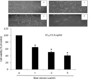
To assess the inhibitory effect of bee venom on cell growth of ovarian cancer SKOV3 and PA-1 cells, we analyzed cell viability by direct cell counting. The cells were treated with several concentrations of bee venom (1, 2, and 5 ㎍/ml) for 24 hr. As shown in Fig. 1 and 2, bee venom in- hibited cell proliferation of ovarian cancer cells in a concentration-dependent manner. Twenty-four hour treatment of bee venom inhibited SKOV3 cell growth with IC50 value of 3.8 ㎍/ml(Fig. 1), and PA-1 cells growth with IC50 values of 2.6 ㎍/ml (Fig. 2), respectively. The magnitude of the in- hibitory effect was much stronger in PA-1 cells than that in SKOV3 cells (Fig. 2). Morphologic observation showed that the cells were gradually reduced in size and changed into a small round single cell shape by the treatment of bee venom in SKOV3 cells (Fig. 1) and PA-1 cells (Fig. 2).
B. Apoptotic cell death by bee venom
To determine the inhibition of cell growth by bee
venom was due to the induction of apoptotic cell
death, we evaluated the changes in the chromatin
morphology of cells by using DAPI staining followed
by TUNEL staining assays, and then the double
labeled cells were analyzed by fluorescence micro-
scope. Conversely well with cell growth inhibition,
DAPI-stained TUNEL-positive cells were significantly
increased in bee venom treated cells. The treatment
of bee venom resulted in about 55-65% or 60-80%
Fig. 1. Effect of bee venom on cell viability in ovarian cancer cells
Concentration-dependent effect of bee venom on the cell viability assay in SKOV3. After treatment of bee venom(1, 2 and 5 ㎍/ml) for 24 hr, the cells were harvested by trypsinization and stained with 0.2%
trypan blue. Relative cell survival rate was deter- mined by counting live and dead cells. The results were expressed as a percentage of viable cells.
Morphologic observation with the treatment of bee venom in SKOV3 cells. Columns, means of three experiments, with triplicates of each experiment; bars, SD. *, p <0.05, significantly different from untreated control cells.
Fig. 2. Effect of bee venom on cell viability in ovarian cancer cells
Concentration-dependent effect of bee venom on the cell viability assay in PA-1. After treatment of bee venom(1,2 and 5 ㎍/ml) for 24 hr, the cells were harvested by trypsinization and stained with 0.2% trypan blue. Rela- tive cell survival rate was determined by counting live and dead cells. The results were expressed as a percentage of viable cells. Morphologic observation with the treatment of bee venom PA-1 cells. Columns, means of three experiments, with triplicates of each experiment;
bars, SD. *, p <0.05, significantly different from untreated control cells.
Fig. 3. Effect of bee venom on apoptotic cell death
The ovarian cancer cells, SKOV3 were treated with bee venom (5 ㎍/ml) for 24 hr, and then labeled with DAPI and TUNEL solution. Total number of cells in a given area was determined by using DAPI nuclear staining (fluorescent microscope). The green color in the fixed cells marks TUNEL-labeled cells. The apoptotic index was determined as the DAPI-stained TUNEL-positive cell number/total DAPI stained cell number(magnifi- cation, 200x). Columns, means of three experiments, with triplicates of each experiment; bars, SD. *, p <0.05, sig- nificantly different from bee venom-untreated control cells.
Fig. 4. Effect of bee venom on apoptotic cell death
The ovarian cancer cells, PA-1 were treated with bee venom (5 ㎍/ml) for 24 hr, and then labeled with DAPI and TUNEL solution. Total number of cells in a given area was determined by using DAPI nuclear staining (fluorescent microscope). The green color in the fixed cells marks TUNEL-labeled cells. The apoptotic index was determined as the DAPI-stained TUNEL-positive cell number/total DAPI stained cell number(magnification, 200x). Columns, means of three experiments, with tripli- cates of each experiment; bars, SD. *, p <0.05, signifi- cantly different from bee venom-untreated control cells.
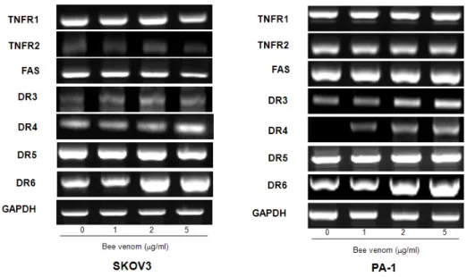
Fig. 5. Effect of bee venom on DRs expression in ovarian cancer cells
Cells were treated with bee venom(1, 2, and 5 ㎍/ml) for 24 hr, and total RNA were extracted and examined for expressions of TNF-R1, TNF-R2, FAS, DR-3, -4, -5, -6, and GAPDH by RT-PCR.
GAPDH was used as an internal control to show equal RNA loading. Each band is representative for three experiments.
induction of apoptotic cell death in SKOV3(Fig. 3) and PA-1(Fig. 4) cancer cells, respectively.
C. Expression of death receptors in ovarian cancer cells by bee venom
Apoptosis can be induced by stimulation of DRs expression. Therefore, to investigate expression of DRs in cancer cells undergoing apoptotic cell death, we performed RT-PCR analysis. RT-PCR analysis showed that bee venom treatment increased DR4 and DR6 mRNA levels in a concentration dependent manner, but TNFR1, TNFR2, FAS, and DR5 ex- pression levels were not changed by bee venom in SKOV3 and PA-1 cells. Expression of DR3 was increased only in PA-1 cells (Fig. 5).
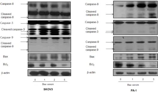
D. Effect of bee venom on the expression of apoptotic regulatory proteins
To figure out the relationship between the induction of apoptotic cell death and increase of DR expression, and the expression of their regulatory proteins by bee venom, expression of apoptotic cell death related proteins was investigated by Western
blots. The expression of anti-apoptotic protein Bcl- 2 was decreased; however, the expression of pro-a poptotic proteins, Bax, cleaved form of caspase-3, -8, and -9 was increased by treatment of bee venom in a concentration dependent manner. How- ever, expression of cleaved caspase-3 was greatly increased in SKOV3, whereas cleaved caspase-8 was significantly increased in PA-1 cancer cells (Fig. 6).
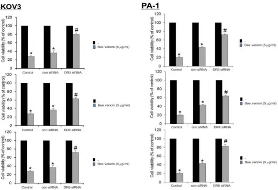
E. Reversed effect of DR siRNAs on bee venom-induced cell growth inhibition
To determine the relationship between DR
expression and cell growth inhibitory effect of bee
venom, we transfected SKOV3 and PA-1 cells with
DR siRNA using a transfection agent. The cells
were transfected with 100 nM siRNA of DRs for 24
hr, and then treated with bee venom (5 ㎍/ml) for
24 hr. Expression of the death receptor (DR3, DR4,
and DR6) at mRNA levels was detected by RT-
PCR. As shown in Fig. 7, transfection of DR3, DR4,
and DR6 siRNA reversed bee venom-induced ex-
pression of DRs. Cell viability was then determined
by direct cell counting. It was also found that knock
down of death receptor generally (DR3, DR4, and
Fig. 6. Effect of bee venom on the expression of apoptosis regulatory proteins
Expression of apoptosis regulatory proteins was determined using Western blot analysis. The ovarian cancer cells were treated with different concentrations of bee venom(1, 2, and 5 ㎍/ml) for 24 hr. Equal amounts of total proteins(50 ㎍/lane) were subjected to 12% or 8% SDS-PAGE. Expression of caspase-8, caspase-3, caspase-9, Bax, Bcl2, and β-actin were detected by Western blotting using specific antibodies.β-actin protein here was used as an internal control. Each band is representative for three experiments.
Fig. 7. Effect of siRNA of DRs on the bee venom-induced cancer cell growth inhibition, DR expression and apoptosis in ovarian cancer cells
Effect of siRNA of DR on the bee venom-induced DR gene expression in ovarian cancer cell. The ovarian cancer cells were transfected with the DR siRNA(100 nM) for 24 hr, the cells were then and treated with bee venom(5 ㎍/ml) for another 24 hr. Total RNA was isolated and RT-PCR was performed to examine gene expression levels of DRs.
Each band is representative for three experiments.
DR6) reversed the cell growth inhibitory effect of bee venom in SKOV3 and PA-1. Especially in PA-1, anti- proliferative effect of DR4 was almost completely reversed, however, the magnitude of reversed effect was relatively small in SKOV3 by DR4 siRNA (Fig. 8). We also evaluated apoptotic cell death by fluorescence microscope. DAPI-stained TUNEL-positive cells increased by bee venom were prevented by transfection with DR siRNA
both in SKOV3 and PA-1 cells (Fig. 9). These results demonstrate that expression of DR3, DR4, and DR6 is strongly correlated with bee venom- induced apoptotic cell death in ovarian cancer cells.
F. Association between death receptor and activation of STAT3
STAT3 phosphorylation is associated with pro-
Fig. 9. Quantification of apoptosis by TUNEL assay
The ovarian cancer cells were transfected with the death receptors iRNA for 24hr, and then treated with bee venom(5㎍/ml) for 24 hr. The apoptotic index was determined as the DAPI-stained TUNEL-positive cell number/
total DAPI stained cell number(magnification, 200x). Columns, means of three ex- periments, with triplicates of each experiment; bars, SD. * : p <0.05, significantly different from untreated control cells. # : p <0.05, significantly different from control siRNA transfected cells.
Fig. 8. Effect of DR3, DR4, or DR6 knockdown on bee venom-induced ovarian cancer cell growth
Ovarian cancer cells were transfected with non targeting control siRNA, DR3, DR4, or DR6 siRNA (100 nM) as described in Materials and Methods for 24 hr. Then, bee venom(5 μg/ml) was treated at 37°C for another 24 hr. The cells were harvested by trypsinization and stained with 0.2% trypan blue. Relative cell survival rate was determined by counting live and dead cells. The results were expressed as a percentage of viable cells.Columns, means of three experiments, with triplicates of each experiment; bars, SD. * : p <0.05, significantly different from untreated control cells. # : p <0.05, significantly different from control siRNA transfected cells.
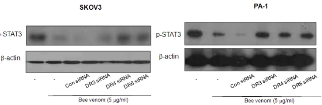
Fig. 11. Ovarian cancer cells were transfected with nontargeting control siRNA, DR3, DR4, or DR6 siRNA(100nM) as described in Materials and Methods for 24hr
Then, bee venom(5μg/ml) was treated at 37°C for another 24hr. Equal amounts of total proteins(50㎍/
lane) were subjected to 8% SDS-PAGE. Expression of STAT3, p-STAT3, JAK2, p-JAK2, and β -actin were detected by Western blotting using specific antibodies. β-actin protein here was used as an internal control. Each band is representative for three experiments.
Fig 10. Effect of bee venom and siRNA of DRs on the activation of JAK2/STAT3 in ovarian cancer cells
The ovarian cancer cells were treated with different concentrations of bee venom(1, 2 and 5 ㎍/ml) for 24 hr. Equal amounts of total proteins(50 ㎍/lane) were subjected to 8% SDS-PAGE. And β-actin were detected by Western blotting using specific antibodies of STAT3, p-STAT3, JAK2, p-JAK2, and β-actin protein here was used as an internal control. Each band is representative for three ex- periments.