INTRODUCTION
Kawasaki disease (KD) is an acute multisystem vasculitis that afflicts mostly young children. Since the first report of KD in 1967 (1), many attempts to determine the etiology of the disease have failed. However, the clinical and epidemi- ologic studies have proposed that KD is closely related to an infectious disease (2, 3). The acute onset of a self-limited co- urse, the prevalent population (rare in <6 months of age and
>5 yr of age), the existence of clusters or epidemics with a wave-like spread, all suggest that KD is related to infectious agents, particularly of viral origin. On the other hand, the elevated levels of the inflammatory indices including the white blood cell (WBC) and neutrophil count and the C-reac- tive protein (CRP) suggest that bacterial agents including superantigens are involved in KD (4). We previously found that the IgM and IgA levels increased at 1 week and 2 weeks after intravenous immunoglobulin (IVIG) treatment with a statistical significance (5). This finding also provides evidence showing that KD is an infection-related disease.
Many therapeutic modalities have been attempted in order to prevent the coronary artery lesions (CAL) as the major com- plication of KD. IVIG therapy is known to decrease the num- bers of CAL, and is now accepted as standard treatment for
KD (6, 7). Therefore, a study on the natural course in KD is unethical and impossible.
The total duration of fever in KD in the era before IVIG therapy was reported to be approximately 1-2 weeks (mean 10 days) regardless of treatments with aspirin or steroids (1, 8).
We evaluated the inflammatory indices including the WBC count and CRP according to the onset of fever in children with KD, and postulated that inflammatory processes in KD reach a peak at the sixth day of fever.
MATERIALS AND METHODS
The subjects of this study were 152 children who had been diagnosed with KD (82 boys and 70 girls) between July 1999 and December 2002 at The Catholic University of Korea, Daejeon St. Mary’s Hospital. The diagnostic criteria were based on the Diagnostic Guidelines of Kawasaki Disease pre- sented by the Japan Kawasaki Disease Research Committee (9). The first day of fever was considered the first day of illness.
The mean age of the patients was 2.4±1.4 yr (2 months- 9.4 yr). One hundred nineteen children met the criteria for KD and 33 cases of an incomplete KD were included. Incom- plete KD patient was defined as those who do not fulfil the
Kyung-Yil Lee, Ji-Whan Han, Ja-Hyun Hong, Hyung-Shin Lee, Joon-Sung Lee, Kyung-Tai-Whang
Department of Pediatrics, College of Medicine, The Catholic University of Korea, Seoul, Korea
Address for correspondence Kyung-Yil Lee, M.D.
Department of Pediatrics, The Catholic University of Korea, Daejeon St. Mary’s Hospital, 520-2 Daeheung-dong, Jung-gu, Daejeon 301-723, Korea Tel : +82.42-220-9541, Fax : +82.42-221-2925 E-mail : leekyungyil@catholic.ac.kr
501 J Korean Med Sci 2004; 19: 501-4
ISSN 1011-8934
Copyright � The Korean Academy of Medical Sciences
Inflammatory Processes in Kawasaki Disease Reach their Peak at the Sixth Day of Fever Onset: Laboratory Profiles According to Duration of Fever
We evaluated the inflammatory indices according to the fever duration in children with Kawasaki disease (KD), and determined duration when the inflammatory pro- cesses in KD reach their peak. Children with KD (n=152) were classified into 7 groups according to fever duration: at the third day or earlier (n=20), fourth (n=33), fifth (n=46), sixth (n=15), seventh (n=15), eighth (n=9), and at the ninth day or later after fever onset (n= 14). The levels of various laboratory indices were determined 3 times: before, 24 hr and 7 days after intravenous immunoglobulin administration (2 g/kg). WBC and neutrophil counts, and C-reactive protein level were the highest at the sixth day. Levels of hemoglobin, albumin, and high density lipoprotein chole- strol were the lowest at the sixth day. Although these indices were not significant statistically between groups, the indices showed either bell-shaped or U-shaped distribution of which peak or trench were at the sixth day. These findiugs showed that the inflammatory processes in KD reach peak on the sixth day of fever onset.
This finding is important because a higher single-dose intravenous immunoglobulin treatment before the peak day may help reduce the coronary artery lesions in KD.
Key Words : Mucocutaneous Lymph Node Syndrome; Kawasaki Disease; Immunoglobulins Intravenous;
Inflammation; C-Reactive Protein; Leukocytes
Received : 3 February 2004 Accepted : 6 May 2004
502 K.-Y. Lee, J.-W. Han, J.-H. Hong, et al.
recommended criteria at presentation regardless of echocardio- graphic findings. All patients with incomplete presentation who did not show elevated CRP (<2.0 mg/dL), and WBC and neutrophil (<10,000/ L and <5,000/ L, respectively) levels with repeated examinations in the earlier days of ill- ness or those who did not show an increased platelet count 7 days after IVIG treatment were excluded (4 cases). Children were treated with IVIG (I.V.-Globulin S, Green Cross, Korea:
5% liquid preparation containing only maltose, IgG:mal- tose=1:2) at a dose of 2 g/kg over 12 hr and a dose of aspirin (30-40 mg/kg) during the febrile period. After obtaining parental consent, the serial examinations were performed three times during admission: before IVIG administration, and 24 hr and 7days after IVIG administration. CAL were defined and classified as follows: ectasia was defined when coronary arterial dilatation with the diameter ≤4 mm was seen or when the diameter was less than 1.5 times than that of adja- cent artery diameter; aneurysm, when dilatation >4 mm ecta- sia with or without, multiple, pyramidal/fusiform aneurysm was present. Twenty-six of the 152 children had CAL (21 in ectasias and 5 in aneurysms). Thirteen children showed a resis- tance to IVIG therapy (a fever for more than 48 hr after initi- ating the IVIG infusion). These children were not excluded
from the study group. The demographic and laboratory data were tabulated. The Ethics Committee on Clinical Research, the Catholic University of Korea, approved this study.
Statistical analyses were done using SPSS 10.0 for Win- dows. The means of all continuous variables were compared using one way ANOVA, Fisher’s extact test and chi-square test. Continuous variables are reported as the mean±Stan- dard deviation. p≤0.05 was considered statistically signifi- cant.
RESULTS
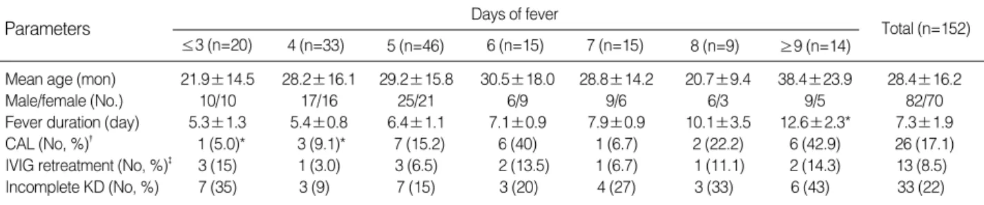
The demographic and clinical characteristics of the patients are summarized in Table 1. There were no significant differ- ences in terms of age (mean 28.4±16.2 months) and sex distribution (male to female ratio, 82:70) among the groups.
The mean total duration of the fever was 7.3±1.9 days. The mean incidence of CAL evaluated within 2-3 weeks of the onset of fever was 17.1%. There was a trend for the incidence of CAL to be higher in the sixth day (40%) and the ≥ninth day groups (42.9%) than the other groups. No significant differences were observed among the groups in the cases of
Parameters Days of fever
≤3 (n=20) 4 (n=33) 5 (n=46) 6 (n=15) 7 (n=15) 8 (n=9) ≥9 (n=14)
Total (n=152)
Mean age (mon) 21.9±14.5 28.2±16.1 29.2±15.8 30.5±18.0 28.8±14.2 20.7±9.4 38.4±23.9 28.4±16.2
Male/female (No.) 10/10 17/16 25/21 6/9 9/6 6/3 9/5 82/70
Fever duration (day) 5.3±1.3 5.4±0.8 6.4±1.1 7.1±0.9 7.9±0.9 10.1±3.5 12.6±2.3* 7.3±1.9
CAL (No, %)� 1 (5.0)* 3 (9.1)* 7 (15.2) 6 (40) 1 (6.7) 2 (22.2) 6 (42.9) 26 (17.1)
IVIG retreatment (No, %)� 3 (15) 1 (3.0) 3 (6.5) 2 (13.5) 1 (6.7) 1 (11.1) 2 (14.3) 13 (8.5)
Incomplete KD (No, %) 7 (35) 3 (9) 7 (15) 3 (20) 4 (27) 3 (33) 6 (43) 33 (22)
Table 1.Clinical and demographic characteristics of Kawasaki disease patients according to fever duration
*Statistically significant (p<0.05) compared to the values at the sixth day; �, Coronary artery lesions; �, Intravenous immunoglobulin retreatment.
Parameters
WBC (/ L, ×103) 14.5±3.5 16.1±4.5 14.4±3.0 17.1±6.3 14.0±4.2 15.0±4.3 14.4±4.7 15.0±4.1 Neutrophil (/ L, ×103) 9.7±1.6 10.3±1.9 9.8±1.2 11.5±1.9 8.6±2.2 8.6±1.7 8.1±1.7 9.6±1.7 Hemoglobin (g/dL) 10.9±0.6 11.3±0.8 10.8±0.7 10.5±0.6 10.8±0.6 10.8±0.6 10.5±0.8 10.9±0.7 Platelet (/ L, ×103) 358±83 387±73 360±77 370±91 423±139 509±123 525±175* 396±105 ESR (mm/hr) 45.6±6.9 43.7±10.2 48.2±8.6 47.0±12.9 46.6±12.0 51.2±10.4 40.8±9.9 46.2±9.8 CRP (mg/dL) 7.5±3.6 6.1±3.8* 11.0±5.9 14.5±7.1 11.2±4.4 6.3±3.3* 7.3±3.4 9.1±5.6
AST (IU/L) 93±83 52±33 54±45 41±27 38±17 39±25 33±15 53±40
ALT (IU/L) 128±100 97±98 84±85 71±48 56±38 31±26 19±11 83±77
LDH (IU/L) 542±201 512±79 490±123 488±83 559±98 580±111 484±67 512±111
CPK (IU/L) 72±25 104±57 105±80 108±111 96±66 74±32 79±61 95±64
T. protein (g/dL) 6.1±0.5 6.5±0.4 6.5±0.5 6.4±0.7 6.4±0.4 6.9±0.5 6.7±0.4 6.5±0.5 Albumin (g/dL) 3.7±0.3 3.8±0.4 3.7±0.4 3.4±0.4 3.6±0.3 3.9±0.4 3.7±0.3 3.7±0.4
T. chol (mg/dL) 127±22 144±19 145±17 141±19 154±24 155±22 163±26 144±21
HDL-chol (mg/dL) 33.6±6.4 30.7±5.8 32.5±8.5 28.1±9.0 33.0±9.2 34.1±6.9 34.8±6.8 32±8.1 Table 2.Laboratory data of Kawasaki disease patients according to fever duration
T. protein, total protein; T. chol, total cholesterol; HDL-chol, HDL-cholesterol. *Statistically significance (p<0.05) compared to the values at sixth day.
Days of fever
≤3 (n=20) 4 (n=33) 5 (n=46) 6 (n=15) 7 (n=15) 8 (n=9) ≥9 (n=14)
Total (n=152)
IVIG retreatment. Twenty-two percent of cases had fever with less than 4 of the diagnostic criteria for KD at presentation (incomplete KD). There was a trend for the incidence of in- complete KD to be higher in the ≤third day (35%), the eighth day (33%) and the ≥ninth day groups (43%).
The laboratory findings obtained before IVIG treatment are summarized in Table 2. WBC and neutrophil counts, CRP and creatine phosphokinase (CPK) values were the highest in the sixth day group, and the last two indices showed a bell- shaped distribution pattern based on the peak sixth day values.
In contrast, albumin, and HDL-cholesterol values were the lowest in the sixth day group, and they showed a pattern of U-shaped distribution. The platelet count and total cholesterol value showed a trend for increase with subsequent days of fever. The hemoglobin, ESR and LDH values did not change significantly. The AST and ALT values were the highest in the ≤third day group, but were not statistically significant.
DISCUSSION
The inflammatory processes of an infection progress to a peak stage, then regress to a convalescence by host immune response. The total duration of fever in most uncomplicated viral infections is approximately 1 week, including measles (5 days) and Epstein-Barr virus infection (6 days). On the other hand, the duration of fever in an untreated bacterial infection differs according to the causative agent. Scarlet fever is known to last 5-7 days. However causative disease caused by intra- cellular organisms such as typhoid fever lasts much longer (>2 weeks).
Early studies revealed that the total duration of KD with- out IVIG therapy was 1-2 weeks (mean 10 days) (1, 8). There- fore, it is postulated that the peak inflammatory process in KD is at the fifth to sixth day after the onset of fever. The results in this study shows that the levels of inflammatory indices reach a peak or nadir on the sixth day of the fever, which agrees with the above postulation. WBC and neutrophil counts, ESR and CRP levels are commonly used as indices of severity of inflammation. CRP increases rapidly within 24 hr in various conditions, like bacterial infections, trauma, tissue necrosis, and malignant neoplasm. It declines rapidly with a resolu- tion of the pathologic conditions (10). ESR is also another acute reactant of inflammation. The decreased albumin or HDL-cholesterol values have been reported in inflammatory diseases including KD (11, 12). Although, this study could not show difference of statistical significance in any of the indices between groups, the distribution patterns of these indices strongly support our postulation. AST and ALT val- ues appeared to be higher in the early days of the natural course in KD. The cases with elevated AST and ALT above two folds the normal values were also higher in the early days of the fever (≤4 day, 20 of 50 cases) than in the later days (≥7 days, 5 of 38 cases). A decreased total cholesterol level in the earli-
er days tends to increase with days of the fever. The platelet count is well known to increase in the convalescent stage in KD. The platelet counts also tended to increase by days of fever in the acute stage.
Recently, two studies reported the relationship between the initial day of the IVIG treatment and the clinical or laboratory outcomes in KD (13, 14). Although the two studies are simi- lar in study design, the results of CAL frequency and labora- tory findings were not identical. Nomura et al. (13) reported that the group receiving IVIG treatment prior to the fifth day of illness showed a higher CAL at 1 month, and no dif- ferences in the laboratory findings except for ALT and AST when compared to those given IVIG after the fifth day. In con- trast, Tse et al. (14) reported a case-control study showing that early IVIG treatment at day 5 or earlier showed a lower inci- dence of CAL at 1 yr, and some differences in the laboratory findings including higher hemoglobin, ALT, and albumin values and lower platelet count. If our postulation is correct, different results of laboratory study between the two groups might result from differences in the number of patients in each different fever day.
Earlier studies have reported that the risk of CAL in KD is associated with some demographic or laboratory factors such as the prolonged fever duration, or an increased CRP (15-17).
IVIG is quite effective for improving the clinical symptoms as well as the laboratory findings including CRP, and for pre- venting coronary complications (6, 7). We previously repored that a high dose IVIG treatment (2 g/kg) lowered the values of various proteins including albumin and lipoproteins, and also values of the inflammatory indices within 24 hr except ESR (5, 18). In addition, we also found that IVIG-resistant patients showed a sustained high values of CRP and WBC on 24 hr after IVIG with a higher risk of CAL (19, 20). If the risk of CAL is associated with the intensity of the inflamma- tion reflected by laboratory indices, early treatment prior to the peak stage of inflammation can help prevent CAL in KD.
There are some limitations in interpreting our results. Be- cause the data had a large number of variables (seven variables) and an uneven distribution of the number of variables, our postulation could not reach statistical significance. A prospec- tive study with a large number of patients would resolve this.
A diagnosis of KD in patients before 5 days illness could not fulfil the diagnostic criteria and those with incomplete KD were included in this study. All patients with incomplete KD were selected on the basis of the laboratory findings as des- cribed earlier as well as the clinical criteria. However, there was no significant difference in laboratory values between the incomplete KD group and typical KD group (data not shown).
In conclusion, it is very important to determine when the inflammatory processes of KD reach a peak as a self-limiting disease. As for the coronary complications in KD, it is believed that the more severe inflammatory processes, which are reflect- ed by prolonged fever duration or higher values of the inflam-
Peak day of Inflammation in Kawasaki Disease 503
504 K.-Y. Lee, J.-W. Han, J.-H. Hong, et al.
matory indices, have a higher risk. Therefore a higher single- dose IVIG treatment before the peak stage of KD may help reduce the intensity of inflammation and the frequency of CAL. Repeated examination of the inflammatory indices can help decide the timing for appropriate IVIG treatment for children with an incomplete presentation in the earlier days, and for evaluating the effects of IVIG treatment.
REFERENCES
1. Kawasaki T. Acute, febrile mucocutaneous syndrome with lymphoid involvement with specific desquamation of the fingers and toes in children. Jpn J Allergy (Arerugi) 1967; 16: 178-222.
2. Sundel RP, Newberger JW. Kawasaki disease. Curr Opin Infect Dis 1992; 5: 664-9.
3. Rowley AH, Gonzalez-Crussi F, Shulman ST. Kawasaki syndrome.
Rev Infect Dis 1988; 10: 1-15.
4. Abe J, Kotzin BL, Jujo K, Melish ME, Glode MP, Kohsaka T, Leung DY. Selective expansion of T cells expressing T-cell receptor variable regions V 2 and V 8 in Kawasaki disease. Proc Natl Acad Sci USA 1992; 89: 4066-70.
5. Lee KY, Lee HS, Hong JH, Han JW, Lee JS, Whang KT. High-dose intravenous immunoglobulin down-regulates the activated levels of inflammatory indices except erythrocyte sedimentation rate in acute stage of Kawasaki disease. J Trop Pediatr 2004; in press.
6. Furusho K, Kamiya T, Nakano H, Kiyosawa N, Shinomiya K, Haya- shidera T, Tamura T, Hirose O, Manabe Y, Yokoyama T. High dose intravenous gamma globulin for Kawasaki disease. Lancet 1984; 2:
1055-8.
7. Newburger JW, Takahashi M, Beiser AS, Burns JC, Bastian J, Chung KJ, Colan SD, Duffy CE, Fulton DR, Glode MP. A single intravenous infusion of gamma globulin as compared with four infusions in the treatment of Kawasaki syndrome. N Engl J Med 1991; 324: 1633-9.
8. Hicks RV, Melish ME. Kawasaki syndrome; Rheumatic complains and analysis of salicylate therapy. Arthritis Rheum 1979; 22: 621-2.
9. Japan Kawasaki Disease Research Committee. Diagnostic guidelines of Kawasaki disease. 4th ed. Tokyo: Japan Kawasaki Disease Research Committee, 1984.
10. Kushner I. C-reactive protein and the acute-phase response. Hosp Pract 1990; 25: 21-8.
11. Sammalkorpi K, Valtonen V, Kerttula Y, Nikkila E, Taskinen MR.
Changes in serum lipoprotein pattern induced by acute infection.
Metabolism 1988; 37: 859-65.
12. Newburger JW, Burns JC, Beiser AS, Loscalzo J. Altered lipid pro- file after Kawasaki syndrome. Circulation 1991; 84: 625-31.
13. Nomura Y, Masuda K, Yoshinaga M, Sameshima K, Miyata K. Pa- tients diagnosed with Kawasaki disease before the fifth day of illness have a high risk of coronary artery aneurysm. Pediatr Intern 2002;
44: 353-7.
14. Tse SML, Silverman ED, McCrindle BW, Yeung RSM. Early treat- ment with intravenous immunoglobulin in patients with Kawasaki disease. J Pediatr 2002; 140: 450-5.
15. Koren G, Lavi S, Rose V, Rowe R: Kawasaki disease: review of risk factors for coronary aneurysms. J Pediatr 1986; 108: 388-92.
16. Beiser AS, Takahashi M, Baker AL, Sundel RP, Newburger JW. A predictive instrument for coronary artery aneurysms in Kawasaki disease. Am J Cardiol 1998; 81: 1116-20.
17. Koyanagi H, Nakamura Y, Yanagawa H. Lower level of serum potas- sium and higher level of C-reactive protein as an independent risk factor for giant aneurysms in Kawasaki disease. Acta Paediatr 1998;
87: 32-6.
18. Lee KY, Han JW, Lee JS, Whang KT. Alteration of biochemical pro- files after high-dose intravenous immunoglobulin administration in Kawasaki disease. Acta Paediatr 2002; 91: 164-7.
19. Lee GB, Lee JW, Lee KY. Prediction of intravenous immunoglobu- lin non-responders in patients with Kawasaki disease. Korean J Pedi- atr 2004; 47: 90-4.
20. Lee KY, Han JW, Lee HS, Hong JH, Hahn SH, Lee JS, Whang KT.
Epidemiologic study of Kawasaki disease at a single hospital in Dae- jeon, Korea (1987 through 2000). Pediatr Infect Dis J 2004; 23: 52-5.