© 2010 The Korean Academy of Medical Sciences.
This is an Open Access article distributed under the terms of the Creative Commons Attribution Non-Commercial License (http://creativecommons.org/licenses/by-nc/3.0) which permits unrestricted non-commercial use, distribution, and reproduction in any medium, provided the original work is properly cited.
pISSN 1011-8934 eISSN 1598-6357
Falciform Ligament Abscess after Omphalitis: Report of a Case
A falciform ligament abscess is a rare type of intra-abdominal abscess. A 2-yr-old male, who had omphalitis two months previously, presented with a fever and right upper quadrant abdominal pain. The ultrasound and CT scan showed an abdominal wall abscess located anterior to the liver, which was refractory to conservative management with percutaneous draninage and antibiotics. On the third recurrence, surgical exploration was performed and revealed an abscess arising from the falciform ligament; the falciform ligament was excised. A follow up ultrasound confirmed complete resolution of the abscess with no further recurrence.
Key Words: Falciform; Ligaments; Abscess; Omphalitis Suk-Bae Moon1, Hae Won Lee2,
Kwi-Won Park1, and Sung-Eun Jung1 Department of Pediatric Surgery1, Seoul National University Children’s Hospital, Seoul; Department of Surgery2, Konkuk University School of Medicine, Seoul, Korea
Received: 6 April 2009 Accepted: 25 June 2009 Address for Correspondence:
Suk-Bae Moon, M.D.
Department of Pediatric Surgery, Samsung Medical Center, 81 Irwondong-gil, Gangnam-gu, Seoul 135-750, Korea Tel: +82.2-3410-0926, Fax: +82.2-3410-6982 E-mail: [email protected]
DOI: 10.3346/jkms.2010.25.7.1090 • J Korean Med Sci 2010; 25: 1090-1092
CASE REPORT
Surgery
INTRODUCTION
The falciform ligament extends from the umbilicus upward to the diaphragm, and laterally to form the hepatic coronary liga- ments. It represents a potential space, and a few cases of falci- form ligament abscess secondary to infectious diseases of the liver and gallbladder have been reported in adults. It is often misdiagnosed as a simple abdominal wall abscess due to the location of the abscess and treated non-operatively, which is usually unsuccessful. Here we report a case of falciform ligament abscess after omphalitis in a child.
CASE REPORT
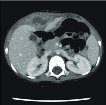
A 2-yr-old male presented with a fever and right upper quadrant abdominal pain. The patient underwent surgery for type three ileal atresia during the newborn period, and had a history of omphalitis two months prior to presentation. On examination the body temperature was 38.6°C and the umbilicus was nor- mal. There was a firm tender mass palpated in the right upper quadrant of the abdomen. The laboratory test results showed a leukocytosis of 18,290/μL with a shift to the left. The abdominal ultrasound and computed tomography (CT) revealed a 3.4×3.4- cm abscess located at the right paramedian abdominal wall ex- tending to the anterior surface of the liver (Fig. 1). The patient was managed with percutaneous drainage and intravenous an- tibiotics. The microbiology examination of the abscess identi- fied Methicillin-resistant Staphylococcus aureus (MRSA). Short- ly after drain removal, the fever and abscess emerged again and
two weeks of intravenous vancomycin and aspiration of the ab- scess were performed two more times. However, after normal- ization of the clinical parameters, a follow-up CT revealed a 3.3×
2.4-cm residual abscess at the same location. At laparotomy, the abscess was found to originate from the falciform ligament; the abdominal wall was clear (Fig. 2). The falciform ligament was completely excised, and the post-operative course was unevent- ful. Pathological examination revealed a fibrosis of the ligament with abscess formation. A follow-up ultrasound 2 weeks after the operation showed complete resolution of the abscess. Now it is 4 months after the operation and he is well and healthy.
DISCUSSION
A soft tissue mass beneath the abdominal wall continuous with a thickened round ligament is a diagnostic feature of a falciform ligament abscess by ultrasound or CT scanning (1). However, because of its rarity and obscure location, a definite radiologi- cal diagnosis of a falciform ligament abscess is difficult. Infec- tions can extend from the liver, gallbladder (2, 3) and umbilicus (4). An infection of a cystic lesion of the falciform ligament has been reported as a cause of a falciform ligament abscess (5). As shown in this case, it is important to suspect a falciform ligament abscess in a patient with a right upper quadrant abscess and a prior history of abdominal infections.
Lipinski et al. (4) reported two cases of a falciform ligament abscess secondary to an omphalitis; contiguous spread of the infection via the round ligament was thought to be the etiology.
In the present case, however, the round ligament was divided
Moon S-B, et al. • Falciform Ligament Abscess and Omphalitis
DOI: 10.3346/jkms.2010.25.7.1090 http://jkms.org 1091
due to the previous operation for ileal atresia, because the su- praumbilical transverse, round ligament-cutting incision was used to provide more wide operative field. The superficial veins of the abdominal wall form a network that radiates out from the umbilicus, and a few small veins named paraumbilical veins connect the network to the portal vein forming a portal-system- ic venous anastomosis (6). This venous network might explain the extension mechanism of the omphalitis into falciform liga- ment abscess in the absence of a round ligament. Moreover, the paucity of the vascular network inside the ligamentous structure might have impaired the venous outflow from the ligament and the MRSA could be colonized easily within the falciform liga- ment to form an abscess. Although the round ligament was ma- nipulated during the operation at the neonatal period, the long time interval between the ileal atresia operation and falciform ligament abscess would preclude the possibility of the abscess as a post-operative complication.
MRSA has been reported to be the most frequent causative agent of omphalitis in children (7). The identification of MRSA, which is consistent with the previously isolated microorganisms from the omphalitis, also supports the speculation that the ab- scess originated from the omphalitis. Delivery at home, low birth weight, use of umbilical catheters, and septic delivery have been known to be risk factors of the omphalitis (8), but the causes of the omphalitis reported here are uncertain. As the omphalitis was cured before the symptoms of the falciform ligament ab- scess became apparent and there had been no abdominal com- plaints before the onset of the omphalitis, we speculate that the omphalitis must have preceded the falciform ligament abscess.
Many readily accessible abscesses are treated successfully with percutaneous drainage and the antibiotics. However, in this patient drainage and antibiotics did not completely treat the abscess. This might be also explained by the paucity of the vascular network that hindered exposure to the circulation and therefore the antibiotics. Previous authors reported successful treatment of the falciform ligament abscess after excision of the ligament (4, 9, 10). Therefore, when a falciform ligament abscess is suspected, surgical excision rather than percutaneous drain- age should be considered for the initial treatment.
We treated a patient with a falciform ligament abscess sec- ondary to a prior omphalitis. The patient was successfully treat- ed with falciform ligament excision. A strong index of suspicion is necessary for early diagnosis and treatment of similar cases.
REFERENCES
1. Mori H, Aikawa H, Hirao K, Futagawa S, Fukuda T, Maeda H, Hayashi K. Exophytic spread of hepatobiliary disease via perihepatic ligaments:
demonstration with CT and US. Radiology 1989; 172: 41-6.
2. de Melo VA, de Melo GB, Silva RL, Aragão JF, Rosa JE. Falciform liga- ment abscess: report of a case. Rev Hosp Clin Fac Med Sao Paulo 2003;
58: 37-8.
3. Sones PJ Jr, Thomas BM, Masand PP. Falciform ligament abscess: ap- pearance on computed tomography and sonography. AJR Am J Roent- genol 1981; 137: 161-2.
4. Lipinski JK, Vega JM, Cywes S, Cremin BJ. Falciform ligament abscess in the infant. J Pediatr Surg 1985; 20: 556-8.
5. Losanoff JE, Kjossev KT. Isolated gangrene of the round and falciform liver ligaments: a rare cause of peritonitis: case report and review of the world literature. Am Surg 2002; 68: 751-5.
6. Snell RS. Clinical anatomy (7th ed). Philadelphia, PA, Lippincott Wil- liams&Wilkins, 2004.
7. Sawardekar KP. Changing spectrum of neonatal omphalitis. Pediatr In- fect Dis J 2004; 23: 22-6.
Fig. 1. The CT scan showing an abscess pocket with thick irregular wall at right paramedian abdominal wall, abutting to the falcifom ligament.
Fig. 2. The falciform ligament abscess was completely excised from the abdominal wall.
Moon S-B, et al. • Falciform Ligament Abscess and Omphalitis
DOI: 10.3346/jkms.2010.25.7.1090
1092 http://jkms.org
8. Cilley RE. Disorders of the umbilicus. In: Grosfeld JL, O’Neill JA Jr, Fonk- alsrud EW, Coran AG, eds. Pediatric Surgery, 6th ed. Philadelphia, PA:
Mosby 2006; 1143-56.
9. Pratap A, Tiwari A, Anchal N, Agrawal CS, Shreshta P, Shakya VC. Falci- form ligament abscess with portal pyemia in a newborn. J Pediatr Surg
2006; 41: 1473-5.
10. Laucks SS 2nd, Ballantine TV, Boal DK. Abscess of the falciform ligament in a child with a ventriculoperitoneal shunt. J Pediatr Surg 1986; 21: 979- 80.