INTRODUCTION
The round window membrane (RWM) located in the pos- terior inferior aspect of the medial tympanic wall deep inside the bony niche suggesting its unique function in the proxim- ity of the inner ear (IE). This niche protects the RWM direct sound transmission and physical trauma, but also poses a threat to the IE, as noxious materials accumulated in the niche may traverse the soft tissue diaphragm, the RWM.
In sound transmission, the RWM acts as a relief organ for the waves of the cochlear fluid evoked by stapedial vibration.
In addition, RWM might play a role in absorption and secre- tion between the middle ear (ME) and IE, and acts as a route for oxygenation of the IE tissue from the ME (1, 2). The RWM is readily permeable to many biological substances and may act as a port of entry for noxious substances from the ME into the IE, resulting in pathological changes in the IE (3, 4).
Histologically, the RWM consists of three basic layers: outer epithelial layer, middle connective tissue layer, and inner meso- thelial layer (1, 5, 6). This triple-layered RWM, called sec- ondary tympanic membrane (TM) is very similar to that of the TM. Interestingly, morphological characteristics of the pars flaccida in particular, markedly resemble those of the
RWM (7, 8), suggesting that they may have shared certain functions such as pressure equilibration. The epithelial layer contain metabolically active cells and the fibroblasts of the middle layer may play an important role in production of collagen fibers and of various active substances, which may be involved in the IE defense system (9).
In inflammatory conditions in ME, there are significant changes in the thickness and morphological characteristics of the RWM. Such alterations may affect the permeability of the RWM, which is an important barrier between the ME and the IE (10, 11). The ME in the rat and man share many similar structural characteristics. Recently, it was also shown that the reaction of the rat ME to Streptococcus pneumoniae closely resembles that in the human ME (12). This animal model for purulent otitis media has been utilized in study- ing the effects of pneumococci on the ME mucosa as well as the outcome of various therapeutic measures (13).
In the present study, we examined the fine structure of the RWM in the different stages of the pneumococcus-evoked otitis media model using rats. This will serve as a structural basis for a continuing studies on the role of the RWM for the protection of IE function in ME infections.
Yong-Joo Yoon, Sten Hellstrom*
Department of Otorhinolaryngology Chonbuk National University Hospital, Chonju, Korea;
Department of Otorhinolaryngology*, University Hospital, University of Umea, Umea, Sweden
Address for correspondence Yong Joo Yoon, M.D.
Department of Otorhinolaryngology Chonbuk National University Hospital, 634-18 Kmam-dong, Chonju 560-180, Korea
Tel : +82.63-250-1988, Fax : +82.63-250-1986 E-mail : [email protected]
230
Ultrastructural Characteristics of the Round Window Membrane During Pneumococcal Otitis Media in Rat
To understand better the pathogenesis of inner ear (IE) damage caused by otitis media (OM), the round window membrane(RWM) structure was investigated in a rat model for pneumococcal otitis media (POM). The RWM of 25 rats were eval- uated light and electron microscopically on 1 day, 3 days, 6 days, 10 days, and 20 days after the unilateral inoculation of type 3 pneumococcus suspension into their middle ear cavities. The thickness of the RWM increased in various stages of the pneumococcus-evoked otitis media, compared with that of the normal. The thickening was most pronounced on day 1, being about 4 to 5 times greater than that of the normal RWM. All layers of the RWM were affected by the pneumococ- cal infection, but the major changes were confined to the subepithelial space close to the basement membrane (BM). Together with alterations to the BM, the most distinct pathological features were characterized by an increase and hypertrophy of fibroblasts in association with abundant collagen fibers. Elastic fibers observed close to the inner mesothelial layer under a high power magnification also in- creased during the experiment. These results will be relevant to a better under- standing of the histologic implication of RWM in stages of acute otitis media involv- ing pneumococcus-evoked otitis media.
Key Words : Round Window Membrane; Pneumococcal Otitis Media
Received : 17 August 2001 Accepted : 25 January 2002
MATERIALS AND METHODS
Healthy male Sprague-Dawley rats (n=25) weighing 200- 250 g were used for the study. All animals exhibited normal TM status. The rats were subjected to anesthesia with an in- travenous injection of sodium methohexital (Brietal�), fol- lowed by an intraperitoneal injection of sodium pentobarbi- tal (Mebumal�). Under an otomicroscope, a ventral vertical midline incision was made in the neck and the tympanic bul- lae were exposed bilaterally. Through the hole in the bulla, the left ME was inoculated with 0.05 mL suspension of type 3 Streptococcus pneumoniae. The right ear served as the control and was inoculated with culture medium only.
The animals were inspected daily under the otomicroscope throughout the experiment. They were divided into 5 rats in each. The groups of animals were killed on 1, 3, 6, 10, and 20 days after inoculation, respectively. Before sacrifice, the TM status was examined and carefully recorded. After an intraperitoneal overdose of Mebumal�, and their animals were decapitated, the temporal bones were roughly trimmed and then fixed by immersion in a solution containing 3%
glutaraldehyde in a 75 mM sodium cacodylate buffer with 4% polyvinylpyrrolidine and 2 mM CaCl2was added. The temporal bones were then carefully dissected and further fixed for 48 hr in the same fixative solution. After several rinses in buffer, the specimens were transferred to a 10%
EDTA solution and decalcified in a microwave oven (Miele M696), at a setting of 150 W, 35℃, until softened (14).
The round window niche with the RWM was now cut out in a single block, which then was dehydrated in a graded series of ethanol to xylene and embedded in an epoxy resin.
The tissues were then cut with an ultramicrotome in 1 m sections for light microscopic examination. The tissue sec- tions were stained with toluidine blue, mounted, and exam- ined under the light microscope. Ultrathin sections (70 nm) were cut in an ultramicrotome and stained with urany1 ace-
tate and lead citrate for transmission electron microscopy. The specimens were studied under JEOL 1200 electron micro- scope.
RESULTS The thickness of the RWM
The control RWM had a convex shape facing the scala tym- pani (Fig. 1). It was thickened at the edge, due to a sudden increase in the middle connective tissue layer. In the central portion of control RWM, the mean thickness was 11.5± 2.1 m (n=25).
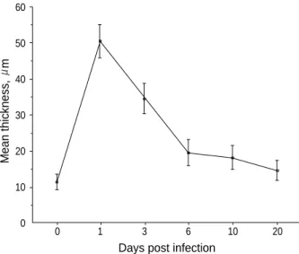
In the pneumococcus-inoculated ears, the RWM was thick- ened considerably. The average thicknesses of each group are summarized in Fig. 2. The change in the thickness of the RWM in pneumococcal otitis media (POM) was most pro- nounced on day 1 when the thickness was 4 to 5-fold greater than that of the control. The alterations were confined mainly to the central portion of the RWM (Fig. 2, 3).
RWM in Pneumococcal Otitis Media
Day 1:All layers of the RWM were markedly thickened, espe- cially in the central part of the RWM. The most prominent thickening was confined to the subepithelial space immedi- ately below the outer epithelial layer. Abundant inflamma- tory cells, mainly macrophages but also polymorphonuclear neutrophils (PMNs) were observed in this area (Fig. 4). Macro- phages were also observed within the outer epithelial layer (Fig. 4). The surface of the outer epithelial cells exhibited an increased number of microvilli, especially at the periphery of the cells. The cytoplasm of the epithelial cells showed an
Fig. 1.The surface is curved as a cone with its apex directed to- wards the scala tympani. ×50. MEC, middle ear cavity; ST, scala tympani.
MEC
ST
Fig. 2.The kinetics of average thickness ( m SD) of the rat RWM in controls (n=25) and on days 1, 3, 6, 10, and 20 in pneumococ- cus-induced otitis media (n=20).
Mean thickness, m
Days post infection 60
50
40
30
20
10
0 0 1 3 6 10 20
extensive vacuolization. Most of the nuclei of the outer epithe- lial cells appeared pale and swollen, with a dark outline. Mi- tochondria, Golgi complex, and endoplasmic reticulum were increased in the dark granulated cells (6). The outer epithe- lium exhibited widened intercellular spaces, and the epithe- lial junctions were often widened to some extent. The en- larged intercellular space was most pronounced towards the basement membrane (BM). The BM showed distinct changes compared with controls and was widened and partly inter- rupted. When in contact with inflammatory cells, the BM was folded (Fig. 4). The fibroblasts were increased in num- bers and accumulated close to leukocytes in the connective tissue layer. The inner epithelium was not so thick as the outer epithelium (Fig. 3).
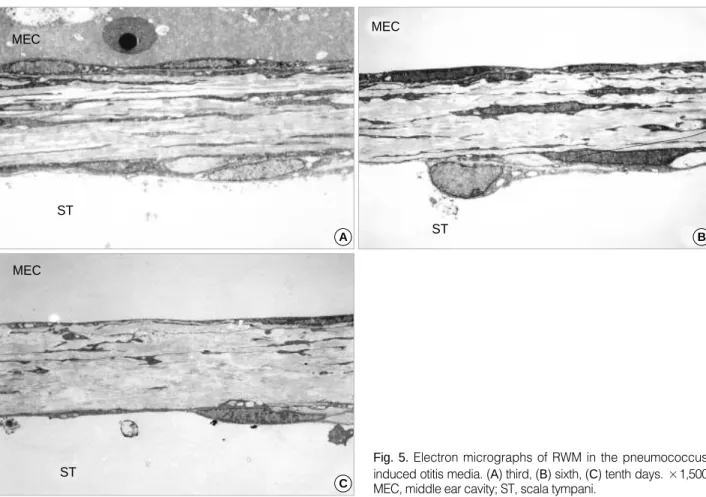
Day 3:The RWM showed severe changes, characteristic of inflammation in the deep connective tissue layer with edema, invasion of inflammatory cells and an increased vasculariza- tion. The thickness of the RWM was slightly decreased com- pared with that of the day 1, in spite of the pronounced in-
flammatory changes (Fig. 5). Inflammatory cells were observed among the outer epithelial cells, as well as in the subepithe- lial space. Many of the epithelial cells were increased in size.
The cytoplasm of outer epithelial cells showed an increased electron density of the ground substance. Swollen mitochon- dria were observed. Adjacent to the BM, the epithelial cells exhibited small round clear vacuoles scattered in the cyto- plasm. Towards the BM, the epithelial cells showed compli- cated infoldings and cytoplasmic processes. The BM seemed thickened but the uninterrupted BM was noticeable in this group. The connective tissue layer contained abundant hyper- trophic fibroblasts with a marked increase in the cytoplasm, whereas fibrocytes seemed to be slightly decreased in num- ber. The purulent effusion in the tympanic cavity was dimin- ished and had fewer inflammatory cells than on the day 1 (Fig. 5).
Day 6: Most specimens showed a general thickening of the outer epithelial layer and an increased vascularization of the subepithelial tissue. PMNs were rarely observed. Macro-
Fig. 3. Light microscopic changes in the thickness of the RWM stained for toluidine blue on normal control (A) and day 1 in pneumococ- cus-induced otitis media (B). The change was most prominent on day 1, which was most evident in the outer epithelium and subepithe- lial space of the RWM. ×200. MEC, middle ear cavity; ST, scala tympani.
A B
MEC
MEC
ST ST
Fig. 4. Electron micrographs of RWM on day 1 of pneumococcus- induced otitis media. (A) The most prominent thickening is confined to the subepithelial space imme- diately below the outer epitheli- um.×1,000. (B) Abundant inflam- matory cells, mainly of macro- phages (M) but also PMNs (P) are present in the outer epithelial layer and subepithelial space. The BM (small arrow heads) shows distinct changes including widen- ing, and is deeply folded and par- tly interrupted (short thick arrow),
×2,500.
A B
ST
M
P
P MEC
phages, evident in the outer epithelial and connective tissue layer on day 3, now appeared enlarged and filled with phago- cytosed material. The epithelial cells, varying in size, exhib- ited a cytoplasm rich in dense granules. The fibroblasts see- med larger with an increased volume of cytoplasm. Collagen fibers had a less irregular distribution in the subepithelial layer and occurred at a higher density close to fibroblasts, but not in the vicinity of fibrocytes (Fig. 5). Although the outer and middle layers were less thick than on the days 1, 3, and 6, the thickening of the inner mesothelial layer still remained. Discontinuity in the mesothelial cell layer was re- markable, suggesting that the perilymph may communicate directly with the middle connective tissue. There was no longer purulent effusion in the tympanic cavity (Fig. 5).
Day 10: The structure of the RWM now started to regain its normal characteristics (Fig. 5). However, in the outer epithe- lium, ciliated epithelial cells appeared in the peripheral part of the RWM close to the junction with the ME mucosa. The mitochondria of epithelial cells showed a marked swelling.
This layer still showed an increased number of intracellular vacuoles and enlarged intercellular spaces.
In the deep part of the connective tissue layer, the inflam- matory cell infiltration was still observed. In contrast to the earlier observations, the number of macrophages was reduced.
PMNs were occasionally found in the subepithelial space. The inner mesothelial cells had a large nucleus and contained many dark granules in their cytoplasm.
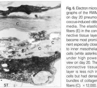
Day 20:On day 20, the structure of the RWM appeared almost normalized except for a minor thickening of the subepithe- lial tissue and the deep connective tissue layer. The thickened BM still remained. The connective tissue layer was less rich in cells but had denser bundles of collagen fibrils. The elastic fibres in the connective tissue layer were more pronounced at this stage of the experiment than any other earlier obser- vations. In particular, close to the inner layer, accumulations of elastin fibrils were observed (Fig. 6).
DISCUSSION
Inoculation of Streptococcus pneumoniae into the ME had an immediate effect on the epithelial lining facing the ME cav- ity, but also caused more slowly developing changes in the connective tissue layer. In fact the inferface area between the outer epithelium and the connective tissue layer showed strik- ing changes including an interrupted BM, which could sig- nificantly affect the permeability of the RWM. Various animal experiments have led to the assumption that RWM response
Fig. 5. Electron micrographs of RWM in the pneumococcus- induced otitis media. (A) third, (B) sixth, (C) tenth days. ×1,500.
MEC, middle ear cavity; ST, scala tympani.
A
C
B MEC
MEC
MEC
ST
ST
ST
and permeability to ME inflammatory conditions can vary according to the different stages of acute otitis media (AOM) (15). Researchers have tried to understand the functional significance of the RWM as a port of entry from the ME to the IE. Various kinds of materials instilled into the ME cav- ity. ME inflammatory conditions, and the presence of bacte- rial toxin have all been shown to induce morphological and functional alterations in the RWM (15, 16).
Albumin, which normally does not pass the RWM, will traverses the RWM under inflamed conditions (10). In experi- ments testing the RWM permeability, these large molecules had difficulty in traversing the connective tissue layer, thus suggesting the involvement of fibroblasts in the permeabili- ty control mechanisms of the RWM.
Streptococcus pneumoniae is the most common bacterial species isolated from patients with AOM. The condition resolves spontaneously within 10-14 days. In the present study, a rat model was used to elucidate the morphological events of the RWM in pneumococcal AOM. The rat AOM model developed by Hermansson et al. (12) is well suited for stud- ies of AOM because of the close resemblance of the clinical course of the pneumococcal infection between man and rat.
A significant difference in the thickness of the RWM was observed between five groups. In the early stage of infection, there was a marked widening of the subepithelial space and hypertrophy of the outer epithelial cells. The thickened RWM may act as an effective barrier protecting the IE from harm- ful agents, such as bacterial products and inflammatory medi- ators. Ikeda et al. (15) reported a reduced permeability reac- tion of the RWM to tetraethylammonium ions in the con- valescent stages of AOM. It is assumed that in convalescent stages of AOM, the RWM permeability is reduced, but that the permeability could be increased in the acute stage of in- flammation. Proliferation of the secretory epithelium and production of dense granules in the proximity of the apical surface in the cytoplasm in the pneumococcus-induced otitis
media may suggest an increased activity of epithelial cells against inflammation. Numerous PMNs and macrophages suggest that the increased RWM permeability in the acute stage may be attributed to bacterial toxins and/or such as cytokines, fibronectin, elastase, and peptides derived from these cells (17, 18).
The BM may be of physiological importance with regard to the permeability as it may act as a filter between the epithe- lial layer and the connective tissue layer (19). The widening and multiple foldings of the BM during the infectious con- dition may indicate strengthened barrier mechanism against bacteria and its byproducts.
Fibroblasts and collagen fibers, the prinicipal elements of the connective tissue layer, may constitute a second line of defence. The elongated cells, arranged in a radial fashion, are in contact with one another and form a network. In inflam- matory conditions and following physical trauma, the fibro- blasts expand. We consider it of utmost importance to clari- fy the role of the fibroblasts in the permeability mechanisms of the RWM.
In contrast to the collagen fibers, elastic fibers are sparse in the normal rat RWM. The elastic fibers could not be dis- tinguished under the light microsope, but were detected by the electron microscope. The elastic fibers were shown to be located predominantly in the region close to the inner meso- thelial layer. It may be assumed that the area of connective tissue layer close to the inner mesothelial layer could be asso- ciated with the perilymph wave and thus involve elasticity.
The high density of elastic fibers toward the mesothelial layer in the convalescent stage of POM suggested that they may participate in the barrier function of the RWM in the later stage of inflammation. With the progression of inflammation, the elastic fibers became progressively easier to visualize in the connective tissue layer. The connective tissue layer of the RWM could be considered the most important layer in aural physiology, as it permits movements of IE fluid, yet main- taining the constant shape and tension. Elastic fibers stretch easily, and when released after stretching, they return almost completely to their original length. The strength and elas- ticity of the RWM are preserved by combination of these fibers. On the other hand, elastic fibers may lose their innate characteristics by elastase (18) secreted from PMLs in the AOM resulting in a sensori-neural hearing loss. However, there is little information so far in this area.
The inner layer of the RWM is composed of mesothelial cells and lacks both microvilli and BM. Lim described that there are numerous underlying micropores and holes for free communication of perilymph between surrounding perilym- phatic tissue and the scala tympani (spiral ligament, basilar membrane, and osseous spiral lamina), but could not point them out in the RWM (2). This indicates an interchange between the RWM and perilymph regarding absorption or secretion, and suggests the possibility of presence of similar micropores and holes on the inner epithelial surface of the
Fig. 6. Electron micro- graphs of the RWM on day 20 pneumo- coccus-induced otitis media. The elastic fibers (E) in the con- nective tissue layer become most promi- nent especially close to inner mesothelial cells (white asteriks) under high power view on day 20. The connective tissue layer is less rich in cells but had dense bundles of collagen fibers (C). ×12,000.
C
C C E E
ST
RWM.
In summary, alterations in the RWM structure were shown to occur in pneumococcus-induced ME inflammation. In- creased knowledge of these dynamic changes in the RWM structure will improve our understanding of how substances pass from the ME to the IE. Further investigations are nec- essary to clarify the functional significance of these structural changes using more advanced methods in the various patho- logical conditions.
The thickening was most pronounced on day 1, being about 4 to 5 times greater than that of the normal RWM.
All layers of the RWM were affected by the pneumococcal infection, but the major changes were confined to the subepi- thelial space close to the BM. Together with alterations in the BM, the most distinct pathological features were charac- terized by an increase and hypertrophy of fibroblasts in asso- ciation with abundant collagen fibers. Elastic fibers observed close to the inner mesothelial layer also increased during the experiment.
REFERENCES
1. Carpenter AM, Muchow D, Goycoolea MV. Ultrastructural studies of the human round window membrane. Arch Otolaryngol Head Neck Surg 1989; 115: 585-90.
2. Nagahara K, Yota T, Naito Y, Ogino F. Oxygenation through the round window membrane and the inner ear function. Auris Naris Larynx (Tokyo) Suppl 1985; 1: 120-2.
3. Anniko M, Hellstrom S, Schmidt SH, Spandow O. Toxic effects on inner ear of noxious agents passing through the round window mem- brane. Acta Otolaryngol (Stockh) Suppl 1989; 457: 49-56.
4. Smith BM, Myers MG. The penetration of gentamicin and neomycin into perilymph across the round window membrane. Otolaryngol Head Neck Surg 1979; 87: 888-91.
5. Belluci RT, Fisher EG, Rhodin J. Ultrastructure of the round win- dow membrane. Laryngoscope 1972; 82: 1021-6.
6. Richardson TL, Ishiyama E, Keels EW. Submicroscopic studies of
the round window membrane. Acta Otolaryngol 1971; 71: 9-21.
7. Lim DJ. Tympanic membrane, Part II. Pars flaccida. Acta Otolaryn- gol 1968; 66: 515-53.
8. Lim DJ. Human tympanic membrane. An ultrastructural observation.
Acta Otolaryngol 1970; 70: 176-86.
9. Leitman DC, Agnost VL, Tuan JJ, Andresen JW, Murad F. Atrial natriuretic factor and sodium nitroprusside increase cyclic GMP in cultured rat lung fibroblasts by activating different forms of guany- late cyclase. Biochem J 1987; 244: 69-74.
10. Goycoolea MV, Paparella MM, Goldberg B, Carpenter AM. Perme- ability of the round window membrane in otitis media. Arch Otolaryn- gol Head Neck Surg 1980; 106: 430-3.
11. Kim CS, Cho TK, Jinn TH. Permeability of the round window mem- brane to horseradish peroxidase in experimental otitis media. Oto- laryngol Head Neck Surg 1990; 103: 918-25.
12. Hermansson A, Emgard P, Prellner K, Hellstrom S. A rat model for pneumococcal otitis media. Am J Otolaryngol 1988; 9: 97-101.
13. Hermansson A, Prellner K, Hellstrom S. Prevention of experimental acute otitis media with penicillin V. Acta Otolaryngol (Stockhol) 1990;
109: 119-23.
14. Hellstrom S, Nilsson M. The microwave oven in temporal bone re- search. Acta Otolaryngol (Stockhol) Suppl 1992; 493: 15-8.
15. Ikeda K, Sakagami M, Morizono T, Juhn SK. Permeability of the round window membrane to middle-sized molecules in purulent oti- tis media. Arch Otolarygol Head Neck Surg 1990; 116: 57-60.
16. Schachern PA, Paparella MM, Goycoolea MV, Goldberg B, Schliev- ert P. The round window membrane following application of staphy- lococcal exotoxin: an electron microscopic study. Laryngoscope 1981; 91: 2007-16.
17. Assoian RK, Fleurdelys BE, Stevenson HC, Miller PJ, Madtes DK, Raines EW, Ross R, Sporn MB. Expression and secretion of type beta transforming growth factor by activated human macrophages.
Proc Natl Acad Sci USA 1987; 84: 6020-4.
18. Janoff A. Elastase in tissue injury. Ann Rev Med 1985; 36: 207-16.
19. Gussen R. Basement membranes in the ear. Ann Otol Rhinol Laryn- gol 1966; 75: 1124-34.
20. Lim DJ. Surface ultrastructure of the cochlear perilymphatic space.
J Laryngol Otol 1970; 84: 413-28.