INTRODUCTION
Extensive burn is not only a skin injury but also a serious systemic illness often accompanied by various complications.
Acute renal failure (ARF) is one of the major complications of burns, carrying an extremely high mortality rate (1).
Although ARF is not commonly encountered in burned patients, this complication merits a special attention in that its outcome is generally very poor and varies depending on the severity and adequacy management of the burn injury.
The quoted incidence of renal failure in burned patients varies widely between 0.5 and 30% and its mortality is as high as between 73 and 100%. The incidence and mortali- ty rate of ARF in burned patients depend on the severity of the burns and on the criteria of renal failure such as free water clearance (greater than - 0.5 mL/min), blood urea nitrogen (above 50 mg/dL) and serum creatinine level (above 2.0 mg/
dL) (2). These criteria have been used to assess the incidence and as an index in treating ARF in burned patients (3). In- creased urinary excretion of protein is one of the most com- mon and easily detected signs of renal pathology. Renal abnormalities may occur both in the quantity and in the composition of urinary proteins (4). Renal pathologies in burns are characterized by the development of extensive
inflammation inducing an intensive acute phase response in the kidney. Urinary malondialdehyde (MDA) is a gross indicator of renal lipid peroxidation (5), and has been shown to increase after burns (6). The present study was aimed to estimate the degree of burn-induced renal damage and the recovery and to determine the treatment response in 12 patients with second- or third-degree burns admitted to the burn center at the Hallym University Hankang Medical Center within 12 hr of insult onset.
MATERIALS AND METHODS Patients
With approvals by the Ethics Committee of the Hallym University Hankang Medical Center and informed consents, 12 patients were selected among flame-burned patients ad- mitted to the burn center at the Hallym University Hankang Medical Center within 12 hr of their injury. The subjects consisted of 10 males and 2 females with an age range from 23 to 65 yr (mean±SD, 37.7±13.6 yr) without chronic illnesses such as hypertension and diabetes mellitus. They had second- or third-degree flame burns covering 20 to 40%
Hyun Kil Kang, Dong Keon Kim, Bong Hwa Lee, Ae Son Om*, Joung Hee Hong�, Hyun Chul Koh�, Chang Ho Lee�, In Chul Shin� Ju Seop Kang�
Department of Surgery, College of Medicine, Hallym University, Pyungchon; *Department of Food and Nutrition, College of Human Ecology and
�Department of Pharmacology & Institute of Biomedical Science, College of Medicine, Hanyang University, Seoul, Korea
Received : 22 January 2001 Accepted : 13 June 2001
Address for correspondence Ju Seop Kang, M.D.
Department of Pharmacology, College of Medicine, Hanyang University, 17 Haengdang-dong, Sungdong-gu, Seoul 133-791, Korea Tel : +82.2-2290-8269, Fax : +82.2-2292-6686 E-mail : [email protected]
598
Urinary N-acetyl- -D-glucosaminidase and Malondialdehyde as a Markers of Renal Damage in Burned Patients
This study was aimed to evaluate renal dysfunction during three weeks after the burn injuries in 12 patients admitted to the Hallym University Hankang Medical Center with flame burn injuries (total body surface area, 20-40%). Parameters assessed included 24-hr urine volume, blood urea nitrogen, serum creatinine, creatinine clearance, total urinary protein, urinary microalbumin, 24-hr urinary N- acetyl- -D-glucosaminidase (NAG) activity, and urinary malondialdehyde (MDA). Statistical analysis was performed using repeated measures ANOVA test. The 24-hr urine volume, creatinine clearance, and urinary protein signifi- cantly increased on day 3 post-burn and fell thereafter. The urine microalbumin excretion showed two peak levels on day 0 post-burn and day 3. The 24-hr uri- nary NAG activity significantly increased to its maximal level on day 7 post-burn and gradually fell thereafter. The urinary MDA progressively increased during 3 weeks after the burn injury. Despite recovery of general renal function through an intensive care of burn injury, renal tubular damage and lipid peroxidation of the renal tissue suggested to persist during three weeks after the burn. There- fore, a close monitoring and intensive management of renal dysfunction is nec- essary to prevent burn-induced acute renal failure as well as to lower mortality in patients with major burns.
Key Words : Burns, Wounds and Injuries; Kidney; Blood Urea Nitrogen; Ceatinine Clearance; Acetyl glucosaminidase, Malondialdehyde
of the total body surface area (TBSA): two with 20%; four with 25%; two with 30%; one with 35%; three with 40%
(mean±SD, 29.6±7.5%). Patients were hospitalized at the burn center and were resuscitated according to the Parkland Formula. The burn wounds were thoroughly and gently cleaned by immersing the patients in a whirlpool bath to which sodium hypochlorite had been added (150:1). The wounds were then debrided by a surgical sponge, rinsed, dried, and 1% silver sulfadiazine cream was applied and then covered with dry coarse mesh gauze. This procedure was done daily in patients. The goal of nutritional support was to maintain a positive nitrogen balance by greater than 6 g/day.
Blood sampling and urine collections
Blood was drawn on the admission day (day 0: mean±SD, 7±6 hr post-injury), and at every 6 a.m. thereafter. Several parameters such as 24-hr urine volume, blood urea nitrogen (BUN), serum and urine creatinine, creatinine clearance, total urine protein, urine microalbumin, urinary N-acetyl- -D-glucosaminidase (NAG) activity, and urinary malondi- aldehyde (MDA) were measured from day 0 to day 3, and on days 7, 14, and 21.
Analytical methods
Seum creatinine, urine creatinine, and blood urea nitrogen (BUN) were quantified by automated chemistry analyzer (SBA-300, Gilford Co, U.S.A.). Total urine protein was quan- tified according to the Braford method (7) and urine microal- bumin was determined by immunoturbidimetry. Urinary NAG activity was measured by a colorimetric procedure as described by Horak et al. (8). Urinary MDA diethyl-thio- barbituric acid complexes were isolated and quantified accord- ing to the method described by Guichardand et al. (9). All
samples were analyzed in duplicate.
Statistics
Data are presented as mean±SD. Statistical analysis was performed using repeated measures ANOVA test in SAS PROC GLM program to compare the patients’characteris- tics and biological values. A p-value less than 0.05 were considered statistically significant.
RESULTS
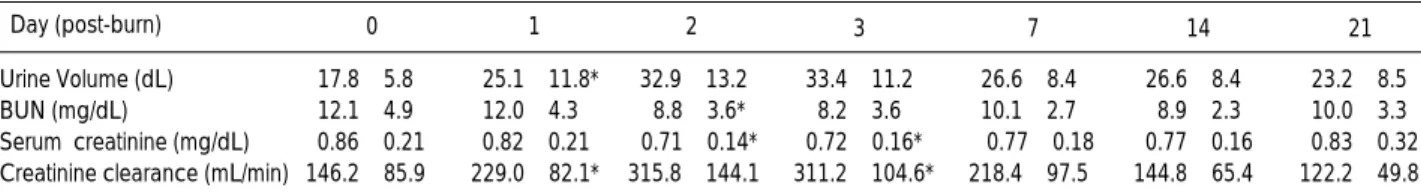
As shown in Table 1, the urine volume was elevated through- out three weeks of post-burn period with a peak level on day 3 post-burn. The BUN and serum creatinine levels were significantly decreased to their nadirs on day 3 post-burn and creatinine clearance was significantly elevated during the 3 days post-burn. The total urinary excretion of protein showed a maximum level on day 3 post-burn, while the mean urinary excretion of microalbumin was at its peak level on admission, and rapidly fell on day 1 post-burn. And then the level increased on days 2 and 3, and was reversed again on day 7 onwards, until on day 21, it was below the level of day 1 (Table 2). By 1 week after the burns the 24-hr urinary NAG activity was progressively elevated to its maximal level and still remained elevated at 5.5-fold level of day 0 on day 21. The 24-hr urinary excretion of MDA was slight- ly decreased on day 1 and thereafter progressively increased to its maximal level on day 21 (Table 3).
DISCUSSION
Severe burn must be managed not only as a dermal injury but also as a serious systemic illness often accompanied by
21 2 7
Day (post-burn) 0 1 3 14
Urine Volume (dL) 17.8±5.8 25.1±11.8* 32.9±13.2� 33.4±11.2� 26.6±8.4� 26.6±8.4 23.2±8.5
BUN (mg/dL) 12.1±4.9 12.0±4.3 8.8±3.6* 8.2±3.6� 10.1±2.7 8.9±2.3 10.0±3.3
Serum creatinine (mg/dL) 0.86±0.21 0.82±0.21 0.71±0.14* 0.72±0.16* 0.77±0.18 0.77±0.16 0.83±0.32 Creatinine clearance (mL/min) 146.2±85.9 229.0±82.1* 315.8±144.1� 311.2±104.6* 218.4±97.5 144.8±65.4 122.2±49.8 Table 1.Various indices of renal function in 12 burned patients during their hospitalization under intensive treatment
Data are mean±SD (n=12) at the consecutive days after burn injury. The normal references values are as follows; urine volume=0.75-2 L/day, BUN=4-18 mg/dL, serum creatinine=0.5-0.9 mg/dL, creatinine clearance=125±23.6 mL/min. The p-values calculated by repeated measures ANOVA test indicate statistically significant differences compared with day 0 post-burn; *p<0.05 and �p<0.01.
21 7
Day (post-burn) 0 1 2 3 14
Total Protein (mg/24 hr) 138.8±65.2 352.2±108.5� 535.6±195.7� 835.2±288.9* 493.7±158.9* 232.0±67.0 200.1±78.2 Microalbumin (mg/24 hr) 65.8±55.2 38.5±25.6* 56.0±44.9 64.0±55.3 29.2±29.8 29.6±50.9 20.7±17.8*
Table 2.Total 24-hr urinary protein and microalbumin in 12 burned patients during their hospitalization under intensive treatment
Legends are the same as in Table 1
various complications (1). ARF is one of the major compli- cations of burns, carrying an extremely high mortality rate.
The reported incidence and mortality rate of ARF in burned patients vary depending on the severity of the burn injury.
ARF occurs either immediately after burn or at a later stage, most often in the third week or later. Although the early form of ARF has become less frequent than before with cur- rent aggressive fluid resuscitation to maintain an adequate cardiac output and urine output, it is still a life-threatening complication, particularly in patients with extensive third- degree burns (10). In this study, the average concentrations of BUN and serum creatinine slightly increased only dur- ing the first 24 hr after the burn injury and thereafter were maintained within normal ranges during the 3-weeks post- burn period. The average 24-hr urine volume seemed to in- crease, but it was not assumed to represent acute and severe renal pathology such as oliguric and polyuric renal failure.
A useful indicator for glomerular filtration rate is endoge- nous creatinine clearance. In this study, average value of endogenous creatinine clearance markedly increased during the first 48 hr after the burn injury and thereafter gradually decreased to the normal level. Collectively, these data showed that our patients underwent and recovered from a transient ARF after the burn injury. Proteinuria following a renal da- mage has been studied most intensively and is still regarded as one of the most sensitive markers for the pathologic con- ditions of the kidney (11). The normal excretion rate of uri- nary protein is less than 150 mg per 24 hr for adults, but values as high as 300 mg per 24 hr may be encountered in apparently healthy adolescents. The normal composition of urinary protein is about 40% of albumin, 40% of tissue proteins originating from renal and other urogenital tissues, 15% of immunoglobulins and their fragments, and remain- ing 5% of other plasma proteins (4). Healthy individuals are known to excrete protein in their urine. The daily amount of excretion is relatively constant. Abnormalities may occur both in the quantity and in the composition of urinary pro- teins. Stress such as burn and heavy physical exercise can cause moderate to severe proteinuria (12). It is now recog- nized that the kidney plays an important role in catabolism of low molecular weight proteins. Low molecular weight proteins readily pass through the glomerular membrane and are largely reabsorbed and catabolized in the renal tubular cells (13). Plasma proteins passing through the glomerular membrane constitute a large proportion of normal urinary
proteins, being the albumin the predominant. The glomeru- lar capillary wall restricts the passage of plasma proteins according to their surface charge and more specially their size. A number of systemic and primary renal diseases may affect one or more glomerular structures and thereby increase the effective permeability of the glomeruar capillary wall to proteins. Under these pathologic conditions, the degree of proteinuria may range from 0.2 to greater than 20 grams per 24 hr. Proteinuria that exceeds 3 to 5 grams per 24 hr provides a direct evidence of an increased effective perme- ability of the glomerular capillary wall, since that amount is greater than that may be filtered by the normal glomerulus and reabsorbed by the renal tubules (4). Increased albumin excretion usually reflects a glomerular disease but when the quantity is small it may be due to an impaired tubular reab- sorption. Thus some albuminuria is commonly found in patients with renal tubular disorders (14). This study showed that the urinary protein excretion in patients sustaining moderate burns was greater than normal with a peak level day 3 post-burn, and then fell to the normal level.
Increased excretion of NAG, an enzyme found in the lyso- somes of proximal renal tubular cells, has been demonstrat- ed to be more specific for renal tubular pathology (15). The molecular weight of NAG is large enough to preclude pas- sage through the normal glomerular basement membrane.
Thus an increased excretion of NAG reflects active tubular damage and has also been reported in patients with glomeru- lonephritis or under nephrotoxic drug treatment (16-18).
The increase in NAG excretion found in diverse renal patholo- gies is consistent with the underlying disease process of injury to the proximal tubular cells. Increased glomerular filtration of protein by itself can seemingly cause tubular pathology, since patients with nephrotic syndrome due to minimal change glomerulonephritis were shown to have large lipid and protein-laden vacuoles in their proximal tubular cells (19, 20). The correlation between albumin excretion and NAG loss might be consistent with protein- uria causing tubular injury, but also could result from the primary disease process in another way. Although our patients received aggressive treatment in a burns unit, the 24-hr uri- nary excretion of NAG increased to a maximal level by 1 week after the burn and was still elevated and remained above five times of the level of day 0 on day 21 after the burn injury.
Burn injury initiates an appreciable oxidative stress and
7 21
Day (post-burn) 0 1 2 3 14
NAG (U/mg of urine creatinine) 15.8±9.5 29.7±16.7* 41.3±29.0* 68.3±26.9� 111.7±59.0� 105.0±50.0� 87.8±40.5� MDA ( mol/24 hrs) 309.2±69.8 226.9±53.7 261.8±50.5 260.8±65.9 271.1±37.2 382.9±74.5� 424.8±80.5� Table 3.24-hr urinary NAG activity and MDA excretion in 12 burned patients during their hospitalization under intensive treatment
Data are mean±SD (n=12) at the consecutive days after burn injury. The p-values calculated by repeated measures ANOVA test indicate statisti- cally significant differences compared with day 0 post-burn (*p<0.05 and �p<0.01) in NAG activity and day 1 post-burn (�p<0.05) in MDA excretion.
inflammation-induced hyper-metabolic state that can lead to severe multiple organ failure (21). Oxidants are major products of inflammation and lipids peroxides have been shown to increase in the plasma of burn animals and patients (6, 13-21). Free radical production is associated with inflam- mation, and circulating lipid peroxides have been shown to increase in burn patients during the first week post injury (6, 21, 22, 24). A close relationship between the intensity of lipid peroxidation and complications after burns has been reported (25, 26). Urinary MDA excretion was greatly in- creased in burn patients which was approximately 20 times higher than normal (27) during the first week post-burn, con- firming that lipid peroxidation is strongly activated after burns (9). Therefore, urinary MDA appears to be a very sen- sitive biochemical parameter and may well be useful in assess- ing renal oxidation status (6, 9). The 24-hr urinary excre- tion of MDA was slightly decreased on day 1 post-burn and thereafter progressively elevated to reach a maximal level on day 21 post-burn in this study, which was a reverse to the findings of other reports (9). It is suggested that renal inflam- mation and tubular injury in burned patients persist during the 3 weeks after the burn injury in spite of early and aggres- sive management.
In conclusion, our results show a persistent renal tubular damage and inflammation in spite of recovery of general renal function as demonstrated by BUN, serum creatinine, and creatinine clearance after a transient acute renal dysfunc- tion. We suggest that an early intensive care of burn-induced renal damage be necessary in order to prevent renal compli- cations as well as to lower the mortality in patients with major burns.
REFERENCES
1. Shinozawa Y, Aikawa N. Complications of burn injury. In: Martyn, JAJ ed. Acute management of the burned patient. Philadelphia, Saunders, 1990: 159-79.
2. Schiavon M, Di Landro D, Baldo M, De Silvestro G, Chiarelli A. A study of renal damages in seriously burned patients. Burns Incl Therm Inj 1988; 14: 107-12.
3. Aikawa N, Shinozawa Y, Ishibiki K, Abe O, Yamamoto S, Motegi M, Yoshii H, Sudoh M. Clinical analysis of multiple organ failure in burned patients. Burns Incl Therm Inj 1987; 13: 103-9.
4. Dennis VW. Investigations of renal function. In: Wyngaarden JB, Smith LH ed. Textbook of Medicine, Vol 1. W.B. Saunders Co, Lon- don, 1985: 507-9.
5. Draper HH, McGirr LG, Hadley M. The metabolism of malondi- aldehyde. Lipids 1986; 2: 305-7.
6. Hiramatsu M, Izawa Y, Hagihara M, Nishigaki I, Yagi K. Serum lipid peroxide levels of patients suffering from thermal injury. Burns 1984; 11: 111-6.
7. Braford MM. A rapid and sensitive method for the quantitation of microgram quantities of protein utilizing the principle protein-dye
binding. Anal Biochem 1976; 72: 248-51.
8. Horak E, Hopfer SM, Sunderman FW Jr. Spectrophotometric assays for urinary N-acetyl- -D-glucosaminidase activity. Clin Chem 1981; 27: 1180-5.
9. Guichardant M, Valette-Talbi L, Cavadini C, Crozier G, Berger M.
Malondialdehyde measurement in urine. J Chromatogr 1994; 655:
112-6.
10. Aikawa N, Wakabayashi G, Ueda M, Shinozawa Y. Regulation of renal function in thermal injury. J Trauma 1990; 30: S174-8.
11. Free AH, Free HM. Urinalysis, critical discipline of clinical sci- ence. CRC Crit Rev Clin Lab Sci 1972; 3: 481-531.
12. Pollak VE, Resce AJ. Maintenance of body protein homeostasis, Pathophysiology, Folich ED, ed., philadelphia: Lippincott W&W, 1972: 195-214.
13. Strober W, Waldman TA. The role of the kidney in the metabolism of plasma proteins. Nephron 1974; 13: 35-66.
14. Lapsley M, Sansom PA, Marlow CT, Flynn FV, Norden AGW. 2- glycoprotein-1 (apolipoprotein H) excretion in chronic renal tubu- lar disorders: a comparison with other protein markers of tubular malfunction. J Clin Pathol 1991; 44: 812-6.
15. Flynn FV, Lapsley M, Sansom PA, Cohen SL. Urinary excretion of
2-glycoprotein-1 (apolipoprotein H) and other markers of tubular malfunction in “non-tubular”renal disease. J Clin Pathol 1992;
45: 561-7.
16. Hultberg B, Ravnskov U. The excretion of N-acetyl- -D-glucosa- minidase in glomerulonephritis. Clin Nephrol 1981; 15: 33-8.
17. Sherman RL, Drayer DE, Leyland-Jones BR, Reidenberg MM. N- acetyl- -D-glucosaminidase and 2-microglobulin. The urinary excretion in patients with renal parenchymal disease. Arch Intern Med 1983; 143: 1183-5.
18. Swedenberg P, Hultberg B, Thysell H. Urinary -hexosamini-dase excretion in polycystic kidney disease. Acta Med Scand 1981; 210:
471-3.
19. Price RG. Urinary N-acetyl- -D-glucosaminidase (NAG) as an indicator of renal disease. Curr Probl Clin Biochem 1979: 150-63.
20. Gibey R, Dupond JL, Alber D, des Floris RL, Henry JC. Predictive value of urinary N-acetyl- -D-glucosaminidase (NAG), alanine- aminopeptidase (AAP) and 2-microglobulin ( 2M) in evaluating nephrotoxicity of gentamicin. Clin Chim Acta 1981; 116: 25-34.
21. Youn Y, LaLonde C, Demling R. Oxidants in the pathology of burn and smoke inhalation injury. Free Radic Biol Med 1992; 12: 409- 15.
22. Woolliscroft JO, Prasad JK, Thomson P, Till GO, Fox IH. Metabol- ic changes in burn patients: detection of adenosine triphosphate de- gradation products and lipid peroxides. Burns 1990; 16: 92-6.
23. Zhang MJ, Wang QF, Gao LX, Jin H, Wang ZY. Comparative observation of the changes in serum lipid peroxides influenced by the supplementation of vitamin E in burn patients and healthy con- trols. Burns 1992; 18: 19-21.
24. Demling RH, Lalonde C. Systemic lipid peroxidation and inflam- mation induced by thermal injury persists into the post-resuscitation period. J Trauma 1990; 30: 69-74.
25. Saez JC, Ward PH, Gunther B, Vivaldi E. Superoxide radical involvement in the pathogenesis of burn shocks. Circ Shock 1984;
12: 229-39.
26. Till GO, Hatherill JR, Tourtelotte WW, Lutz MJ, Ward PA. Lipid peroxidation and acute lung injury after thermal trauma to skin. Am J Pathol 1985; 119: 376-84.
27. Knight JA, Smith SE, Kinder VE, Pieper RK. Urinary lipoperox- ides quantified by liquid chromatography, and determination of ref- erence values for adults. Clin Chem 1988; 34: 1107-10.