INTRODUCTION
A number of injectable materials have popularly been used to treat soft tissue defects, although fat injections or grafts are most popular. Micronized acellular dermal matrix (Alloderm), an injectable material, is a highly versatile intermediate-length implant with many applications. However, though cell in- growths and revascularization of implanted micronized Allo- derm are an important factor for lasting the length of results, this process unfortunately does not appear to occur in every- one (1). To improve results after injecting micronized Allo- derm, the authors admixed micronized Alloderm with adi- pose stem cells (ASCs). ASCs have been used in adipose tis- sue engineering (2, 3), because ASCs, harvested sufficiently from adipose tissue, are known to possess the ability of high proliferation and strong differentiation to adipocytes (4) and endothelial cells (5). The authors considered that adipocytes differentiated from ASCs in micronized Alloderm would make the injected micronized Alloderm a more acceptable soft tissue substitute, and the differentiated endothelial cells would enhance implantation of the injected soft tissue con- structs.
To develop an injectable soft tissue filler, the authors tried to produce adipose tissue equivalents using ASCs and micro-
nized Alloderm as scaffold by introducing the concept of tissue engineering.
MATERIALS AND METHODS Isolation and multiplication of human ASCs
Human ASCs were isolated from adipose tissue obtained by abdominoplasty via enzymatic digestion. Briefly, after remov- ing visible fibrous tissue and vessels, adipose tissue was fine- ly minced and enzymatically digested in dulbeccos modified eagles medium (DMEM)/F-12 media (Gibco, Gaithersburg, MD, U.S.A.) containing 0.1% type I collagenase (Sigma Chemical Co., St. Louis, MO, U.S.A.), 1% fatty acid free bovine serum albumin (Sigma), and 1× Penicillin-Strepto- mycin (Gibco) for 30 min at 37℃at adipose tissue to a dis- sociation medium ratio of 2 g/1 mL. The digested tissue was filtered through cotton gauze, and the filtrate suspension was centrifuged at 1,000 rpm for 5 min, cell pellet were then resuspended in DMEM/F-12 media (Gibco) supplemented with 10% bovine calf serum (Hyclone, Logan, UT, U.S.A.) and 1× penicillin-streptomycin (Sigma). ASCs were plated at 104cells/mL and passaged two or three times.
104
Gyeol Yoo and Jin Soo Lim
Department of Plastic Surgery, the Catholic University of Korea, Seoul, Korea
Address for correspondence Jin Soo Lim, M.D.
Department of Plastic Surgery, College of Medicine, the Catholic University of Korea, St. Vincent’s Hospital, 93-6 Ji-dong, Paldal-gu, Suwon 442-723, Korea
Tel : +82.31-249-7206, Fax : +82.31-241-0005 E-mail : prsdrlim@yahoo.com
DOI: 10.3346/jkms.2009.24.1.104
Tissue Engineering of Injectable Soft tissue Filler: Using Adipose Stem Cells and Micronized Acellular Dermal Matrix
In this study of a developed soft tissue filler, adipose tissue equivalents were con- structed using adipose stem cells (ASCs) and micronized acellular dermal matrix (Alloderm). After labeling cultured human ASCs with fluorescent green protein and attaching them to micronized Alloderm (5××105cells/1 mg), ASC-Alloderm complex- es were cultured in adipogenic differentiation media for 14 days and then injected into the dorsal cranial region of nude male mice. The viabilities of ASCs in micronized Alloderm were determined at 1, 4, 7, and 14 days, and complexes, which had been cultured for 14 days and implanted in vivo for 2 months, were histologically evaluated by light, confocal, and scanning electron microscopy. The viabilities represented that ASCs in micronized Alloderm were alive during the culture period. ASC-Allo- derm complexes cultured for 14 days contained round cells with large lipid vesicles by light microscopy and many spherical cells by SEM. ASCs in implanted ASC- Alloderm complexes harvested from mice at 2 months postinjection were histologi- cally found to have differentiated into adipocytes which had green fluorescence dye. Micronized Alloderm may be found useful as scaffold for human ASCs when constructing fat tissue for three-dimensional soft tissue filling. The present study suggests that ASC-Alloderm complexes can be used as injectable three-dimen- sional soft tissue fillers.
Key Words : Mesenchymal Stem Cells; Alloderm; Tissue Engineering
Received : 16 August 2007 Accepted : 29 April 2008
Labeling and the induction of differentiation of human ASCs
PKH67 green fluorescent cell linker kits (Sigma) were used for labeling ASCs. Briefly, ASCs were trypsinized and washed once with DMEM/F-12 medium without serum. 2
×107cells suspended in medium were placed in 15 mL cen- trifuge tubes, and centrifuged (400× g) for 5 min to form loose pellets. Medium was then carefully aspirated to leave no more than 25 μL of residual medium on pellets. The cells were then resuspended in this residual medium, and 1 mL of Diluent C was added. Immediately prior to staining, a 2×
staining solution of PKH67 was prepared in polypropylene tube by diluting 4 μL of 1 mM dye stock in 1 mL of Dilu- ent C. Staining was initiated by rapidly adding a 2× con- centrated cell suspension to the 2× dye solution. Staining was stopped after 5 min by adding an equal volume (2 mL) of fetal bovine serum over a period of 1 min followed by an equal volume (4 mL) of complete medium containing 10%
serum. Cells were then centrifuged and washed three times with 10 mL of complete medium. Stained cells were cultured as monolayers in adipogenic differentiation media (Zen-Bio Co, Research Triangle, NC, U.S.A.) for 14 days. ASCs cul- tured in adipogenic differentiation media were visualized and photographed under an inverted microscope (Axiovert 200 M, Zeiss, Gottingen, Germany) equipped with a color digital camera. A fluorescein isothiocyanate fluorescent fil- ter set was used to visualize labeled cells.
In vitro culture and the induction of differentiation of ASC- Alloderm complex
Micronized acellular dermal matrix (micronized Alloderm) was purchased from ShebaTM(Hans Biomed Co., Seoul, Korea).
Briefly, micronized Alloderm in a syringe was hydrated with 6 mL of DMEM/F-12 media (Gibco) for 30 min, and cen- trifuged at 1,000 rpm for 5 min. Supernatant was aspirated off, and 3mL media was added.
ASC-Alloderm complexes were made by mixing human ASCs and micronized Alloderm at 5×10⁴cells/mg of mic- ronized Alloderm. After incubating with rocking overnight, 10 μL aliquots of the complex (10 mg Alloderm with ASCs) were divided into each well of ten 24-well culture plates con- taining 1 mL DMEM/F-12 media supplemented with 10%
bovine calf serum, and 100 U/mL penicillin-streptomycin.
To induce differentiation, media were replaced with adipo- genic differentiation media 24 hr after plating, and plates were incubated for 14 days in an incubator (37℃, 5% CO2).
Differentiation media were changed every 3 days.
Estimating cell viabilities of ASCs in cultured ASC- Alloderm complexes
At 1, 4, 7, and 14 days after seeding ASCs in micronized
Alloderm, 200 μL of sodium 3′-[1-(phenylamino-carbonyl)- 3,4-tetrazolium]-bis (4-methoxy-6-nitro) benzene sulfonic acid hydrate labeling reagent (XTT; Boehringer Mannheim GmbH, Mannheim, Germany) with electron-coupling rea- gent were added to each well (described above) and incubat- ed for 24 hr. The absorbances of media in wells were deter- mined at 490 nm and 690 nm by spectrophotometry. Cell viabilities were expressed by calculating the difference bet- ween ‘the defference between two absorbances of media of sample in well’ and ‘the difference between two absorbances of DMEM/F-12 medium containing XTT reagent’.
In vivo implantation of ASC-Alloderm complex
Cultured human ASCs were labeled using PKH67 green fluorescent cell linker kits (Sigma) to determine whether adi- pose formation in complexes originated from seeded cells or from surrounding perivascular tissue. Subsequently, ASCs were mixed with micronized Alloderm as described above.
ASC-Alloderm complexes were cultured in adipogenic differ- entiation media for 2 weeks, then 50 mg of ASC-Alloderm complexes were gently injected subcutaneously into the dor- sal cranial region of nude male mice using 1 mL syringe with 18 G needle. Among the 8 mice used, 4 mice were injected with only micronized Alloderm, and 4 mice were injected with ASC-Alloderm complex. Two months later, the four mice were sacrificed and the injected tissues were excised.
The excised masses were weighed and then fixed in 10% for- malin or OCT compound.
Histological examination
ASC-Alloderm complexes were obtained after 14 days of in vitro culture, and injected tissues were obtained from nude mice 2 months after implantation.
For hematoxylin and eosin (H&E) staining, samples were fixed in 10% formalin, prepared as paraffin blocks, sectioned at 6 μm, and stained with H&E.
For Oil-red O staining, injected ASC-Alloderm complexes excised from nude mice at 2 months after implantation (frozen in dry ice and stored at -80℃) were placed in a cryomold with OCT (Tissue-Tek�, Sakura Finetek Inc., Torrance, CA, U.S.A.).
Ten-micrometer sections were cut using a microtome, placed on slides, and stained with Oil-red O. Nuclei were then coun- terstained with hematoxylin, and sections were then cover- slipped. The slides were examined under a confocal micro- scope (MRC 1024 MP, Bio-Rad, Hercules, CA, U.S.A.).
Scanning electron microscope (SEM)
ASC-Alloderm complexes obtained after 14 days of in vitro culture were treated with a fixative containing 3% glutaralde- hyde and 4% paraformaldehyde in 0.1 M cacodylate buffer (all pH 7.2) for 1 hr at room temperature. Samples were rinsed
. .
with 0.1 M cacodylate buffer (3×, 5 min), post-fixed with 1% osmium tetroxide in 0.1 M cacodylate buffer for 2 hr, dehydrated in ethanol, subjected to critical point drying, and sputter coated. The samples were examined in a JSM 5410 LV (Jeol LTD, Tokyo, Japan) at 5 kV.
Statistics
Results are expressed as mean±standard deviation. Sta- tistical analysis was performed by one-way analysis of vari- ance (one-way ANOVA) using SPSS Ver. 10 for Windows.
Multiple comparison of Scheffe was used for comparing the differences for these different culture periods. Differences were considered significant at p<0.05.
RESULTS Monolayer culture of ASCs
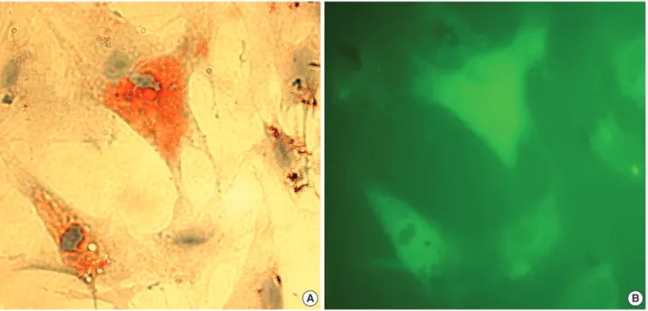
After ASCs had been cultured as monolayers in differenti- ation media for 14 days, most were enlarged and contained many lipid droplets, which were stained by Oil-red O (Fig.
1A), showed a green fluorescence (Fig. 1B).
In vitro culture of ASC-Alloderm complex
ASCs viabilities were; 0.61±0.19, 0.71±0.09, 0.79± 0.15, and 0.86±0.14 after culture for 1, 4, 7, and 14 days (one-way ANOVA, F=2.939, p=0.065). Although these rates showed an increasing tendency with culture time (Fig. 2),
no significant difference was observed for these different cul- ture periods (p>0.05).
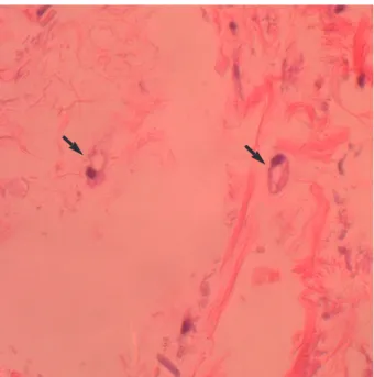
By H&E histological examination, some ASCs in mic- ronized Alloderm were observed to have differentiated and to contain large lipid vesicles after 14 days (Fig. 3).
The micronized Alloderm resembled pieces of dried tree roots by SEM (Fig. 4A). After culture for 14 days, many cells were covered the micronized Alloderm, and some spherical cells suspected of being adipocytes were observed (Fig. 4B).
In vivo implantation of ASC-Alloderm complex
All animals remained healthy throughout the experimen-
Fig. 1. Microscopic findings of adipose stem cells (ASCs) cultured in a monolayer for 14 days. (A) Some ASCs had differentiated into adipocytes, which contained many lipid droplets as visualized by Oil-red O staining. (B) ASCs were well labeled with PHK67 green fluo- rescent dye (×400).
A B
Absorbance (A490-A690)
1 0.8 0.6 0.4 0.2
0.0 1 day 4 day 7 day 14 day
Incubation time
Fig. 2. Changes in ASCs viabilities over 14 days in culture. ASCs were cultured in vitro after mixing then with micronized Alloderm (5×10⁴cells/1 mg of Alloderm). Cell viabilities were determined using XTT colorimetric assays. Values represent mean±stan- dard deviation (error bar).
tal period. At two months after implantation, injected tis- sues were easily identified in the cranial region. The weight of injected tissues of micronized Alloderm injected group and ASC-Alloderm complex injected group was 51.4±5.8 mg and 54.6±3.7 mg, respectively, but there was no sig- nificant difference between the weights of two groups (p>
0.05). An H&E histological examination revealed that some cells and small capillary had infiltrated the micronized der- mal matrix in controls (Fig. 5A), whereas markedly increased
numbers of large signet-ring cells and large capillaries were observed in ASC-Alloderm complex injected mice (Fig. 5B).
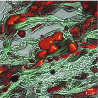
Moreover, in injected ASC-Alloderm complexes, signet-ring cells were stained with Oil-red O and some cells showed weak green fluorescent under the confocal microscope (Fig. 6).
DISCUSSION
Soft tissue defects range from the furrow of rhytid to a major loss of subcutaneous tissue secondary to congenital malforma- tion, trauma, or extirpation. The reconstruction of such de- fects is the province of plastic surgery. Over the years, various methods of fat injection or graft have been used, and various materials evolved to treat such defects. However, these treat- ment have many limitations, such as donor-site morbidity and deformity, unsatisfactory and unpredictable results, and complications resulting from the toxicity of implants (6).
For abrogating these limitations, adipose tissue engineering recently has been proposed as an alternative for current treatment (7). The choice of the types of seeding cells and scaffold used in tissue engineering determines the success of adipose tissue engineering (7).
The adipose stem cells (ASCs) may be an ideal autologous cell source for adipose tissue engineering (8). ASCs are much more resistant to mechanical damage and ischemic conditions than mature adipocytes (9). Moreover, ASCs can be readily harvested from excised human subcutaneous fat or liposuc- tion samples, and proliferate rapidly and differentiate into mature adipocytes both in vitro and in vivo (4, 10, 11). Many studies have recently done using ASCs in adipose tissue engi- neering because ASCs have been demonstrated as a good cell
Fig. 4. Scanning microscopic findings after 14 day of in vitro culture. (A) Micronized Alloderm alone. (B) ASC-Alloderm complex. Many cells were found to cover the micronized Alloderm, and some spherical adipocyte-like cells were observed.
A B
400×5 kV 400×5 kV
10 μ 10 μ
Fig. 3. Microscopic findings of in vitro cultured ASC-Alloderm com- plex for 14 days. ASCs in micronized Alloderm differentiated to round cells (arrows) with large lipid vesicles (H&E stain, ×400).
source for adipose tissue engineering because of their high proliferation, strong differentiation, and less invasive pro- curement (2, 3). In the present study, as in previous studies, human ASCs were found to differentiate readily to adipocytes in monolayer (2-dimensional culture), in 3-dimensional in vitro cultures, and in vivo.
Alloderm has been used as a good scaffold for skin and oral mucosa in tissue engineering, and it has been reported to orga- nize the arrangement of seeded keratinocytes and cultured keratinocyte sheets into three-dimensional epidermal struc- ture that has several layers, and to recreate the original rete ridges at the interface of the dermis (12). The Alloderm has been found to produce composite oral mucosa equivalent con- sisting of a stratified epidermis on a dermal matrix using cul- tured oral keratinocytes and Alloderm (13, 14). In the present study, micronized Alloderm was used as a scaffold of ASCs for adipose tissue engineering. In the previous study, a num- ber of different scaffolds have been investigated for adipose tissue engineering applications, e.g., polyglycolic acid (PGA) (10), poly (lactic-co-glycolic acid) (PLGA) (15), and hyaluron- ic acid (16). In addition, injectable materials, such as, fibrin (17), matrigel (18), and alginate gel (19) have also been stud- ied as scaffolds for adipose tissue engineering. Injectable scaf- folds can widen the scope of the engineered adipose tissue.
This injectable engineered adipose tissue would be easily use- ful in softening wrinkles, augmenting depressed scars, and reconstructing nipples. As micronized Alloderm have been used for soft tissue augmentation of facial rhytids, scars, and deformities in injectable form (20), micronized Alloderm has succeeded to this characteristic when they would be used as a scaffold for adipose tissue engineering. In the present study, XTT cell viability testing showed that ASCs remained viable in micronized Alloderm in vitro. Moreover, these adopted a round morphology and contained large lipid vesicles after 14 days in culture, which suggests that micronized Alloderm is a suitable ASCs scaffold for adipose tissue engineering purposes.
Fig. 5. Microscopic findings after 2 months of in vivo implantation. (A) Micronized Alloderm alone. Some fibroblast-like cells and few small capillaries were observed in the micronized Alloderm. (B) ASC-Alloderm complex. Many signet-ring cells and large capillaries were found (H&E stain, ×400).
A B
Fig. 6. Confocal microscopic findings of injected ASC-Alloderm complexes excised at 2 months after implantation. Many signet-ring cells were stained by Oil-red O and some cells showed weak green fluorescence (Oil-red O stain, ×400).
10 μm
ASC-Alloderm complexes after 2 months in vivo contained many signet-ring cells and large capillaries, whereas mic- ronized Alloderm alone contained some fibroblast-like cells and few small capillaries. Moreover, many signet-ring cells in ASC-Alloderm complexes were stained positively by Oil- red O and also showed green fluorescence indicating that the observed differentiated adipocytes originated from inject- ed ASC-Alloderm rather than surrounding fibrovascular tis- sues. Moreover, implanted ASC-Alloderm complex is softer than micronized Alloderm alone because of this adipose tis- sue development.
In conclusion, the present study suggests that micronized Alloderm provides a suitable scaffold for ASCs for adipose tissue engineering purpose, and that ASC-Alloderm complex has potential as a softer, more natural injectable soft tissue filler.
REFERENCES
1. Maloney BP, Murphy BA, Cole HP 3rd. Cymetra. Facial Plast Surg 2004; 20: 129-34.
2. Rodriguez AM, Elabd C, Delteil F, Astier J, Vernochet C, Saint-Marc P, Guesnet J, Guezennec A, Amri EZ, Dani C, Ailhaud G. Adipocyte differentiation of multipotent cells established from human adipose tissue. Biochem Biophys Res Commun 2004; 315: 255-63.
3. Hong L, Peptan IA, Colpan A, Daw JL. Adipose tissue engineering by human adipose-derived stromal cells. Cells Tissues Organs 2006;
183: 133-40.
4. Zuk PA, Zhu M, Mizuno H, Huang J, Futrell JW, Katz AJ, Benhaim P, Lorenz HP, Hedrick MH. Multilineage cells from human adipose tissue: implications for cell-based therapies. Tissue Eng 2001; 7:
211-28.
5. Fraser JK, Wulur I, Alfonso Z, Hedrick MH. Fat tissue: an under- appreciated source of stem cells for biotechnology. Trends Biotech- nol 2006; 24: 150-4.
6. Billings E Jr, May JW Jr. Historical review and present status of free fat graft autotransplantation in plastic and reconstructive surgery.
Plast Reconstr Surg 1989; 83: 368-81.
7. Patrick CW Jr. Tissue engineering strategies for adipose tissue repair.
Anat Rec 2001; 263: 361-6.
8. De Ugarte DA, Ashjian PH, Elbarbary A, Hedrick MH. Future of fat as raw material for tissue regeneration. Ann Plast Surg 2003;
50: 215-9.
9. von Heimburg D, Hemmrich K, Zachariah S, Staiger H, Pallua N.
Oxygen consumption in undifferentiated versus differentiated adi- pogenic mesenchymal precursor cells. Respir Physiol Neurobiol 2005; 146: 107-16.
10. Cho SW, Kim SS, Rhie JW, Cho HM, Choi CY, Kim BS. Engineer- ing of volume-stable adipose tissues. Biomaterials 2005; 26: 3577-85.
11. Zuk PA, Zhu M, Ashjian P, De Ugarte DA, Huang JI, Mizuno H, Alfonso ZC, Fraser JK, Benhaim P, Hedrick MH. Human adipose tissue is a source of multipotent stem cells. Mol Biol Cell 2002; 13:
4279-95.
12. Hickerson WL, Compton C, Fletchall S, Smith LR. Cultured epi- dermal autografts and allodermis combination for permanent burn wound coverage. Burns 1994; 20 (Suppl 1): S52-5.
13. Izumi K, Takacs G, Terashi H, Feinberg SE. Ex vivo development of a composite human oral mucosal equivalent. J Oral Maxillofac Surg 1999; 57: 571-7.
14. Izumi K, Terashi H, Marcelo CL, Feinberg SE. Development and characterization of a tissue-engineered human oral mucosa equiva- lent produced in a serum-free culture system. J Dent Res 2000; 79:
798-805.
15. Patrick CW Jr, Chauvin PB, Hobley J, Reece GP. Preadipocyte seed- ed PLGA scaffolds for adipose tissue engineering. Tissue Eng 1999;
5: 139-51.
16. Halbleib M, Skurk T, de Luca C, von Heimburg D, Hauner H. Tis- sue engineering of white adipose tissue using hyaluronic acid-based scaffolds. I: in vitro differentiation of human adipocyte precursor cells on scaffolds. Biomaterials 2003; 24: 3125-32.
17. Schoeller T, Lille S, Wechselberger G, Otto A, Mowlavi A, Piza- Katzer H, Mowlawi A. Histomorphologic and volumetric analysis of implanted autologous preadipocyte cultures suspended in fibrin glue: a potential new source for tissue augmentation. Aesthetic Plast Surg 2001; 25: 57-63.
18. Kawaguchi N, Toriyama K, Nicodemou-Lena E, Inou K, Torii S, Kitagawa Y. De novo adipogenesis in mice at the site of injection of basement membrane and basic fibroblast growth factor. Proc Natl Acad Sci USA 1998; 95: 1062-6.
19. Yoo G, Yea BH, Rhie JW, Kwon H, Wee SS, Ahn ST. Growth and Differentiation of Preadipocytes in Alginate and Collagen Gels. J Korean Soc Plast Reconstr Surg 2000; 27: 386-92.
20. Cheng JT, Perkins SW, Hamilton MM. Collagen and injectable fillers.
Otolaryngol Clin North Am 2002; 35: 73-85.