Mi Kyung Lee
1, Soon Ja Kang
2,
Mortimer Poncz
3, Ki-Joon Song
4and Kwang Sook Park
4,51Department of Genetics, College of Life Science 2Department of Science Education
College of Education Ewha Womans University Seoul 120-750, Korea
3Division of Hematology, Children’s Hospital of Philadelphia PA 19104, USA
4Department of Microbiology and Bank for Pathogenic Viruses
Division of Brain Korea 21 Program for Biomedical Science College of Medicine, Korea University
Seoul 136-705, Korea
5Corresponding author: Tel: 82-2-920-6164; Fax: 82-2-923-3645; E-mail: [email protected] Accepted 11 April 2007
Abbreviations: DA, dopamine; MMP, mitochondrial membrane potential; PI, propidium iodide; PS, phosphatidyl serine
Abstract
Dopamine (DA) is an oxidant that may contribute to the degeneration of dopaminergic neurons. The present study demonstrates that DA-induced cytotoxicity in human-derived neurotypic cells, SH-SY5Y, is prevented by resveratrol, one of the major antioxidative constituents found in the skin of grapes. SH-SY5Y cells, a neuroblastoma cell line, treated with DA at 300 and 500 µM for 24 h underwent apoptotic death as determined by characteristic morphological features, including nuclear conden sation, and loss of mitochondrial membrane poten -tial (MMP). Flow cytometric analysis using Annexin V showed that DA can induce significant and severe apoptosis. Exposure to resveratrol (5 µM) for 1 h prior to the DA treatment attenuated DAinduced cyto -toxicity, and rescued the loss of MMP. To investigate the apoptotic signaling pathways relevant to the restoration of DA-induced apoptosis by resveratrol, we carried out quantitative analysis of Bcl2, cas -pase-3, and cleaved poly ADP-ribose polymerase (PARP) by immunoblot analysis. Resveratrol pre -treatment led to a decrease in cleavage of PARP, an
increase in the Bcl2 protein, and activation of cas -pase-3. These results suggest that DA may be a potential oxidant of neuronal cells at biologically relevant concentrations. Resveratrol may protect SH-SY5Y cells against this cytotoxicity, reducing intracellular oxidative stress through canonical signal pathways of apoptosis and may be of bio -logical importance in the prevention of a dopaminergic neurodegenerative disorder such as Parkin -son disease.
Keywords: antioxidant; apoptosis; dopamine; neuro-blastoma; neurodegenerative diseases; resveratrol
Introduction
Parkinson’s disease (PD) is a common neurode-generative disease, characterized by a selective loss of dopaminergic neurons in the substantia nigra. Many factors are speculated to operate in the me-chanism of cell death of nigrostriatal dopaminergic neurons in PD, including oxidative stress and cyto-toxicity of reactive oxygen species (ROS), distur-bances of intracellular calcium homeostasis, exoge-nous and endogeexoge-nous toxins, and mitochondrial dys-function. An endogenous neurotransmitter dopamine (DA) is thought to be a major source of oxidative stress to these neural cells (Olanow, 1993;Offen et al., 1997). DA contains an unstable catechol moiety, and it can oxidize spontaneously to form ROS, free radicals, and quinones (Cohen and Heikkila, 1974; Graham, 1978; Hastings, 1995). In the human subs-tantia nigra, the DA oxidation products may further polymerize to form another neurotoxin, neurome-lanin (Jellinger et al., 1993). These oxidation products can damage cellular components such as lipids, proteins, and DNA (Halliwell, 1992).
One of the plausible ways to prevent the cell death induced by oxidative stress is dietary or pharmacological intake of antioxidants. Lai and Yu (1997) showed that DA-induced apoptosis is protec-ted by some antioxidants such as glutathione and N-acetyl-L-cysteine. One family of naturally occurring
compounds possessing free radical scavenging pro-perties is the polyphenols. It is found in fruit, vegeta-bles and plant-derived beverages and may have important roles as dietary components via cytopro-tective actions in many organs (Youdim and Joseph, 2001). Resveratrol (3,5,4'-tihydroxy-trans-stilbene), a
Resveratrol protects SH-SY5Y neuroblastoma cells from
apoptosis induced by dopamine
polyphenolic phytoalexin found in the skin of grapes, has been reported to possess a wide range of bio-logical and pharmacobio-logical activities including oxidant, inflammatory, antimutagenic, and anti-carcinogenic effects (Jang et al., 1997; Soleas et al., 1997; Surh, 1999). In previous studies, antioxidant properties of resveratrol attenuated hippocampal cell death and intracellular ROS formation (Bastianetto et al., 2000). Neuroprotective activities against exci-totoxic brain damage (Virgili and Contestabile, 2000) and anti-apoptotic effects by caspase activation (Nicolini et al., 2001) suggest a putative neuropro-tective action for resveratrol.
In the present study, we have examined the po-ssible protective effect of resveratrol against apop-tosis induced by DA in human derived neurotypic cell, SH-SY5Y. Our results indicate that DA indeed triggers apoptosis and this deleterious effect can be attenuated by resveratrol.
Materials and Methods
Materials
MEM, FBS and penicillin and streptomycin were obtained from Gibco BRL (Grand Island, NY). Re-sveratrol, dopamine, DMSO, Hoechst 33254, pro-pidium iodide (PI) were obtained from Sigma (St. Louis, MO). DA and resveratrol were dissolved in sterilized H2O and in DMSO, respectively. Annexin V
was from BD Biosciences (Palo Alto, CA). Mono-clonal antibodies against caspase-3, Bcl-2, cleavage form of PARP were obtained from Cell Signaling Technology (Danvers, MA). Bicinchoninic acid pro-tein assay kit and ECL chemiluminescence system were obtained from Pierce (Rockford, IL).
Cell culture
Catecholaminergic neuroblastoma SH-SYSY cells were grown in MEM supplemented with 10% FBS, 100 U/ml penicillin, and 100 g/ml streptomycin. Cells were maintained in a humidified atmosphere of 5% CO2, at 37oC. The cells were plated at an
appro-priate density according to each experimental scale.
Cell viability assay
Cell viability was determined using Cell Titer 96 Aqu-eous Non-Radioactive Cell Proliferation Assay, availa-ble from Promega Corp (Madison, WI). MTS (3-(4,5- dimethylthiazol-2-yl)-5-(3-carboxymethoxyphenyl)-2-(4-sulfophenyl)-2H-tetrazo-lium inner salt) conversion assay was performed according to the protocol sup-plied by the manufacturer. In brief, a mixture of MTS and PMS (phenazine methosulfate) was added to the treated SH-SY5Y cells cultured in 96-well plates
and incubated in the CO2 incubator for 3 h. Plates
were read with a microplate reader at a wavelength of 490 nm. Results were expressed as the per-centage of MTS reduction, assuming that the ab-sorbance of control cells was 100%. For each ex-periment, all treatments were performed in triplicate wells.
Nuclear staining for assessment of apoptosis SH-SY5Y cells were stained with the chromatin- specific dye Hoechst 33324 (Molecular Probes, Eugene, OR). The SH-SY5Y cells on the chamber culture slide were incubated for 2 days, and then treated with or without the resveratrol and DA at different concentrations. SH-SY5Y cells were fixed for 10 min with 3.5% paraformaldehyde in PBS at room temperature. After twice rinses with PBS, the cells were stained with 10 g/ml Hoechst 33324 in PBS for 10 min at 37oC. Mounted slides were analyzed under a fluorescence microscope (Axiovert 135, ZEISS, Germany) using a mercury lamp through a 360-370 nm bandpass filter. Light emitted from Hoechst dye was collected through a 420 nm longpass filter.
Measurement of mitochondrial membrane potential The level of mitochondrial membrane potential (MMP) was determined by flow cytometry and the fluore-scent dye rhodamine 123. The rhodamine 123 data were recorded using an FL-1 photomultiplier. Sample data (10,000 cells) were used to prepare histograms of the Cell Quest data analysis program (Becton Dickinson).
Flow cytometric analysis using Annexin V and PI SH-SY5Y cells were centrifuged to remove the me-dium, washed with PBS and stained with Annexin V-FITC and PI in binding buffer (10 mM Hepes, 140 mM NaCl, 2.5 mM CaCl2). Ten thousand events
were collected on each sample. Stained cells were analyzed using a FACScalibur (Becton Dickinson, Mountain View, CA) in the FL1-H and FL2-H channels.
Immunoblot analysis
Blotting was performed essentially as described previously (Schulz et al., 1999). The cells treated with or without resveratrol and DA, were washed once with ice-cold PBS and then lysed using ice- cold RIPA buffer and protease inhibitor cocktail (Sigma) at 4oC for 30 min. Cell lysates were cen-trifuged at 13,000 rpm for 25 min, and the protein concentrations were determined by the bicinchoninic acid method (Pierce, Rockford, IL) using BSA as
standard. The protein lysates were separated by 12% SDS-PAGE and transferred to nitrocellulose membrane (Bio-Rad, CA). Protein expression was detected using primary antibodies and secondary antibodies conjugated with HRP.
Ressults
Resveratrol protected SH-SY5Y cells against DA-induced cytotoxicity
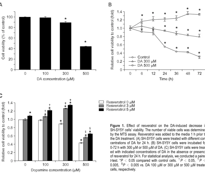
DA concentrations from 0 to 500 M were tested and demonstrated that DA is capable of induce metabolic interruption in SH-SY5Y cells in a dose range of 300-500 M (Figure 1A). DA decreased cell viability concentration over time (Figure 1B). Prior studies have shown that resveratrol at up to 10 M slightly promotes cell proliferation, while higher concentration resulted in significant apoptosis (Stervbo et al., 2006). We now found that 5 M
resveratrol slightly but significantly increased cell proliferation and DA-induced apoptotic effects were markedly attenuated by the co-presence of 3 or 5 M resveratrol (Figure 1C).
Resveratrol attenuated DA-induced apoptotic cell death
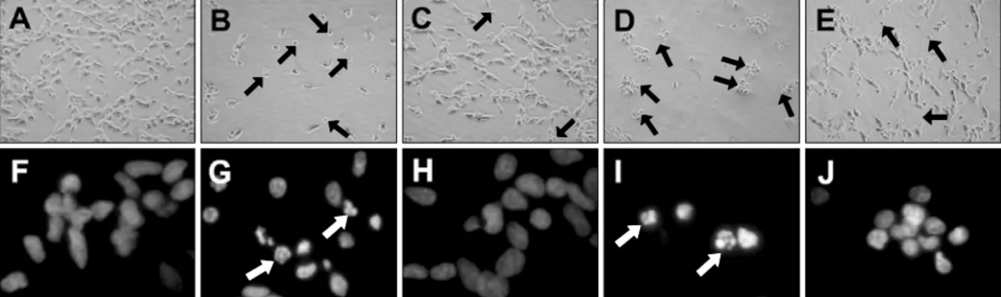
SH-SY5Y cells treated for 24 h with DA at con-centrations of 300 and 500 M exhibited morphological alterations such as cell shrinkage and mem -brane blebbing that are normally associated with the occurrence of apoptotic cell death. Compared to the normal SH-SY5Y cells (Figure 2A), cells treated with DA round up, detached from the bottom, and ag-gregated as assessed by phase-contrast microscopy (Figure 2B and D). Resveratrol pretreatment miti-gated these pathologic morphological features (Figure 2C and E). The presence of apoptotic cells was further confirmed by measurement of nuclear
Figure 1. Effect of resveratrol on the DA-induced decrease in SH-SY5Y cells’ viability. The number of viable cells was determined by the MTS assay. Resveratrol was added to the media 1 h prior to the DA treatment. (A) SH-SY5Y cells were treated with different con-centrations of DA for 24 h. (B) SH-SY5Y cells were incubated for 0-72 h with 300 M or 500 M of DA. (C) SH-SY5Y cells were treat-ed with indicattreat-ed concentrations of DA in the absence or presence of resveratrol for 24 h. For statistical analysis, we conducted a paired t-test. *P < 0.05 compared with control cells. †P < 0.05, ‡P< 0.005,§P < 0.005 vs. DA 100 M or 300 M or 500 M treated cells, respectively.
§ §
condensation using Hoechst 33342 (Hanrott et al., 2006). Treatment with 300 and 500 M DA signi-ficantly increased the proportion of condensed nuclei (Figure 2G and I). This incidence of cells with nuclear condensation was significantly reduced in DA-treated cells by 5 M resveratrol treatment (Figure 2H and J). In addition, DNA fragmentation analysis was carried out, suggesting that DA-indu-ced apoptosis was significantly reversed by resvera-trol (data not shown).
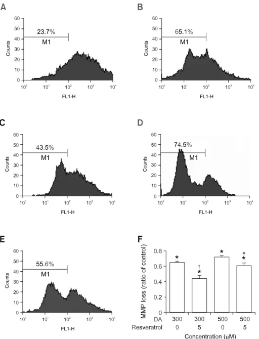
Resveratrol had a protective effect on DA-induced dissipation of the mitochondrial membrane potential The Bcl-2 family has a regulatory role in mitochon-drial permeability transition pore opening and has an anti-apoptotic property (Crompton, 2000). Upon tochondrial permeability transition pore opening, mi-tochondria lose their membrane potential across the inner membrane. Disruption of the MMP has been recognized in DA-induced apoptosis (Tang et al., 2005a). To examine if DA-induced apoptosis and its rescue by resveratrol involve an MMP pathway in SH-SY5Y cells, measurement of MMP was carried out using rhodamine 123 (Tang et al., 2005b). Pre-conditioning with DA alone induced the rapid depo-larization of the MMP. In 300 and 500 M DA- conditioned media, the degrees of depolarization were 65.1% and 74.5%, respectively (Figure 3B and D). However, preconditioning with resveratrol at 5
M had protective effects with 43.5% and 55.6% of MMP at 300 and 500 M DA, respectively (P < 0.005, each when compared to without resveratrol). These results indicate that DA-induced dissipation of the MMP can be effectively blocked by the pretreat-ment with resveratrol.
DA-induced cell death is attenuated in resveratrol-treated cells
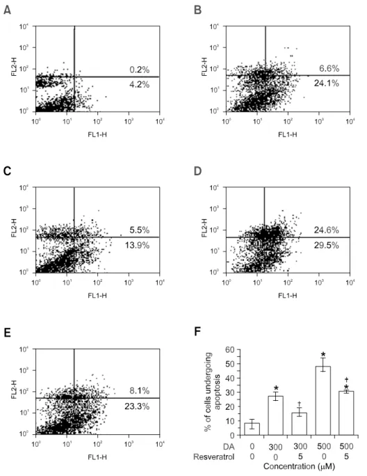
To quantify cell death induced by DA, we have utili-zed flow cytometric analysis of externalization of phosphatidyl serine using Annexin V/PI. As shown in Figure 4, cells stained negative for both Annexin V and PI (PS-/PI-) were live cells as shown in the lower left quadrant (R3). Annexin V positive and PI-ne-gative (PS+/PI-) stained cells undergo early stages of apoptosis, where the plasma membrane are still intact and exclude PI (lower right quadrant, R4). In late stages of apoptosis, dying cells can no longer exclude PI and the upper right region (R2) displayed both Annexin V-positive and PI-positive (PS+/PI+). PI positive and Annexin V-negative (PS-/PI+) stained cells in the upper left region (R1) were necrotic cells. Our data confirms that DA was capable of inducing cell death in SH-SY5Y cells. The increases of cell death over control in 300 M DA treated cells were 6.6% in R2, and 24.1% in R4, respectively. Exposure to DA resulted in a significant increase in cell death (30.7%, P < 0.01 vs control) in SH-SY5Y cells. Si-milarly, 500 M DA-treated cells also exhibited a marked increase in cell death (54.1%, P < 0.01 vs control). In contrast, resveratrol-pretreated SH-SY5Y cells showed significant resistance to DA-induced cytotoxicity because approximate 19.4% and 31.4% of the cells were affected, respectively (Figure 4, P < 0.01 vs. DA-only treated cells).
Resveratrol influenced the DA-induced apoptotic signaling pathway
To gain further insight into the way in which resve-ratrol is neuroprotective, we studied its effect on the possible caspases activated during the process of
Figure 2. Microscopic analysis of resveratrol on DA-induced cell death. The figures show the phase-contrast photographs of SH-SY5Y cells after DA treatment in the absence or presence of resveratrol. The black arrows indicate typical apoptotic cells with round shape. Nuclear condensation was de-termined by Hoechst 33342 staining. The white arrows indicate apoptotic cells demonstrating partially condensed nuclei: (A, F) No treatment; (B, G) DA 300 µM; (C, H) DA 300 M + resveratrol 5 M; (D, I) DA 500 M; (E, J) DA 500 M + resveratrol 5 M.
apoptosis. Caspase-3 is activated by multiple pro-teolytic cleavage of its 32 kDa precursor form to generate an enzymatically active p12/p17 complex, which has been used to monitor the activation of caspase-3 (Nicholson et al., 1995). Treatment of SH- SY5Y cells with 300 and 500 M DA induced the cleavage of caspase-3, demonstrated by the appea-rance of p17 fragments detected by an antibody for the cleaved form of caspase-3 and full-length cas-pase-3 (Figure 5). The increase in cascas-pase-3 clea-vage was associated with increased poly ADP- ribose polymerase (PARP) cleavage, a down stream substrate of caspase-3, and the extent of DA- induced PARP cleavage was also rescued by
res-veratrol (Figure 5).
We also examined the expression of Bcl-2 protein. Treatment of SH-SY5Y cells with DA decreased the expression of Bcl-2, which was reversed by resve-ratrol pretreatment (Figure 5). The partial restoration of Bcl-2 by resveratrol implies the possible involve-ment of other antiapoptotic proteins in regulating the death and survival in SH-SY5Y cells. These results suggested that resveratrol treatment shifted the balance between positive and negative regulators of apoptosis towards cell survival through canonical apoptosis signaling pathways.
Figure 3. Effect of resveratrol pre-conditioning on DA-induced dis-sipation of mitochondrial mem-brane potential. MMP alteration was measured by flow cytometry using rhodamine 123 staining. The number in M1 indicates the per-centage of cells with a reduced MMP level. SH-SY5Y cells were in-cubated in drug-free medium (A) or medium containing 300 M or 500 M DA (B, D); or cells were pre-incubated with 5 M resveratrol and then exposed to DA for 24 h (C, E). (F) A bar graph summary of the averages of rhodamine 123 da-ta collected from three independent flow cytometry experiments is shown. Bars represent the stand-ard error of the mean. For stat-istical analysis, we conducted a Student paired t-test. *P<0.001 compared with control cells. †P < 0.005, ‡P < 0.005 vs. DA 300 M or 500 M treated cells.
Discussion
Previous studies indicate that DA, although it is an essential neurotransmitter under physiological condi-tions, may participate in neurodegenerative proce-sses as a neurotoxin (Luo et al., 1998). Oxidative stress has been proposed as a major source of dopaminergic neuronal degeneration in PD, and one of the possible causes for oxidative stress is ROS generated during normal DA metabolism (Junn and Mouradian, 2001). The oxidative products of DA may function as stimuli to activate caspases and subsequent apoptosis, similar to other environmental stresses including UV radiation, ionizing radiation
and heat shock (Verheij et al., 1996). In contrast, polyphenols possesses free radical scavenging pro-perties and thus, they may have cytoprotective ac-tions in several organs such as kidney and brain (Skrzydlewska et al., 2005; Jang et al., 2006). Resveratrol is a known polyphenolic compound na-turally present in grapes, wine, and peanuts (Ulrich et al., 2005), and it has shown to exert antioxidant, anti-inflammatory, antimutagenic, and anticarcino-genic effects (Jang et al., 1997; Soleas et al., 1997; Surh, 1999).
Our data showed that DA significantly induces the cell death in time and dose dependent manner (Figure 1A and B) and DA-induced cytotoxicity was
Figure 4. Effect of resveratrol on DA-induced apoptosis in SH-SY5Y cells measured by flow cytometry. Flow cytometric analysis of SH- SY5Y cells stained with Annexin V and PI performed 24 h after cells were exposed to the experimental condition. The number in the lower right quadrant denotes the percent-age of early apoptotic cells, and the number in the upper right quad-rant denotes the percentage of late apoptotic cells. The results shown in (F) are the standard error of the mean for three independent expe-riments. (A): no treatment; (B): 300
M DA; (C): 300 M DA + 5 M re-sveratrol; (D): 500 M DA; and (E): 500 M DA + 5 M resveratrol. For statistical analysis, we con-ducted a Student paired t-test. *P
<0.01 compared with control cells. †P <0.001, ‡P < 0.01 vs. DA 300 M or 500 M treated cells.
partially restored by 3-5 M resveratrol (Figure 1C). The cytoprotective effects of resveratol appeared to be better by low concentration resveratrol than by high concentration resveratrol of 10-25 M (data not shown). In previous study, resveratrol was actually shown to have dual functions at different concen-trations: High concentrations of resveratrol (40-160
M) induce apoptosis, whereas cell proliferation increased at low concentrations of resveratrol (up to 10 M) (Stervbo et al., 2006).
In the present study, DA caused apoptotic cell death with significant morphological nuclear changes (Figure 2), consistent with previous in vitro studies (Emdadul Haque et al., 2003). To examine the role of DA for SH-SY5Y cell apoptosis, change of MMP was traced in the experimental conditions. Mainten-ance of MMP is necessary for production of energy (ATP) and preservation of cellular homeostasis, and is a critical primary determinant of cell survival (Akao et al., 2003). We confirm that DA causes dissipation of MMP and that inhibition of MTS conversion and disruptions in mitochondrial functions may be invol-ved in the mechanisms of DA-induced apoptosis in SH-SY5Y cells. We now show that this effect is inhibited by pretreatment with resveratrol (Figure 3), and that the protective effect of resveratrol against DA-induced apoptosis might be related to the pro-duction of ATP and the preservation of cellular ho-meostasis.
A key event during the early stage of apoptosis is that PS of the inner leaflet of the cell membrane appears on the outer leaflet. Annexin V preferentially binds to PS and can be used to detect expression of PS on the surface of apoptotic cells. PI diffuses into necrotic cells (Nicolini et al., 2001),and was used in these experiments as an indicator of lost of cell membrane integrity. Membrane permeability by dual
staining with Annexin V and PI revealed that high concentrations of DA (300 and 500 M) significantly damaged plasma membrane integrity. These dele-terious effects on SH-SY5Y cells can be reversed by resveratrol (Figure 4).
The decrease in MMP is a necessary step for in-ducing cytochrome c release and caspase-3 activa-tion in many apoptotic events (Perkins et al., 2000; Qiu et al., 2000). Caspases play an important role in the apoptotic process by two distinct pathways: the death receptor pathway and the mitochondrial path-way (Markus, 2000). Whichever pathpath-way is involved, caspase-3 acts as an apoptotic executor. Caspase-3 activates DNA fragmentation factor, which in turn activated endonucleases to cleave nuclear DNA, and ultimately leads to cell death (Wang and Xu, 2005). In this study, treatment with DA led to an increase in caspase-3 activity, but resveratrol pre-treatment effectively suppressed the caspase-3 acti-vation. Therefore, resveratrol may act upstream of caspase-3 to block apoptosis.
By virtue of measuring the proteolytic cleavage of nuclear enzyme PARP that is the down-stream tar-get of caspase-3, apoptosis signaling event between DA and resveratrol was tested. Proteolytic cleavage of PARP has been shown to occur during 2,2',5, 5'-tetrachlorobiphenyl-induced apoptosis in human neuronal SK-N-MC cells (Hwang et al., 2001), and staurosporine-induced apoptosis in dopaminergic neurons (Kim et al., 1999). On activating caspase-3, DA treatment induced the cleavage of PARP. The activation of caspase-3 as well as the cleavage of PARP that follow DA treatment was also prevented completely by the co-incubation with resveratrol (Figure 5).
Bcl-2 is a key member of the anti-apoptotic Bcl-2 family that plays a key role in regulating mitochon-drial-mediated apoptotic cell death (Marchetti et al., 1996; Yang et al., 1997; Tsujimoto, 1998). Bcl-2 has been shown to attenuate caspase-3 activation (Sa-wada et al., 2000; Yamakawa et al., 2000), and over- expression of Bcl-2 can protect neuronal cells from neurotoxic insult (Borg and London, 2002; Howard et al., 2002; Saito et al., 2003). In this study, the Bcl-2 protein decreased in the SH-SY5Y cells treated with DA, whereas it increased in cells pretreated with resveratrol. These results suggest that DA induces apoptosis through canonical apoptosis signal, and it is possible for resveratrol to suppress apoptotic cell death signals induced by DA.
In summary, DA caused apoptosis in SH-SY5Y cells through the induction of oxidative stress path-ways, and this apoptotic effect can be ameliorated by resveratrol pretreatment in a specific, low dose range. The elucidation of intracellular signal in res-ponse to DA-induced oxidative stress and their
mo-Figure 5. Effect of resveratrol on the DA-induced apoptotic signaling. SH-SY5Y cells were incubated with 300 M or 500 M DA for 24 h in the presence or absence of 5 M resveratrol and harvested for Immunoblot analysis. The protein of Bcl-2, caspase-3, cleaved form of caspase-3 and PARP were determined using immunoblot analysis with corresponding antibodies. Actin was used as an internal control. Each blot is representative of three independent experiments.
dulation by resveratrol may provide additional insights into the molecular basis of the neuroprotective effects of this antioxidant phytochemical. Whether DA contributes significantly to the development of PD, needs further studies as does whether resvera-tol or related polyphenolic compounds may have a role in prevention of the advancement of PD, remains to be determined.
References
Akao M, O'Rourke B, Teshima Y, Seharaseyon J, Marba'n E. Mechanistically distinct steps in the mitochondrial death path-way triggered by oxidative stress in cardiac myocytes. Circ Res 2003;92:186-94
Bastianetto S, Zheng WH, Quirion R. Neuroprotective abilities of resveratrol and other red wine constituents against nitric ox-ide-related toxicity in cultured hippocampal neurons. Br J Pharmacol 2000;131:711-20
Borg J, London J. Copper/zinc superoxide dismutase over-expression promotes survival of cortical neurons exposed to neurotoxins in vitro. J Neurosci Res 2002;70:180-9
Cohen G, Heikkila RE. The generation of hydrogen peroxide, superoxide radical, and hydroxyl radical by 6-hydroxydop-amine, dialuric acid, and related cytotoxic agents. J Biol Chem 1974;249:2447-52
Crompton M. Bax, Bid and the permeabilization of the mi-tochondrial outer membrane in apoptosis. Curr Opin Cell Biol 2000;12:414-9
Emdadul Haque M, Asanuma M, Higashi Y, Migazaki I, Tanaka K, Ogawa N. Apoptosis-inducing neurotoxicity of dopamine and its metabolites via reactive quinone generation in neuro-blastoma cells. Biochim Biophys Acta 2003;1619:39-52 Graham DG. Oxidative pathways for catecholamines in the genesis of neuromelanin and cytotoxic quinones. Mol Pharma-col 1978;14:633-43
Halliwell B. Reactive oxygen species and the central nervous system. J Neurochem 1992;59:1609-23
Hanrott K, Gudmunsen L, O'Neill MJ, Wonnacott S. 6-hydrox-ydopamine-induced apoptosis is mediated via extracellular auto-oxidation and caspase 3-dependent activation of protein kinase Cdelta. J Biol Chem 2006;281:5373-82
Hastings TG. Enzymatic oxidation of dopamine: the role of prostaglandin H synthase. J Neurochem 1995;64:919-24 Howard S, Bottino C, Brooke S, Cheng E, Giffard RG, Sapolsky R. Neuroprotective effects of bcl-2 overexpression in hippo-campal cultures: interactions with pathways of oxidative damage. J Neurochem 2002;83:914-23
Hwang SG, Lee HC, Lee DW, Kim YS, Joo WH, Cho YK, Moon JY. Induction of apoptotic cell death by a p53-independent pathway in neuronal SK-N-MC cells after treatment with 2,20,5,50-tetrachlorobiphenyl. Toxicology 2001;165:179-88 Jang M, Cai L, Udeani GO, Slowing KV, Thomas CF, Beecher CW, Fong HH, Farnsworth NR, Kinghorn AD, Mehta RG, Moon
RC, Pezzuto JM. Cancer chemopreventive activity of resvera-trol, a natural product derived from grapes. Science 1997;275:218-20
Jang YH, Lee YC, Park NH, Shin HY, Mun KC, Choi MS, Lee MY, Kim AR, Kim JM, Lee SR, Park HR. Polyphenol (-)-epi-gallocatechin gallate protection from ischemia/reperfusion-in-duced renal injury in normotensive and hypertensive rats. Transplant Proc 2006;38:2190-4
Jellinger K, Kienzl E, Rumpelmair G, Riederer P, Stachelberg-er H, Ben-Shachar D, Youdim MB. Neuromelanin and nigros-triatal dopamine neuron degeneration. J Neurochem 1993; 59:1168-71
Jiang H, Zhang L, Kuo J, Kuo K, Gautam SC, Groc L, Rodriguez AI, Koubi D, Hunter TJ, Corcoran GB, Seidman MD, Levine RA. Resveratrol-induced apoptotic death in human U251 glioma cells. Mol Cancer Ther 2005;4:554-61
Junn E, Mouradian MM. Apoptotic signaling in dopamine-in-duced cell death: the role of oxidative stress, p38 mi-togen-activated protein kinase, cytochrome c and caspases. J Neurochem 2001;78:374-83
Kim JE, Oh JH, Choi WS, Chang II, Sohn S, Krajewski S, Reed JC, O´Malley KL, Oh YJ. Sequential cleavage of poly(ADP-ri-bose)polymerase and appearance of a small Bax-immunor-eactive protein are blocked by Bcl-X(L) and caspase inhibitors during staurosporine induced dopaminergic neuronal apoptosis. J Neurochem 1999;72:2456-63
Lai CT, Yu PH. Dopamine- and L-beta-3,4-dihydroxyphenylal-amine hydrochloride (L-Dopa)-induced cytotoxicity towards catecholaminergic neuroblastoma SH-SY5Y cells. Effects of oxidative stress and antioxidative factors. Biochem Pharma-col 1997;53:363-72
Luo Y, Umegaki H, Wang X, Abe R, Roth GS. Dopamine in-duces apoptosis through an oxidation-involved SAPK/JNK activation pathway. J Biol Chem 1998;273:3756-64 Marchetti P, Hirsch T, Zamzami N, Castedo M, Decaudin D, Susin SA, Masse B, Kroemer G. Mitochondrial permeability transition triggers lymphocyte apoptosis. J Immunol 1996; 157:4830-6
Markus GG. Caspases: key players in programmed cell death. Curr Opin Struct Biol 2000;10:649-55
Mohan J, Gandhi AA, Bhavya BC, Rashmi R, Karunagaran D, Indu R, Santhoshkumar TR. Caspase-2 triggers Bax-Bak-de-pendent and -indeBax-Bak-de-pendent cell death in colon cancer cells treated with resveratrol. J Biol Chem 2006;281:17599-611 Nicholson DW, Ali A, Thornberry NA, Vaillancourt JP, Ding CK, Gallant M, Gareau Y, Griffin PR, Labelle M, Lazebnik YA, Munday NA, Raju SM, Smulson ME, Yamin T-T, Yu VL, Miller DK. Identification and inhibition of the ICE/CED-3 protease necessary for mammalian apoptosis. Nature 1995;376:37-43 Nicolini G, Rigolio R, Miloso M, Bertelli AAE, Tredici G. Anti-apoptotic effect of trans-resveratrol on paclitaxel-induced apoptosis in the human neuroblastoma SH-SY5Y cell line. Neurosci Lett 2001;302:41-4
Offen D, Ziv I, Panet H, Wasserman L, Stein R, Melamed E, Barzilai A. Dopamine-induced apoptosis is inhibited in PC12 cells expressing Bcl-2. Cell Mol Neurobiol 1997;17:289-304
Olanow CW. A rationale for monoamine oxidase inhibition as neuroprotective therapy for Parkinson's disease. Mov Disord 1993;8 Suppl 1:S1-7
Perkins CL, Fang G, Kim CN, Bhalla KN. The role of Apaf-1, caspase-9, and bid proteins in etoposide- or paclitaxel-in-duced mitochondrial events during apoptosis. Cancer Res 2000;60:1645-53
Qiu JH, Asai A, Chi S, Saito N, Hamada H, Kirino T. Proteasome inhibitors induce cytochrome c-caspase-3-like protease- mediated apoptosis in cultured cortical neurons. J Neurosci 2000;20:259-65
Saito A, Hayashi T, Okuno S, Ferrand-Drake M, Chan PH. Overexpression of copper/zinc superoxide dismutase in trans-genic mice protects against neuronal cell death after transient focal ischemia by blocking activation of the Bad cell death sig-naling pathway. J Neurosci 2003;23:1710-8
Sawada M, Nakashima S, Banno Y, Yamakawa H, Takenaka K, Shinoda J, Nishimura Y, Sakai N, Nozawa Y. Influence of Bax or Bcl-2 overexpression on the ceramide-dependent apoptotic pathway in glioma cells. Oncogene 2000;19:3508-20 Schulz JB, Weller M, Moskowitz MA. Caspases as treatment targets in stroke and neurodegenerative diseases. Ann Neurol 1999;45:421-9
Skrzydlewska E, Augustyniak A, Michalak K, Farbiszewski R. Green tea supplementation in rats of different ages mitigates ethanol-induced changes in brain antioxidant abilities. Alcohol 2005;37:89-98
Soleas GJ, Diamandis EP, Goldberg DM. Resveratrol: a mole-cule whose time has come? And gone? Clin Biochem 1997; 30:91-113
Stervbo U, Vang O, Bonnesen C. Time- and concentration- de-pendent effects of Resveratrol in HL-60 and HepG2 cells. Cell Prolif 2006;39:479-93
Surh YJ. Molecular mechanisms of chemopreventive effects of selected dietary and medicinal phenolic substances. Mutat Res 1999;428:305-27
Tang XQ, Zhi JL, Cui YU, Feng JQ, Chen PX. Hydrogen per-oxide preconditioning protects PC12 cells against apoptosis induced by dopamine. Life Sci 2005a;78:61-6
Tang XQ, Feng JQ, Chen J, Chen PX, Zhi JL, Cui Y, Guo RX, Yu HM. Protection of oxidative preconditioning against apopto-sis induced by H2O2 in PC 12 cells: mechanisms via MMP, ROS, and Bcl-2. Brain Res 2005b;1057:57-64
Tewari M, Quan LT, O'Rourke K, Desnoyers S, Zeng Z, Beidler DR, Poirier GG, Salvesen GS, Dixit VM. Yama/CPP32 beta, a mammalian homolog of CED-3, is a CrmA-inhibitable pro-tease that cleaves the death substrate poly (ADP-ribose) polymerase. Cell 1995;81:801-9
Tsujimoto Y. Role of Bcl-2 family proteins in apoptosis: apopto-somes or mitochondria? Genes Cells 1998;3:697-707 Ulrich S, Wolter F, Stein JM. Molecular mechanisms of the che-mopreventive effects of resveratrol and its analogs in carcinogenesis. Mol Nutr Food Res 2005;49:452-61
Verheij M, Bose R, Lin XH, Yao B, Jarvis WD, Grant S, Birrer MJ, Szabo E, Zon LI, Kyriakis JM, Haimovitz-Friedman A, Fuks Z, Kolesnick RN. Requirement for ceramide-initiated SAPK/ JNK signalling in stress-induced apoptosis. Nature 1996; 380:75-9
Virgili M, Contestabile A. Partial neuroprotection of in vivo ex-citotoxic brain damage by chronic administration of the red wine antioxidant agent, trans-resveratrol in rats. Neurosci Lett 2000;281:123-6
Wang XJ, Xu JX. Salvianic acid A protects human neuro-blastoma SH-SY5Y cells against MPP+-induced cytotoxicity. Neurosci Res 2005;51:129-38
Yamakawa H, Ito Y, Naganawa T, Banno Y, Nakashima S, Yoshimura S, Sawada M, Nishimura Y, Nozawa Y, Sakai N. Activation of caspase-9 and -3 during H2O2-induced apoptosis of PC12 cells independent of ceramide formation. Neurol Res 2000;22:556-64
Yang J, Liu X, Bhalla K, Kim CN, Ibrado AM, Cai J, Peng TI, Jones DP, Wang X. Prevention of apoptosis by Bcl-2: release of cytochrome c from mitochondria blocked. Science 1997; 275:1129-32
Youdim K, Joseph J. A possible emerging role of phytochem -icals in improving age-related neurological dysfunctions: a multiplicity of effects. Free Radic Biol Med 2001;30:583-94