759
C ardiac allograft vasculopathy (CAV) is the leading cause of late graft failure and mortality in heart transplanta- tion.1,2 Based on the registry of the International Society for Heart and Lung Transplantation, CAV is detected angiograph- ically in 32% of patients within 5 years of transplantation.
2
CAV is a progressive disorder that may affect both the epicar- dial vessels and the microcirculation of the heart.
1–3
Although earlier studies on CAV have focused on epicardial disease, recent studies have called attention on the importance of microvascular disease.
4–8Functionally, microvascular dis- ease is measured using coronary flow reserve (CFR with usual threshold <2.5) or the recently described index of microcir- culatory resistance (IMR).
3,9–14Compared with CFR, IMR is a more specific and a more reproducible measure of micro- vascular function.
15We have previously shown that IMR improves significantly during the first posttransplant year, whereas fractional flow reserve (FFR), a marker of epicardial coronary physiology, worsens.
3Clinical Perspective on p 768
At this time, the determinants and functional consequences of early microvascular dysfunction defined using the IMR have not been established. For this study, we sought to deter- mine whether a history of acute rejection during the first posttransplant year would be a strong determinant of early microvascular dysfunction. Our second objective was to deter- mine whether the presence of microvascular dysfunction was associated with worse ventricular function at 1 year. Finally, in an exploratory analysis, we sought to determine whether the presence of early microvascular dysfunction would be associated with a higher likelihood of death, retransplant, or early allograft vasculopathy.
Methods Study Design
This study is a retrospective cohort study designed to determine the determinants and clinical correlates of microvascular dysfunction in heart transplant recipients. Patients enrolled in the study were trans- planted between January 2001 and June 2008 at Stanford University Medical Center. Informed consent was obtained from all patients
© 2012 American Heart Association, Inc.
Circ Heart Fail is available at http://circheartfailure.ahajournals.org DOI: 10.1161/CIRCHEARTFAILURE.111.962787 Received May 1, 2012; accepted May 18, 2012.
From the Department of Medicine (F.H., K., J.Z., C.W.N., T.A.V., F.A.G., M.S., A.S., I.S., B.V., S.A.H., W.F.F.) and Department of Cardiovascular Surgery (T.D.), Stanford University, Stanford, CA; and Medical Director of Heart Transplantation, Permanente Medical Group, Santa Clara, CA (D.W.).
Correspondence to François Haddad, MD, FRCPC, Division of Cardiovascular Medicine, Stanford University, Stanford, CA. E-mail [email protected]
Background—Microvascular dysfunction is emerging as a strong predictor of outcome in heart transplant recipients. At this time, the determinants and consequences of early microvascular dysfunction are not well established. The objective of the study was to determine the risk factors and functional correlates associated with early microvascular dysfunction in heart transplant recipients.
Methods and Results—Sixty-three heart transplant recipients who had coronary physiology assessment, right heart catheterization, and echocardiography performed at the time of their first annual evaluation were included in the study.
Microvascular dysfunction was assessed using the recently described index of microcirculatory resistance. The presence of microvascular dysfunction, predefined by an index of microcirculatory resistance >20, was observed in 46% of patients at 1 year. A history of acute rejection and undersized donor hearts were associated with microvascular dysfunction at 1 year, with odds ratio of 4.0 (1.3–12.8) and 3.6 (1.2–11.1), respectively. Patients with microvascular dysfunction had lower cardiac index (3.1±0.7 versus 3.5±0.7 L/min per m
2; P=0.02) and mild graft dysfunction measured by echocardiography- derived left and right myocardial performance indices ([0.54±0.09 versus 0.43±0.09; P<0.01] and [0.47±0.14 versus 0.32±0.05; P<0.01], respectively). Microvascular dysfunction was also associated with a higher likelihood of death, graft failure, or allograft vasculopathy at 5 years after transplant (hazard ratio, 2.52 [95% CI, 1.04–5.91]).
Conclusions—A history of acute rejection during the first year and smaller donor hearts were identified as risk factors for early microvascular dysfunction. Microvascular dysfunction assessed using index of microcirculatory resistances at 1 year was also associated with worse graft function and possibly worse clinical outcomes. (Circ Heart Fail. 2012;5:759-768.) Key Words: coronary physiology ◼ microcirculation ◼ coronary artery disease ◼ heart transplantation ◼ heart function
by guest on June 30, 2016 http://circheartfailure.ahajournals.org/
Downloaded from
according to Stanford Medical Center’s Human Investigation Review Board before coronary physiology measurements. Patients included in the analysis were enrolled as part of National Institutes of Health–
funded trials (1 K23 HL072808-01A1, 1 PO1-AI50153, and 5 R01 HL093475-02). Data collection was performed by 2 research associ- ates trained in data extraction.
Patient Sample
The study sample consisted of 63 adult heart transplant patients who underwent assessment of microvascular function and echocardiogra- phy as part of their first annual posttransplant evaluation at Stanford University Medical Center.
Patients with evidence of acute rejection, significant CAV, or graft failure at the time of their annual evaluation were excluded from the study. Acute rejection was defined as an event that led to an acute augmentation of immunosuppression.16 Both cellular (International Society for Heart and Lung Transplantation grade ≥2R) and noncel- lular rejections with hemodynamic compromise (decrease in relative left ventricular ejection fraction [LVEF] >25%) were considered significant rejections. Graft failure was diagnosed if patients met the Framingham Criteria for Congestive Heart Failure and required hospitalization for heart failure management.17 We excluded acute rejections at the time of the annual evaluation to allow analysis of the relationship between microvascular dysfunction and graft func- tion, independently of an acute rejection episode. Significant epicar- dial CAV was predefined by a luminal stenosis >50% by quantitative angiography.18 Of the 67 patients considered for the study, 4 patients were excluded for the following reasons: 2 patients had evidence of acute cellular rejection, 1 patient had evidence of graft failure without evidence of rejection, and 1 patient had evidence of signifi- cant epicardial disease with 60% luminal stenosis in the left anterior descending artery.
The study sample represents 19% of adult patients transplanted during the study period (n=337). Compared with patients excluded from the study, there was no significant difference in recipient age (51±12 versus 50±14 years; P=0.39), recipient race (black race 14%
versus 11%; P=0.45), donor age (33±13 versus 32±12; P=0.66), ischemic time (216±50 versus 217±53 minutes; P=0.89), rejection rate during first year (35% versus 29%; P=0.35), or prevalence of diabetes mellitus during the first posttransplant year (32% versus 28%; P=0.53). By design of the study, no patient in the study had significant coronary artery disease defined compared with a preva- lence of 7.0% at the first annual evaluation in the sample not part of the study.
Immunosuppressive Regimen
Induction therapy was used in all patients and consisted of daclizum- ab, an anti–interleukin-2 monoclonal antibody, or OKT3, a mouse antibody directed against the CD3 antigen that is closely associated with the T-cell receptor (5 patients). Maintenance immunosuppres- sion consisted of a calcineurin inhibitor (cyclosporine or tacrolimus) and either mycophenolate mofetil or sirolimus. Corticosteroid ther- apy (methylprednisone) was initiated immediately postoperatively and progressively tapered over 1-year posttransplant in the absence of rejection. Cytomegalovirus prophylaxis consisted of valganciclo- vir for a total of 6- to 12-month duration of prophylaxis in patients with evidence of seropositive donor or recipient status. Intravenous cytomegalovirus immunoglobulin therapy was given in seronegative recipients of seropositive donors.
Echocardiography
Digitized echocardiographic studies were analyzed by a reader (F.H.) blinded to the hemodynamic data using quantitative criteria in ac- cordance with the published guidelines of the American Society of Echocardiography.19 Echocardiographic studies were obtained within 24 hours of invasive measurements for each patient. Left ventricular internal dimension in diastole, as well as left ventricular septal wall thickness and left ventricular posterior wall thickness in diastole, was measured in the parasternal long-axis view using a 2-dimensional
echocardiographic method and averaged over 3 measures. In measur- ing the septal wall thickness, careful attention was taken to exclude the right ventricular septal bands; similarly, careful attention was taken not to include chordae in the measurement of the posterior wall.
Small left ventricular dimension was defined as left ventricular internal dimension (LVID) <2.4 cm/m2 for women and <2.2 cm/m2 for men according to the criteria established by the American Society of Echocardiography.19 Concentric ventricular remodeling was iden- tified in the presence of a relative wall thickness (2 left ventricular posterior wall thickness in diastole/left ventricular internal dimension in diastole) ≥0.42.19 Left ventricular hypertrophy was defined by a ventricular mass >96 g/m2 for women or >116 g/m2 for men accord- ing to the threshold values established by the American Society of Echocardiography.19 LVEF was calculated using the biplane Simpson method of disk. Myocardial performance indices were calculated, as previously described, as the ratio of isovolumic relaxation and contrac- tion times divided by the ejection time.20 To measure the myocardial performance index, all time intervals were averaged over 3 consecu- tive cycles. Myocardial performance index represents a measure of both systolic and diastolic function.21 A higher value of the myocardial performance index is associated with worse ventricular function.20,21 Normal values of myocardial performance indices have been previ- ously established; for the right ventricle, normal values of right ven- tricular myocardial performance index (RVMPI) are 0.28±0.04, and for the left ventricle, normal values of left ventricular myocardial performance index have been previously reported to be 0.38±0.05.20,21 Tissue Doppler parameters were not available for data analysis.
Coronary Physiology Measurements
Microcirculatory disease was quantified using the IMR (Figure 1).
To measure IMR, a 0.014-inch coronary pressure wire (Radi Medical Systems) was calibrated outside of the body and then advanced through a 6F guiding catheter to position the pressure sensor at the ostium of the guiding catheter where equal pressure readings by the guiding catheter and the pressure wire were confirmed.3,10 The wire was then advanced in the distal portion of the left anterior descending artery. The shaft of the pressure wire acts as a proximal thermistor, and the pressure sensor acts as a distal thermistor. Room tempera- ture saline was injected down the left anterior descending artery in 3-mL aliquots 3×, and the resting mean transit time of the saline was recorded and averaged. Maximal hyperemia was then induced by ad- ministration of intravenous adenosine (140 μg/kg per min) via a cen- tral venous line, and the hyperemic mean transit time was determined by averaging the transit times after 3 injections of 3 mL of saline.
The IMR was calculated by dividing pressure by flow—in this case, the distal pressure by the inverse of the hyperemic mean transit time or, more simply, distal pressure multiplied by the hyperemic mean transit time. A threshold value of 20 mm Hg seconds for IMR was used to define microvascular dysfunction based on prior work by our group in heart transplantation.3,13 The threshold value corresponded to the median value of patients without a history of hemodynamically compromising rejection and defines patients with lower versus higher microvascular dysfunction. In a subgroup of 28 patients (44%), mi- crovascular function was also available at baseline. CFR by thermodi- lution was calculated by dividing the resting mean transit time by the hyperemic mean transit time (Figure 1). A CFR threshold value <2.5 was considered abnormal based on previously published physiologi- cal and outcome studies.14,22
Epicardial physiology was measured using FFR. FFR was measured by dividing the mean distal pressure by the mean aortic pressure during maximal hyperemia. Significant epicardial CAV was defined by a luminal stenosis of >50% by quantitative angiography in the left main or primary vessel.18
Clinical Definitions and Combined Outcome
Donor-recipient mismatch was defined clinically as a 20% weight dif- ference between the recipient and the donor at the time of transplant.
Diabetes mellitus was defined according to the American Diabetes Association criteria as a fasting glucose >7 mmol/L (126 mg/dL) requiring at least 3 months of hypoglycemic or insulin therapy.23
Glomerular filtration rate was estimated using the Modification of Diet in Renal Disease Study equation.24
For the purposes of an exploratory analysis, patients were followed for up to 5 years for the combined outcome of death, graft failure, or significant allograft vasculopathy. Graft failure was diagnosed if patients met the Framingham Criteria for Congestive Heart Failure and had evidence of new-onset systolic dysfunction with LVEF <45%
and at least 25% relative change from baseline.17 Significant epicar- dial CAV was defined by a luminal stenosis of >50% by quantitative angiography by a reader blinded to the other clinical variables.
Statistical Methods
Results are expressed as mean±SD for continuous variables or as number of cases and percentage for categorical variables. Comparison of groups was performed using Student t test for continuous vari- ables and χ2 test or Fisher exact Test, as appropriate, for categori- cal variables. For the χ2test, the P value reported corresponds to the Pearson χ2 without continuity correction. Logistic regression analy- sis was used to determine the factors independently associated with microvascular dysfunction at 1 year. We used a stepwise regression analysis combining forward selection and backward elimination;
variables with P<0.30 were entered in the regression and variables with P>0.5 were removed from the model. In the subgroup of 29 patients who had both baseline and 1-year values of microvascular function, comparison of baseline and 1-year values of IMR, cardiac index, pulmonary capillary wedge pressure (PCWP), and right atrial pressure (RAP) was made using paired t test. Logistic regression analysis was used to determine the factors independently associated with change in cardiac index, PCWP, or RAP from baseline to 1 year.
For the purpose of an exploratory outcome analysis, Cox propor- tional hazard analysis was performed to determine the hazard ratio of factors associated with the combined outcome of death, graft failure, or epicardial allograft vasculopathy. Because of the small number of events, multivariable survival analysis was not performed. Kaplan- Meier survival curve was used to represent the survival of patients with or without microvascular dysfunction. P<0.05 was considered to be statistically significant. Statistical analysis was performed us- ing the PASW software (PASW 18.0 Inc, Chicago, IL).
Results Patient Characteristics
The mean age at transplant was 51±12 years, and the major- ity of patients (79%) were men (Table 1). Eighteen patients (29%) were transplanted for ischemic cardiomyopathy, donor age was 33±13years, and mean ischemic time was 216±50 minutes. Donor-recipient size mismatch defined by >20%
weight difference between donor and recipient (donor less than recipient) was present in 12 patients (19%). All patients were treated with triple immunosuppressive therapy (predni- sone, cyclosporine, or tacrolimus and mycophenolate mofetil or sirolimus), and the majority received statin therapy (96%).
The mean cyclosporine level at the first annual evaluation was 156±61 ng/mL (n=44), and the mean tacrolimus level was 7.5±4.1 ng/mL (n=19).
Rejection during the first posttransplant year was docu- mented in 22 patients (35%), 7 (32%) of whom had evidence of associated systolic graft dysfunction at the time of rejec- tion (relative change in LVEF >25% from baseline). Of the 7 patients with rejection and hemodynamic compromise, 3 were diagnosed with noncellular rejection (International Society for Heart and Lung Transplantation grade <2R).
At 1-year posttransplant, the average LVID in diastole was 4.6±0.4 cm and 2.3±0.3 cm/m
2when indexed to body surface area, the relative wall thickness was 0.43±0.06, and the indexed left ventricular (LV) mass was 82±18 g/m
2. Small indexed LV dimension based on the American Society of Echocardiography criteria was observed in 36 patients (57%). The majority of patients with weight mismatch had smaller donor hearts based on indexed LVID at 1 year (75%), but 48% of patients without weight mismatch also had small indexed LV size at 1 year.
Figure 1. Common measures of coronary physiology in clinical practice. Epicardial coronary physiology is usually estimated using frac- tional flow reserve (FFR) and is calculated during maximal hyperemia conditions as the ratio of distal pressure (Pd) to mean aortic pres- sure (Pa). Coronary flow reserve (CFR) assesses both the epicardial and microvasculature physiology and is calculated as the ratio of hyperemic to basal flow, which can be simplified as the ratio of basal to hyperemic mean transit time. Index of microcirculatory resistance (IMR) assesses more specifically the microcirculation and is calculated as the product of Pd and mean transit time. Pw indicates coronary wedge pressure.
Concentric LV remodeling defined by a relative wall thickness
>0.42 was seen in 28 patients (44%), whereas LV hypertro- phy by indexed mass criteria was present in 7 patients (11%).
LVEF was 61±8%, and all patients had an LVEF >45% at the time of their annual evaluation. Left ventricular myocardial performance index was 0.46±0.08 (reference, 0.39±0.05), and RVMPI was 0.39±0.13 (reference, 0.28±0.04). On right heart catheterization, systolic blood pressure was 123±16 mm Hg, mean RAP was 6±5 mm Hg, mean pulmonary arterial pressure (mPAP) was 21±8 mm Hg, mean PCWP was 11±5 mm, Hg and cardiac index was 3.4±0.8 L/min per m
2.
Coronary Physiology Measures at 1 Year
The average FFR was 0.86±0.06 (median, 0.87), the aver- age IMR was 23±17 (median, 19), and the average CFR was
3.4±1.9 (median, 2.9). Four patients in the study had FFR values <0.75 in the absence of severe focal stenosis, sugges- tive of diffuse disease (Figure 2).
Microvascular dysfunction, predefined by an IMR>20, was observed in 29 patients at 1 year (46%). There was no significant difference between FFR of patients with and with- out microvascular dysfunction (0.87±0.07 versus 0.85±0.06;
P=0.87). When using a CFR threshold of 2.5 to classify
microvascular dysfunction, there was a concordance rate of 75% between microvascular dysfunction defined by CFR or IMR (Figure 2).
Factors Associated With Early Microvascular Dysfunction
A higher proportion of patients with microvascular dysfunc- tion had a history of acute rejection during the first post- transplant year (P=0.028). Smaller LV ventricles based on indexed Left ventricular internal dimension in diastole were also more common among patients with microvascular dys- function (P=0.20) (Table 1). There was a trend for an associa- tion between sirolimus-based therapy and a lower incidence of microvascular dysfunction (32% versus 17%; P=0.17).
There was no significant difference in body mass index, obe- sity (body mass index >30), prevalence of diabetes mellitus, cyclosporine drug levels, concentric left ventricular remod- eling, or left ventricular hypertrophy between patients with or without microvascular dysfunction (Table 1). On logistic multivariable analysis that included covariates with P<0.3, both acute rejection and smaller left ventricular size were independently associated with microvascular dysfunction at 1 year (Table 2).
Functional Correlates of Microvascular Dysfunction
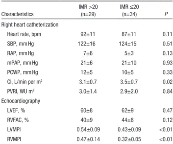
Patients with microvascular dysfunction (IMR >20) had lower cardiac index and higher values of myocardial performances indices, suggestive of impaired ventricular function (Table 3 and Figure 3). There was, however, no significant differences between groups for PCWP, RAP, LVEF, or right ventricular fractional area change.
On multivariable linear regression analysis, microvascu- lar dysfunction (IMR >20) was an independent covariate for cardiac index, as well as RVMPI and left ventricular myocardial performance index, but not LVEF. The covari- ates considered in the model were based on both statistical and clinical significance and included IMR >20, rejection history, diabetes mellitus, donor age, heart rate, and sys- tolic blood pressure. For cardiac index, IMR >20 was the only independent variable in the regression equation, with a
P=0.03 and a coefficient of determination R2=0.16. For left ventricular myocardial performance index, IMR >20 was the only independent variable in the regression equation, with a P<0.01 and a coefficient of determination R
2=0.29. For RVMPI, IMR >20 was an independent variable in the regres- sion equation with a P<0.01, whereas history of rejection in the first year had a P=0.086 and a coefficient of determi- nation R
2=0.37. For LVEF, no variable was retained in the regression equation.
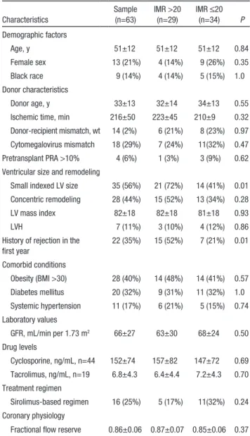
Table 1. Comparison of Factors Potentially Associated With Early Microvascular Dysfunction
Characteristics
Sample (n=63)
IMR >20 (n=29)
IMR ≤20 (n=34) P Demographic factors
Age, y 51±12 51±12 51±12 0.84
Female sex 13 (21%) 4 (14%) 9 (26%) 0.35
Black race 9 (14%) 4 (14%) 5 (15%) 1.0
Donor characteristics
Donor age, y 33±13 32±14 34±13 0.55
Ischemic time, min 216±50 223±45 210±9 0.32 Donor-recipient mismatch, wt 14 (2%) 6 (21%) 8 (23%) 0.97 Cytomegalovirus mismatch 18 (29%) 7 (24%) 11(32%) 0.47 Pretransplant PRA >10% 4 (6%) 1 (3%) 3 (9%) 0.62 Ventricular size and remodeling
Small indexed LV size 35 (56%) 21 (72%) 14 (41%) 0.01 Concentric remodeling 28 (44%) 15 (52%) 13 (34%) 0.28
LV mass index 82±18 82±18 81±18 0.93
LVH 7 (11%) 3 (10%) 4 (12%) 0.86
History of rejection in the first year
22 (35%) 15 (52%) 7 (21%) 0.01
Comorbid conditions
Obesity (BMI >30) 28 (40%) 14 (48%) 14 (41%) 0.57 Diabetes mellitus 20 (32%) 9 (31%) 11 (32%) 1.0 Systemic hypertension 11 (17%) 6 (21%) 5 (15%) 0.74 Laboratory values
GFR, mL/min per 1.73 m2 66±27 63±30 68±24 0.50 Drug levels
Cyclosporine, ng/mL, n=44 152±74 157±82 147±72 0.69 Tacrolimus, ng/mL, n=19 6.8±4.3 6.4±4.4 7.2±4.3 0.70 Treatment regimen
Sirolimus-based regimen 16 (25%) 5 (17%) 11(32%) 0.24 Coronary physiology
Fractional flow reserve 0.86±0.06 0.87±0.07 0.85±0.06 0.37 IMR indicates index of microcirculatory resistance; PRA, panel reactive antibody; LV, left ventricular; LVH, left ventricular hypertrophy; BMI, body mass index; GFR, glomerular filtration rate.
*Defined according to the American Society of Echocardiography guidelines for small LV size.
Change in Microvascular Dysfunction at Baseline and 1 Year and Its Relationship to Ventricular Function
In 28 patients, microvascular function and hemodynamics were available at both baseline and 1 year. Baseline stud- ies were performed on average at 4 weeks after transplant.
Patients at baseline had a lower hemoglobin level (102±12 versus 118±16 g/L; P<0.001) and a lower heart rate (79±11 versus 85±11 beats per minute; P=0.038). The average IMR decreased during the first posttransplant year, with some patients showing greater change in microvascular function than others (27±15 versus 19±8; P=0.01) (Figure 4). The average cardiac index was 3.5±0.5 L/min per m
2at baseline and 3.3±0.9 L/min per m
2at 1 year (P=0.36) (Figure 4). The average stroke volume index was 45±9 mL/m
2at baseline and 40±12 mL/m
2at 1 year (P=0.059). The average PCWP was 13±7 mm Hg at baseline and 12±6 mm Hg at 1 year (P=0.23).
When analyzing the relationship between the dynamic change in IMR and cardiac index, stroke volume index, PCWP, and RAP, a significant correlation was found between change in IMR and change in cardiac index (r=−0.57; P< 0.001) or change in stroke volume index (r=−0.57; P=0.001) (Figure 4, lower panel). No relationship was found between change in IMR and change in PCWP (P=0.97) or change in IMR and change in RAP (P=0.74). To determine whether a change in cardiac index or stroke volume was independently associated with a change in IMR, we conducted a multivariable model with a change in microvascular function, history of rejection, history of hemodynamically compromising rejection, diabetes
mellitus, donor age, and relative change in hemoglobin as the potential independent variables. We found that a change in cardiac index was independently associated with a change in IMR (P=0.001) and a history of hemodynamically com- promising rejection (P=0.032) with an R
2of 0.43. Similarly, a change in stroke volume index was also associated with a change in IMR (P=0.003) and a history of hemodynamically compromising rejection (P=0.014) with an R
2of 0.44.
Exploratory Outcome Analysis
Patients were followed for up to 5 year after heart transplanta- tion for outcome analysis. Because of the small sample size, our outcome analysis was only intended to be exploratory. The mean follow-up time was 3.5±0.5 years, and the combined end
Figure 2. Relationship between index of microcirculatory resistance (IMR) and fractional flow reserve (FFR) (left panel) and IMR and cor- onary flow reserve (CFR) (right panel). The left panel shows that in the majority of patients, microvascular dysfunction occurs in patients with FFR >0.75. The right panel emphasizes the relationship between IMR and CFR using usual threshold to microvascular dysfunction.Table 2. Unadjusted and Multivariable Correlates of Microvascular Dysfunction (IMR >20)
Unadjusted Correlates Multivariable Correlates
OR (95% CI) P OR (95% CI) P
Acute rejection first year
4.13 (1.37–12.48) 0.011 4.23 (1.33–13.41) 0.015
LV size mismatch 3.17 (1.12–9.00) 0.029 3.25 (1.07–9.84) 0.036 Sirolimus therapy 0.44 (0.13–1.45) 0.17 … …
IMR indicates index of microcirculatory resistance; OR, odds ratio; LV, left ventricular.
Table 3. Functional Characteristics Associated With Microvascular Dysfunction
Characteristics
IMR >20 (n=29)
IMR ≤20
(n=34) P
Right heart catheterization
Heart rate, bpm 92±11 87±11 0.11
SBP, mm Hg 122±16 124±15 0.51
RAP, mm Hg 7±6 5±3 0.13
mPAP, mm Hg 21±6 21±10 0.93
PCWP, mm Hg 12±5 10±5 0.33
CI, L/min per m2 3.1±0.7 3.5±0.7 0.02
PVRI, WU m2 3.0±1.4 2.9±2.0 0.84
Echocardiography
LVEF, % 60±8 62±9 0.47
RVFAC, % 40±9 44±8 0.12
LVMPI 0.54±0.09 0.43±0.09 <0.01
RVMPI 0.47±0.14 0.32±0.05 <0.01
IMR indicates index of microcirculatory resistance; bpm, beats per minute;
SBP, systolic blood pressure; RAP, right atrial pressure; mPAP, mean pulmonary arterial pressure; PCWP, pulmonary capillary wedge pressure; CI, cardiac index; PVRI, pulmonary vascular resistance index; LVEF, left ventricular ejection fraction; RVFAC, right ventricular fractional area change; LVMPI, left ventricular myocardial performance indices; RVMPI, right ventricular myocardial performance indices.
point was reached in 22 patients. Six patients died, 3 as a result of progressive graft failure, 2 as a result of acute graft failure, and 1 secondary to sudden cardiac death. Twelve patients had evidence of symptomatic graft dysfunction for >3 months of duration, and 4 patients had evidence of significant allograft vasculopathy on coronary angiography in the absence of heart failure (1 patient had a luminal stenosis of the left anterior
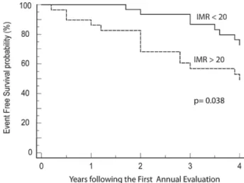
descending artery of 60% and 3 others had luminal stenosis of >70%, 1 patient undergoing coronary artery stenting). On univariable analysis, IMR >20 was significantly associated with outcome on univariable analysis (Table 4). Figure 5 illus- trates the Kaplan-Meier survival curve associated with IMR
>20. A history of hemodynamically compromising rejection during the first year was the strongest factor associated with
Figure 3. Differences in cardiac index and left ventricular myocardial performance indices (LVMPI) between patients with or without microvascular dysfunction (index of microcirculatory resistance [IMR] >20).Figure 4. Change in index of microcirculatory resistance (IMR) and cardiac index (CI) from baseline to 1 year. The lower panels show the relationship between change in CI and change in IMR, as well as the relationship between change in indexed stroke volume (SVI) and change in IMR.
the combined end point. Other factors significantly associated with outcome included rejection history, diabetes mellitus, RAP, and PCWP.
Intraobserver Variability in Measurements of Echocardiographic Indices
To determine intraobserver variability, a blinded reader (F.H.) repeated the measurements on 30 studies. There was good concordance between measures of LV and right ventricular parameters. For LVID, the difference in abso- lute measurement was 0.6±1.3 mm, with an intraclass cor- relation coefficient of 0.94. For LVEF, the difference in absolute measurement was 1.4±2.6%, with an intraclass correlation coefficient of 0.85. For left ventricular myocar- dial performance index, the average difference in absolute measurement was 0.02±0.05, with an intraclass correlation coefficient of 0.91. For RVMPI, the average difference in absolute measurement was 0.03±0.04, with an intraclass correlation coefficient of 0.89.
Discussion
Our study is the first to assess the clinical correlates of early microvasculopathy using IMR, a more specific index of microvascular function. We have found that a history of rejection during the first year and smaller indexed LV dimen- sions were more common among patients with microvascu- lar dysfunction. Early microvascular dysfunction was also associated with mild lower cardiac index, echocardiographic evidence of mild graft dysfunction, and a higher incidence of death, graft failure, or significant CAV on long-term follow-up.
CAV continues to limit the long-term survival of patients with cardiac transplantation.
2,25Although CAV may affect both the epicardial vessels and the microvasculature,
microvascular dysfunction often occurs in the absence of epicardial disease.
1,2,22,25In our study, the majority of patients with evidence of early microvascular dysfunction (86%) had no evidence of impaired epicardial physiology based on FFR, a measure of epicardial physiology. In terms of evolution, microvascular function improves on average from between baseline to 1 year, whereas epicardial physiology measured by FFR worsens as was previously shown in the Physiologic Investigation for Transplant Arteriopathy (PITA) II trial.
3Different methods have been developed to assess the microvasculature, with each method having its own advan- tages and disadvantages.
14A pathology-based system for grading microvasculopathy based on light microscopy has been recently proposed by Hiemann et al
4based on the his- tological characteristics of endothelium, wall, and lumen.
Stenotic microvasculopathy was defined as a ratio of lumi- nal radius to wall thickness <1. Functional assessment of the microvasculature is clinically based on assessing both endothelial-dependent and endothelial-independent vaso- dilation.
4–6,14,22,25,26Endothelial-dependent vasodilation is usually assessed using acetylcholine, which acts on the endothelium, whereas endothelial-independent assay mainly involves agents that act on vascular smooth muscles cells, usually with adenosine. In terms of indices, although CFR has been the most commonly used index, IMR has the advan- tage of being more specific for the microvasculature and less dependent on hemodynamics. In our study, we assessed the microvasculature using IMR, an endothelium-independent vasodilation with adenosine. Our study confirms that micro- vascular dysfunction based on CFR and IMR is often but not always concordant (75% of cases). In fact, several patients with normal microvasculature function based on IMR may have CFR <2.5; similarly, several patients with IMR >20 have CFR >2.5.
Consistent with the study of Osto et al,
27we have found that rejection is more common among patients with microvascular disease. Osto et al
27have recently found that, in the absence of significant epicardial CAV, rejection score was the only inde- pendent correlate of microvascular dysfunction defined using
Microvascular dysfunction at 1 y (IMR >20) 2.52 (1.04–5.91) 0.04Microvascular dysfunction at 1 y (CFR <2.5) 1.3 (0.6–3.0) 0.52
Diabetes mellitus 2.4 (1.03–5.6) 0.04
History of rejection during the first year 2.1 (0.9–4.8) 0.09 History of hemodynamically compromising
rejection during the first year
12.6 (4.2–38.4) <0.01
Right atrial pressure per 5 mm Hg 1.8 (1.2–2.8) <0.01 Pulmonary capillary wedge pressure per 5 mm Hg 1.5 (1.04–2.0) 0.03 Cardiac index per 0.5 L/min per m2 decrease 1.4 (0.99–2.1) 0.06
LVEF per 5% decrease 1.1 (0.9–1.4) 0.39
LVEF <55% 1.7 (0.6–4.7) 0.28
Left ventricular size mismatch 1.6 (0.7–3.7) 0.33
Donor age >40 y 1.7 (0.7–4.0) 0.21
IMR indicates index of microcirculatory resistance; HR, hazard ratio; CFR, coronary flow reserve; LVEF, left ventricular ejection fraction.
Figure 5. Kaplan-Meier survival curve of patients with evidence of early microvascular dysfunction compared with patients with- out evidence of microvascular dysfunction. IMR indicates index of microcirculatory resistance.
CFR during the first 5 years of posttransplant. Compared with the study of Osto et al,
27we defined microvascular dysfunction invasively using IMR, and every patient was assessed system- atically during the first posttransplant year. The association between rejection and microvascular dysfunction underscores the importance of immune mechanisms in CAV.
1This associa- tion could be even stronger with antibody-mediated rejection, which is known to target the endothelium of small vessels.
28,29At our center, complement split product C4d staining is not routinely performed on biopsy, and it is difficult for us to spe- cifically study this association.
The association between LVID and microvascular dys- function is novel and needs to be confirmed in future stud- ies. In our study, our classification of smaller donor size at 1 year was based on the criteria of the American Society of Echocardiography.
19This definition differs from the usual definition of undersized donor hearts, which was based on a 20% weight difference between donor and recipients but has the advantage of being based on direct measurements of ventricular size.
30Prior studies have shown that under- sized donor hearts based on weight differences were asso- ciated with a higher likelihood of mortality, especially in patients with increased pulmonary vascular resistance.
30Theoretically, smaller donor hearts could have rarefac- tion of the microvasculature, which could contribute to increased shear stress.
31Previous studies in heart transplan- tation and systemic hypertension have also shown that left ventricular hypertrophy was associated with microvascular dysfunction.
27,31Functionally, microvascular dysfunction was associ- ated with evidence of mild graft dysfunction based on both cardiac index and right and left myocardial performance index. Myocardial performance index represents an index of global systolic and diastolic function and is measured as the ratio between isovolumic and relaxation time divided by ejection time. In patients with normal microvascular func- tion, both left and right myocardial performance indices were close to the reference range of healthy volunteers.
20,21The association between microvascular function and graft function is further supported by the fact that a dynamic change in IMR was an independent correlate of a change in cardiac index between baseline and 1 year. These findings are consistent with the work of Weis et al
25who have shown that the presence of endothelium-independent microvascu- lar dysfunction predicted deterioration of left ventricular systolic function both at rest and during exercise (n=17).
Of importance, causal relationship may not be directly inferred from our findings, and future longitudinal studies with larger sample size analyzing the dynamic relationship between microvascular function and development of graft failure are needed.
Our exploratory analysis also suggests the importance of microvascular function. Because of the small sample size, multivariable analysis could not be performed without over- fitting the model. This finding is consistent with prior studies that have called attention to the importance of microvas- cular disease in heart transplantation. Hiemann et al
4have recently found that stenotic microvascular disease detected
on endomyocardial biopsy, epicardial coronary disease, and diabetes mellitus was an independent correlate of posttrans- plant mortality. Earlier pathological studies by Billingham et al
32have also found an association between microvascular disease and sudden cardiac death in heart transplantation.
Studies analyzing the clinical correlates of CFR-derived measures of microvascular function have found variable relationships with outcome. In the study by Hollenberg et al,
5,6endothelial-dependent microvascular response to ace- tylcholine assessed by CFR but not endothelial-independent response by adenosine (CFR based) was related to the devel- opment of epicardial CAV or cardiac death. In the study by Kübrich et al,
8an association between microvascular endothelium-independent dysfunction assessed by CFR and adverse outcome was found on unvariable analysis but not on multivariable analysis.
Our study has several clinical implications. First, because of the interrelationship among microvascular function, rejec- tion, and graft function, powering the studies adequately to prove the independent predictive value of microvascular dys- function will require a large sample size. Second, future stud- ies are necessary to investigate whether early treatment of patients with evidence of microvascular dysfunction but with- out evidence of early CAV on intravascular ultrasound imag- ing will improve outcome. A study by Sinha et al
33showed that sirolimus-based therapy initiated early after transplant was associated with improved coronary artery physiology involving both the epicardial vessel and the microvascu- lature. Prior studies have shown that treatment with mam- malian targets of rapamycin inhibitors, such as sirolimus or everolimus, decreases the progression of CAV.
34,35Whether the assessment for early microvascular dysfunction should be made at 6 months or 1 year is also a subject of future research.
This study has several limitations. First, the small sam- ple size limits our ability to conduct multivariable analysis without overfitting our model. Also, the cohort did not rep- resent consecutive patients undergoing heart transplantation.
Furthermore, although IMR is a more reproducible marker of microvascular function than CFR, IMR does not take into account all factors involved in microvascular physiol- ogy, such as true back pressure, coronary capacitance, or tissue volume supplied by the artery.
14Furthermore, future studies will also be needed to better understand the relation- ship between IMR and microvasculopathy detected in biopsy specimens.
4Conclusions
Microvascular dysfunction at 1-year posttransplant is asso-
ciated with lower cardiac index and echocardiographic
indices of graft dysfunction. Changes in microvascular func-
tion between baseline and 1 year were also associated with
changes in cardiac index. History of rejection and smaller
donor hearts assessed using echocardiography are key cor-
relates of microvascular dysfunction. Future multicenter
studies will be needed to validate these findings and to deter-
mine predictive role of early microvascular dysfunction on
long-term outcomes after heart transplantation. Furthermore,
Lung and Blood Institute, Bethesda, MD.
Disclosures
None.
References
1. Schmauss D, Weis M. Cardiac allograft vasculopathy: recent develop- ments. Circulation. 2008;117:2131–2141.
2. Stehlik J, Edwards LB, Kucheryavaya AY, Benden C, Christie JD, Dob- bels F, Kirk R, Rahmel AO, Hertz MI. The Registry of the International Society for Heart and Lung Transplantation: Twenty-eighth Adult Heart Transplant Report–2011. J Heart Lung Transplant. 2011;30:1078–1094.
3. Fearon WF, Hirohata A, Nakamura M, Luikart H, Lee DP, Vagelos RH, Hunt SA, Valantine HA, Fitzgerald PJ, Yock PG, Yeung AC. Discordant changes in epicardial and microvascular coronary physiology after cardi- ac transplantation: Physiologic Investigation for Transplant Arteriopathy II (PITA II) study. J Heart Lung Transplant. 2006;25:765–771.
4. Hiemann NE, Wellnhofer E, Knosalla C, Lehmkuhl HB, Stein J, Hetzer R, Meyer R. Prognostic impact of microvasculopathy on survival after heart transplantation: evidence from 9713 endomyocardial biopsies. Cir- culation. 2007;116:1274–1282.
5. Hollenberg SM, Klein LW, Parrillo JE, Scherer M, Burns D, Tamburro P, Bromet D, Satran A, Costanzo MR. Changes in coronary endothelial function predict progression of allograft vasculopathy after heart trans- plantation. J Heart Lung Transplant. 2004;23:265–271.
6. Hollenberg SM, Klein LW, Parrillo JE, Scherer M, Burns D, Tamburro P, Oberoi M, Johnson MR, Costanzo MR. Coronary endothelial dysfunc- tion after heart transplantation predicts allograft vasculopathy and car- diac death. Circulation. 2001;104:3091–3096.
7. Kobashigawa JA, Tobis JM, Starling RC, Tuzcu EM, Smith AL, Valan- tine HA, Yeung AC, Mehra MR, Anzai H, Oeser BT, Abeywickrama KH, Murphy J, Cretin N. Multicenter intravascular ultrasound validation study among heart transplant recipients: outcomes after five years. J Am Coll Cardiol. 2005;45:1532–1537.
8. Kübrich M, Petrakopoulou P, Kofler S, Nickel T, Kaczmarek I, Meiser BM, Reichart B, von Scheidt W, Weis M. Impact of coronary endothelial dysfunction on adverse long-term outcome after heart transplantation.
Transplantation. 2008;85:1580–1587.
9. Fearon WF, Aarnoudse W, Pijls NH, De Bruyne B, Balsam LB, Cooke DT, Robbins RC, Fitzgerald PJ, Yeung AC, Yock PG. Microvascular re- sistance is not influenced by epicardial coronary artery stenosis severity:
experimental validation. Circulation. 2004;109:2269–2272.
10. Fearon WF, Balsam LB, Farouque HM, Caffarelli AD, Robbins RC, Fitzgerald PJ, Yock PG, Yeung AC. Novel index for invasively assessing the coronary microcirculation. Circulation. 2003;107:3129–3132.
11. Fearon WF, Farouque HM, Balsam LB, Caffarelli AD, Cooke DT, Rob- bins RC, Fitzgerald PJ, Yeung AC, Yock PG. Comparison of coronary thermodilution and Doppler velocity for assessing coronary flow reserve.
Circulation. 2003;108:2198–2200.
12. Fearon WF, Potena L, Hirohata A, Sakurai R, Yamasaki M, Luikart H, Lee J, Vana ML, Cooke JP, Mocarski ES, Yeung AC, Valantine HA.
Changes in coronary arterial dimensions early after cardiac transplanta- tion. Transplantation. 2007;83:700–705.
13. Hirohata A, Nakamura M, Waseda K, Honda Y, Lee DP, Vagelos RH, Hunt SA, Valantine HA, Yock PG, Fitzgerald PJ, Yeung AC, Fearon WF.
Changes in coronary anatomy and physiology after heart transplantation.
Am J Cardiol. 2007;99:1603–1607.
14. Pries AR, Habazettl H, Ambrosio G, Hansen PR, Kaski JC, Schächinger V, Tillmanns H, Vassalli G, Tritto I, Weis M, de Wit C, Bugiardini R. A re- view of methods for assessment of coronary microvascular disease in both clinical and experimental settings. Cardiovasc Res. 2008;80:165–174.
15. Ng MK, Yeung AC, Fearon WF. Invasive assessment of the coronary mi- crocirculation: superior reproducibility and less hemodynamic dependence
tory of congestive heart failure: the Framingham study. N Engl J Med.
1971;285:1441–1446.
18. Mehra MR, Crespo-Leiro MG, Dipchand A, Ensminger SM, Hiemann NE, Kobashigawa JA, Madsen J, Parameshwar J, Starling RC, Uber PA.
International Society for Heart and Lung Transplantation working for- mulation of a standardized nomenclature for cardiac allograft vasculopa- thy-2010. J Heart Lung Transplant. 2010;29:717–727.
19. Lang RM, Bierig M, Devereux RB, Flachskampf FA, Foster E, Pellikka PA, Picard MH, Roman MJ, Seward J, Shanewise JS, Solomon SD, Spen- cer KT, Sutton MS, Stewart WJ; Chamber Quantification Writing Group;
American Society of Echocardiography’s Guidelines and Standards Committee; European Association of Echocardiography. Recommen- dations for chamber quantification: a report from the American Society of Echocardiography’s Guidelines and Standards Committee and the Chamber Quantification Writing Group, developed in conjunction with the European Association of Echocardiography, a branch of the Europe- an Society of Cardiology. J Am Soc Echocardiogr. 2005;18:1440–1463.
20. Tei C, Ling LH, Hodge DO, Bailey KR, Oh JK, Rodeheffer RJ, Tajik AJ, Seward JB. New index of combined systolic and diastolic myo- cardial performance: a simple and reproducible measure of cardiac function–a study in normals and dilated cardiomyopathy. J Cardiol.
1995;26:357–366.
21. Tei C, Dujardin KS, Hodge DO, Bailey KR, McGoon MD, Tajik AJ, Seward SB. Doppler echocardiographic index for assessment of global right ventricular function. J Am Soc Echocardiogr. 1996;9:838–847.
22. Weis M, Peter-Wolf W, Mazzilli N, Olbrich HG, Schacherer C, Wiemer J, Burger W, Hartmann A. Variations of segmental endothelium-depen- dent and endothelium-independent vasomotor tone after cardiac trans- plantation (qualitative changes in endothelial function). Am Heart J.
1997;134(2 Pt 1):306–315.
23. Saudek CD, Herman WH, Sacks DB, Bergenstal RM, Edelman D, Da- vidson MB. A new look at screening and diagnosing diabetes mellitus. J Clin Endocrinol Metab. 2008;93:2447–2453.
24. Levey AS, Coresh J, Greene T, Marsh J, Stevens LA, Kusek JW, Van Lente F; Chronic Kidney Disease Epidemiology Collaboration. Express- ing the Modification of Diet in Renal Disease Study equation for estimat- ing glomerular filtration rate with standardized serum creatinine values.
Clin Chem. 2007;53:766–772.
25. Weis M, Hartmann A, Olbrich HG, Hör G, Zeiher AM. Prognostic sig- nificance of coronary flow reserve on left ventricular ejection fraction in cardiac transplant recipients. Transplantation. 1998;65:103–108.
26. Wolford TL, Donohue TJ, Bach RG, Drury JH, Caracciolo EA, Kern MJ, Miller LW. Heterogeneity of coronary flow reserve in the exami- nation of multiple individual allograft coronary arteries. Circulation.
1999;99:626–632.
27. Osto E, Tona F, Angelini A, Montisci R, Ruscazio M, Vinci A, Tarantini G, Ramondo A, Gambino A, Thiene G, Caforio AL, Gerosa G, Iliceto S.
Determinants of coronary flow reserve in heart transplantation: a study performed with contrast-enhanced echocardiography. J Heart Lung Transplant. 2009;28:453–460.
28. Uber WE, Self SE, Van Bakel AB, Pereira NL. Acute antibody-me- diated rejection following heart transplantation. Am J Transplant.
2007;7:2064–2074.
29. Michaels PJ, Espejo ML, Kobashigawa J, Alejos JC, Burch C, Takemoto S, Reed EF, Fishbein MC. Humoral rejection in cardiac transplantation:
risk factors, hemodynamic consequences and relationship to transplant coronary artery disease. J Heart Lung Transplant. 2003;22:58–69.
30. Patel ND, Weiss ES, Nwakanma LU, Russell SD, Baumgartner WA, Shah AS, Conte JV. Impact of donor-to-recipient weight ratio on survival after heart transplantation: analysis of the United Network for Organ Sharing Database. Circulation. 2008;118(14 Suppl):S83–S88.
31. Hoenig MR, Bianchi C, Rosenzweig A, Sellke FW. The cardiac micro- vasculature in hypertension, cardiac hypertrophy and diastolic heart fail- ure. Curr Vasc Pharmacol. 2008;6:292–300.
32. Billingham ME. Graft coronary disease: the lesions and the patients.
Transplant Proc. 1989;21:3665–3666.
33. Sinha SS, Pham MX, Vagelos RH, Perlroth MG, Hunt SA, Lee DP, Va- lantine HA, Yeung AC, Fearon WF. Effect of rapamycin therapy on coro- nary artery physiology early after cardiac transplantation. Am Heart J.
2008;155:889.e1–889.e6.
34. Raichlin E, Bae JH, Khalpey Z, Edwards BS, Kremers WK, Clavell AL, Rodeheffer RJ, Frantz RP, Rihal C, Lerman A, Kushwaha SS.
Conversion to sirolimus as primary immunosuppression attenuates the
progression of allograft vasculopathy after cardiac transplantation. Cir- culation. 2007;116:2726–2733.
35. Raichlin E, Prasad A, Kremers WK, Edwards BS, Rihal CS, Lerman A, Kushwaha SS. Sirolimus as primary immunosuppression is associ- ated with improved coronary vasomotor function compared with cal- cineurin inhibitors in stable cardiac transplant recipients. Eur Heart J.
2009;30:1356–1363.