Korean J Intern Med 2020;35:897-905 https://doi.org/10.3904/kjim.2018.326
Background/Aims: Atrial arrhythmia (AA) occasionally occurs after lung trans-plantation (LT); however, risk factors for AA and their impact on clinical outcomes are inconsistent. We aimed to investigate the incidence, predisposing factors, and clinical outcomes of AA after LT.
Methods: We retrospectively evaluated 153 consecutive patients who underwent LT between January 2010 and August 2016. An AA episode was defined as a docu-mented atrial fibrillation (AF), atrial flutter, or atrial tachycardia on 12-lead elec-trocardiography or episodes lasting ≥ 30 seconds on telemetry monitoring.
Results: The mean follow-up time was 22.0 ± 19.1 months. Postoperative AA oc-curred in 46 patients (30.1%) after LT. Patients with postoperative AA were older, had larger body surface area, and had an increased incidence of paroxysmal AF prior to transplantation, idiopathic pulmonary fibrosis, and postoperative trache-ostomy than patients without AA. Preoperative right atrial pressure (RAP) (odds ratio [OR], 1.19; p = 0.005) and longer periods of mechanical ventilation (OR, 1.03; p = 0.008) were found to be independent risk factors for AA after surgery. Devel-opment of AA was a significant predictor of long-term overall mortality (hazard ratio, 2.75; p = 0.017).
Conclusions: Patients with elevated preoperative RAP and long-term ventilator care had a higher risk of AA after LT. Further, AA after LT was associated with poor long-term survival.
Keywords: Lung transplantation; Atrial fibrillation; Atrial f lutter; Tachycardia,
ectopic atrial; Survival
Clinical significance of postoperative atrial
arrhythmias in patients who underwent lung
transplantation
Byung Gyu Kim1,*,†, Jae-Sun Uhm2,*, Pil-Sung Yang3, Hee Tae Yu2, Tae-Hoon Kim2, Boyoung Joung2, Hui-Nam Pak2, Song Yee Kim4, Moo Suk Park4, Jin Gu Lee5, Hyo Chae Paik5, and Moon-Hyoung Lee2
INTRODUCTION
Lung transplantation (LT) has been performed increasingly over the past 2 decades [1,2]. Atrial arrhythmias (AA), including atrial fibrillation (AF), atrial flutter (AFL), and atrial tachycardia (AT), are highly prevalent complications after
LT, with a reported incidence ranging from 25% to 45% depending on their definition and methods of detection [3-9]. At present, there is inconsistent evidence supporting the association between mortality after LT and post-operative AA [3,6,10,11]. In addition, dif-ferent risk factors have been reported
1Division of Cardiology, Department
of Internal Medicine, Inje University Seoul Paik Hospital, Seoul; 2Division
of Cardiology, Department of Internal Medicine, Severance Hospital, Yonsei University College of Medicine, Seoul; 3Division of
Cardiology, Department of Internal Medicine, CHA Bundang Medical Center, CHA University, Seongnam;
4Division of Pulmonology,
Department of Internal Medicine and 5Department of Thoracic and
Cardiovascular Surgery, Severance Hospital, Yonsei University College of Medicine, Seoul, Korea
Received : September 7, 2018 Revised : December 11, 2018 Accepted : January 18, 2019 Correspondence to Moon-Hyoung Lee, M.D. Division of Cardiology, Department of Internal Medicine, Severance
Cardiovascular Hospital, Yonsei University College of Medicine, 50-1 Yonsei-ro, Seodaemun-gu, Seoul 03722, Korea
Tel: +82-2-2228-8443 Fax: +82-2-2227-7732 E-mail: [email protected]
*Theseauthors contributed
equally to this study.
†Current affiliation: Division
of Cardiology, Department of Internal Medicine, Inje University Sanggye Paik Hospital, Seoul, Korea
with inconsistent results [3,7,11]. An increasing number of LT recipients at an advanced age, bilateral nature of LT, development of donor selection criteria, donor management protocols, and postoperative management are some current noteworthy changes, especially after the implementation of the lung allocation score in 2005 [1,12], which necessitated re-evaluation of these arrhyth-mias. Therefore, the purpose of this study was to assess the incidence of AA in patients who underwent LT and to determine its risk factors and impact on clinical out-comes in the contemporary era.
METHODS
Patient population
This was a single-centered, retrospective cohort analy-sis. The study was approved by the Institutional Review Board of Severance Hospital (IRB number: 2013-0522-035) and was conducted in compliance with the Declara-tion of Helsinki. Informed consent and a critical event committee were exempted by the board due to this study’s retrospective design. Lung transplant patients, who underwent either single or bilateral LT, between January 2010 and August 2016, were eligible for the study. Patients who underwent heart-lung transplanta-tion or were lost to follow-up were excluded from this study.
Data collection
Baseline characteristics, operative and postoperative clinical variables, and clinical outcomes during the fol-low-up period were captured from electronic medical records. Baseline demographics collected included: age, gender, comorbidities, history of AF, preoperative med-ications, indications for LT, electrocardiography (ECG), echocardiographic data, right heart catheterization data (if available), and laboratory results. Operation notes were reviewed to obtain details of the surgery such as transplant type (single or bilateral), surgical procedure, and intraoperative complications. Preoperative echocar-diographic data included left ventricular ejection frac-tion (LVEF), size of the cardiac chambers, right atrial pressure (RAP), right ventricular systolic pressure, and E/E’ ratio. LVEF and left atrial volume were determined using the biplane Simpson’s method. RAP was
estimat-ed by evaluating the inferior vena cava during respira-tion. Right ventricular systolic pressure was calculated by adding the estimated RAP to the pressure gradient between the right atrium and right ventricle (4 × peak tricuspid regurgitant jet velocity2). Further, to determine
if postoperative clinical features predicted postopera-tive AA, data on the duration of mechanical ventilation needed and cardiac markers were collected as well.
Study outcomes
All patients completed continuous ECG monitoring in the surgical intensive care unit (ICU). The outcomes collected included development of AA (including early and late postoperative AA), postoperative length of ICU stay, length of hospital stay, postoperative occurrences of stroke, and all-cause mortality. Early postoperative AA was defined as the first documentation of AF, AFL, or AT lasting ≥ 30 seconds via either a 12-lead ECG or rhythm strips obtained from the ECG monitoring with-in 30 days of transplantation. Late AA was defwith-ined as the occurrence of AA, after 30 days of transplantation, at any time during the follow-up period. ECGs were analyzed by two independent cardiologists (B.G.K. and J.S.U.). Progress notes, emergency room records, and consul-tation notes were also intensively reviewed to determine the physician’s assessment of arrhythmia or tachycardia. Onset time of postoperative AA was defined as the num-ber of days from the date of LT to the date of the first ep-isode of AA. Lengths of ICU stay and hospital stay were calculated from the date of LT to the date of transfer to the general ward and discharge, respectively. Postop-erative occurrence of stroke was defined as occurrence of a focal neurological deficit confirmed from abnormal findings of brain imaging studies by a neurologist after surgery. All-cause mortality included any death after LT. Treatment strategies for AA episodes were stratified into rate control (β-blockers, non-dihydropyridine calcium channel blockers, and digoxin) and rhythm control (an-ti-arrhythmic drugs and electrical cardioversion). Deci-sions regarding treatments were at the discretion of the transplant surgeons or consulting cardiologists.
Statistical analysis
Continuous variables were presented as mean ± stan-dard deviation and categorical variables were presented as number and percentage. Mean differences between
groups were tested using the Student’s t test for nor-mally distributed data or the Mann-Whitney U test for skewed data. Categorical variables were analyzed us-ing the Pearson’s chi-square test or Fisher’s exact test. Uni- and multivariate analyses using logistic regression models were constructed to validate predictors of AA. Variables with p < 0.1 from the univariate analyses were considered as potential risk factors of postoperative AA in the multivariate analyses. Log-rank and Kaplan-Mei-er tests wKaplan-Mei-ere used to compare survival between groups. Multivariate analysis using Cox proportional hazards models was performed to identify whether postopera-tive AA development was one of the independent risk factors for mortality. All statistical analyses were carried out in SPSS version 23.0 (IBM Corp., Armonk, NY, USA). All tests were two-sided, and the results were considered statistically significant at p < 0.05.
RESULTS
Patient demographics
In total, 153 patients (age, 51.2 ± 13.6 years; men, 58.8%) who underwent LT were evaluated. Bilateral and single LT was performed in 141 (92.2%) and 12 (7.8%) patients, respectively. The major indications for LT were (in or-der of frequency) idiopathic pulmonary fibrosis (IPF), graft-versus-host disease after hematopoietic stem cell transplantation, lymphangioleiomyomatosis, bronchi-ectasis, and chronic obstructive pulmonary disease. Less frequent diagnoses included idiopathic pulmonary hy-pertension, miliary tuberculosis, pulmonary capillary
hemangioma, Langerhans cell histiocytosis, and inter-stitial lung diseases associated with systemic rheumatic diseases. Comorbidities of the patients included hyper-tension (20.9%), diabetes (17.6%), dyslipidemia (9.8%), and coronary artery disease (7.2%). All patients had sinus rhythm at the time of surgery, but five patients (3.3%) had a history of paroxysmal AF prior to the transplant. All patients were treated with standard immunosup-pressive medications, such as tacrolimus, azathioprine, and prednisolone, after surgery.
Incidence, timing, and types of AA
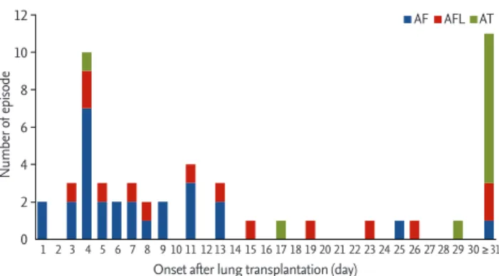
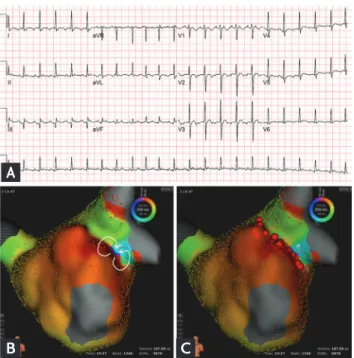
The mean follow-up period post-transplantation was 22.0 ± 19.1 months. During the follow-up period, AA de-veloped in 46 of the 153 of patients (30.1%). Early post-operative AA occurred in 40 patients (26.1%) with AA, of whom 26 (17.0%) had AF, 11 (7.2%) had AFL, and three (2.0%) had AT. There were no significant differences in the onset time among patients with AF, AFL, and AT (7.2 ± 5.3, 11.3 ± 6.9, and 17.3 ± 10.3 days, respectively; p = 0.195). The occurrence of early postoperative AA peaked on the postoperative day (POD) 4. In 67.5% of patients, AA de-veloped within POD 10, and in 90.0% of patients before POD 20 (Fig. 1). The conversion of AA into sinus rhythm was observed in all patients during hospitalization and the mean duration of early postoperative AA was 2.6 ± 2.6 days (range, 1 to 11). Late AA occurred in 11 patients (7.2%), of whom one (0.7%) had AF, two (1.3%) had AFL, and eight (5.2%) had AT. The median interval from LT to onset of late AA was 64 days (interquartile range, 45 to 95). AF and AT were the most common AA in early and late postoperative periods (65.0% and 72.7%, respec-tively) (Fig. 1). Conversion of late AA into sinus rhythm was also observed in all patients with a duration of 8.2 ± 10.4 days (range, 1 to 32). Five patients developed both early postoperative and late AA: three patients with early AF and one patient with early AFL developed AT: one patient with early AF developed AFL in the late postop-erative period (Fig. 2) [13].
Risk factors for AA
Differences in patients’ characteristics according to the development of AA are presented in Table 1. Patients with AA were significantly older, had larger body sur-face area (BSA), and had frequent incidences of AF be-fore transplantion, IPF, and postoperative tracheostomy
Figure 1. Incidence and time to onset of atrial arrhythmias
after lung transplantation. AF, atrial fibrillation; AFL, atrial flutter; AT, atrial tachycardia.
12 10 8 6 4 2 0 1 2 3 4 5 6 7 8 9 10 11 12 13 14 15 16 17 18 19 20 21 22 23 24 25 26 27 28 29 30 ≥ 31 Number of episode
Onset after lung transplantation (day)
than those without AA. No significant differences were observed between patients with and those without AA in preoperative echocardiographic data (including LVEF, left atrial size, RAP, right ventricular systolic pressure, and E/E’), and cardiac catheterization data (including mean pulmonary artery pressure, mean right atrial pres-sure, and mean pulmonary capillary wedge pressure). Predictors of AA at any time after LT are shown in Table 2. Longer periods of mechanical ventilation (hazard ra-tio [HR], 1.03; 95% confidence interval [CI], 1.01 to 1.06; p = 0.008) and preoperative RAP (HR, 1.19; 95% CI, 1.06 to 1.35; p = 0.005) were found to be significant risk factors for AA after LT, when adjusted for other risk factors.
Treatment strategies for AA
Forty of the 46 patients (87.0%) received treatment for AA. The remaining six patients, with early postoperative AA (three AF, one AFL, and two AT), were not treated as their heart rates remained stable and the AA was
spon-taneously converted to sinus rhythm within 1.8 ± 1.5 days. Rate control treatment, using β-blockers, calcium channel blockers (diltiazem or verapamil), and digoxin, was used in the majority of patients (39 of 40, 97.5%). Six of the 40 patients (16.7%) were treated with combined antiarrhythmic medical therapy (amiodarone), and two patients underwent electrical cardioversion due to he-modynamic instability. Non-vitamin K-dependent oral anticoagulants were administered to five patients with an AF lasting for > 2 days and a CHA2DS2-VASc score ≥ 2.
Clinical impact of AA
Length of ICU stay and hospital stay, as well as in-hos-pital mortality, were not significantly different between patients with and without AA (Table 1). Stroke developed in six patients (3.9%) after LT, and the incidence was sim-ilar between patients with and without AA. Long-term overall mortality was significantly higher in patients who developed AA in comparison to their counterparts (Fig. 3). Landmark analyses were performed to evaluate survival 30 days after LT. Incidence of all-cause mortali-ty was higher in the patients with early postoperative AA than in those without AA (Fig. 4). Further, patients who developed late AA or both early and late AA had greater rate of all-cause mortality than those without AA. (Fig. 4).
The major causes of death in all LT recipients includ-ed infection (63.0%) and multi-organ failure syndrome (16.1%). There was no difference in causes of death between the patients with and without AA (infection: 64.0% vs. 62.1%, p = 0.872; multi-organ failure syndrome: 16.0% vs. 13.8%, p = 0.826). All-cause mortality and cause of death were summarized in Table 3.
Postoperative AA, including early and late AA, was found to be an independent predictor of overall mor-tality, even after adjustment for other covariates. An E/E’ > 15 and longer periods of mechanical ventilation were also identified as independent risk factors associated with increased mortality after LT (Table 4).
DISCUSSION
The major findings of this study were as follows: (1) AA was common after LT; (2) elevated preoperative RAP and long-term ventilator care were independent predictors for the development of AA after LT; and (3) all-cause
Figure 2. A case of a 62-year-old man who was treated for
atrial tachycardia with radiofrequency catheter ablation. (A) Electrocardiography showing atrial tachycardia. (B) Three-di-mensional activation map reveals figure-of-eight intra-atrial reentrant tachycardia (white arrows) with the slow conduction zone at the anastomosis line. (C) Radiofrequency catheter ablation was performed along the ridge (red balls). Adaptad from Uhm et al. [13]. aVR, augmented Vector right; aVL, aug-mented Vector left; aVF, augaug-mented Vector foot.
A
Table 1. Baseline characteristics of patients with and without postoperative atrial arrhythmia
Characteristic AA (n = 46) No AA (n = 107) p value
Age at transplant, yr 57.0 ± 10.7 48.8 ± 14.0 < 0.001
Male sex 32 (69.6) 58 (54.2) 0.077
Body surface area, m2 1.65 ± 0.19 1.56 ± 0.24 0.042
Body mass index, kg/m2 21.7 ± 4.3 20.9 ± 3.7 0.275
Comorbidities
Hypertension 12 (26.1) 20 (18.7) 0.302
Diabetes 12 (26.1) 15 (14.0) 0.073
Dyslipidemia 7 (15.2) 8 (7.5) 0.149
Smoking 22 (47.9) 41 (38.3) 0.273
Coronary artery disease 6 (13.0) 5 (4.7) 0.088
Stroke 1 (2.2) 1 (0.9) 0.512
Atrial fibrillation 4 (8.7) 1 (0.9) 0.029
Serum creatinine, mg/dL 0.7 ± 0.3 0.7 ± 0.4 0.336
Indication for transplant
Idiopathic pulmonary fibrosis 34 (73.9) 56 (52.8) 0.015
GVHD 3 (6.5) 10 (9.4) 0.755
COPD 3 (6.5) 4 (3.8) 0.433
Bronchiectasis 2 (4.3) 5 (4.7) 0.921
Lymphangioleiomyomatosis 1 (2.2) 8 (7.5) 0.278
Other 3 (6.5) 23 (21.5) 0.024
Type of lung transplantation
Bilateral 43 (93.5) 98 (91.6) 0.487
Echocardiography findings
Ejection fraction, % 64.3 ± 9.8 64.3 ± 11.4 0.981
Left atrial volume index, mL/m2 19.1 ± 7.2 18.0 ± 7.2 0.596
RAP, mmHg 8.7 ± 5.2 7.2 ± 3.9 0.075
RVSP, mmHg 53.6 ± 27.6 49.2 ± 21.6 0.332
E/E’ 10.6 ± 4.5 10.5 ± 4.1 0.889
Right-sided heart catheterization data
Mean PAP, mmHg 29.1 ± 14.4 28.2 ± 10.4 0.752
Mean RAP, mmHg 6.8 ± 3.7 8.0 ± 5.0 0.275
Mean PCWP, mmHg 10.8 ± 5.3 11.2 ± 6.7 0.756
Pre- and postoperative data
Preoperative PCPS 10 (22.2) 22 (20.8) 0.840
Preoperative MV 16 (35.6) 30 (28.3) 0.376
Postoperative TnT 0.810 ± 0.891 0.491 ± 0.264 0.056
Postoperative tracheostomy 35 (76.1) 45 (42.5) < 0.001
Perioperative MV duration, day 27.2 ± 20.5 17.8 ± 21.8 0.020
Length of postoperative ICU stay, day 14.5 ± 10.0 13.1 ± 12.1 0.541
Length of hospital stay, day 63.5 ± 55.1 47.8 ± 37.3 0.085
In-hospital mortality 4 (8.7) 5 (4.7) 0.242
New onset of stroke 3 (6.5) 3 (2.8) 0.360
Values are presented as mean ± SD or number (%).
AA, atrial arrhythmia; GVHD, graft-versus-host disease; COPD, chronic obstructive pulmonary disease; RAP; right atrial pressure; RVSP, right ventricular systolic pressure; PAP, pulmonary artery pressure; PCWP, pulmonary capillary wedge pres-sure; PCPS, percutaneous cardiopulmonary support; MV, mechanical ventilation; TnT, troponin T; ICU, intensive care unit.
mortality after LT was higher in patients with AA than in those without AA.
Potential mechanisms of AA after LT
Our data shows that AA occurred most frequently in the early postoperative period (within 30 days) and its frequency declined after 1 month. AF was the most commonly occurring AA in the early postoperative pe-riod, whereas late AA was composed of mainly AT and AFL. These observations are consistent with previously reported data [4,5,14]. AF could be occurring frequent-ly during the earfrequent-ly postoperative period due to local inflammation, myocardial injury, sympathetic activa-tion, fluid shift, and electrolyte imbalances [15-18]. Ad-ditionally, the operative technique during LT, such as sutured anastomosis between the donor’s pulmonary
Table 3. All-cause mortality and cause of death in patients with and without atrial arrhythmia
Variable AA (n = 46) (n = 107)No AA p value 30-day mortality 3 (6.5) 2 (1.9) 0.323 90-day mortality 8 (13.0) 11 (10.3) 0.780 1-yr mortality 23 (50.0) 25 (23.4) 0.001 All-cause mortality 25 (54.3) 29 (27.1) 0.001 Cause of death Sepsis 16 (64.0) 18 (62.1) Bleeding 3 (12.0) 4 (13.8) Cardiac death 2 (8.0) 2 (6.9) Multi-organ failure 4 (16.0) 4 (13.8) Unknown 0 1 (3.4)
Values are presented as number (%). AA, atrial arrhythmia.
Table 2. Uni- and multivariate analyses for predictors of postoperative atrial arrhythmia
Risk factor Univariate analysis Multivariate analysis
OR (95% CI) p value OR (95% CI) p value
Age at transplant, /1 yr 1.05 (1.02–1.09) 0.002 0.97 (0.92–1.03) 0.327 Body surface area, /1 m2 8.35 (1.10–63.59) 0.040 3.09 (0.11–84.47) 0.504 History of atrial fibrillation 10.44 (1.13–96.18) 0.038 5.24 (0.39–71.11) 0.213 Idiopathic pulmonary fibrosis 2.41 (1.13–5.17) 0.024 2.30 (0.62–8.63) 0.216
RAP 1.09 (0.99–1.19) 0.060 1.19 (1.06–1.35) 0.005
MV duration, /1 day 1.03 (1.01–1.05) 0.004 1.03 (1.01–1.06) 0.008
OR, odds ratio; CI, confidence interval; RAP, right atrial pressure; MV, mechanical ventilation.
Figure 3. Kaplan-Meier curves for survival in patients with
and without atrial arrhythmias after lung transplantation. AA, atrial arrhythmia.
100 80 60 40 20 0 6 12 18 24 30 36 Survival (%) Time (mon) Long rank p < 0.001 No. at risk No AA AA 107 46 76 23 72 19 67 16 53 13 42 10 32 4 No AA AA
Figure 4. Landmark analysis for survival after the first 30
days post transplantation. AA, atrial arrhythmia. aLog lank
p = 0.001. 100 80 60 40 20 0 030 day 6 12 18 24 30 36 Survival (%) Time (mon) No. at risk No AA Only early AA Only late AA Both AA 107 35 6 5 76 18 3 2 72 15 3 1 67 13 2 1 53 11 1 1 42 9 0 1 32 3 0 1 No AA
Only early postoperative AA Only late AA Both early and late AA
vein cuffs and the recipient’s left atrium, can provide an electroanatomic substrate for potential AA [4,19]. However, it could also serve as an antral electrical iso-lation of the pulmonary veins since the suture line is similar with the line ablation created by the Cox-Maze operation and can theoretically provide an antiar-rhythmogenic effect for AF in the long-term. Later oc-currences of AFL or AT than AF after LT could be due to reentry from the anastomosis after healing. Azadani et al. [14] reported a greater proportion of atypical pat-tern of AFL in recipients who underwent LT and pro-posed that atypical AFL stems from the suture line of the left atrium in contrast to typical AFL, which arises from the right atrium around the tricuspid annulus. In an electrophysiological study, See et al. [4] showed cases of postoperative AT related to macro-reentry that originated from suture lines, and focal AT that arose from the pulmonary vein anastomoses. Taken togeth-er, these results suggest that surgical suture lines can help prevent AF, and can also act as substrate for the development of AFL or AT after scarring. Additional electrophysiological studies must be carried out to confirm this theory and better understand the mech-anism of AA after LT.
Risk factors for AA after LT
In this study, we identified that elevated RAP and a prolonged period of mechanical ventilation period were independent predictors of AA, after adjusting for confounding variables. Mechanical ventilation
in-duces intermittent positive pressure during respirato-ry cycles, which may raise intrathoracic pressure and right atrial pressure. Regardless, due to the application of additional positive end expiratory pressure, right atrial pressure may remain higher throughout the re-spiratory cycle [20]. Such elevated right atrial pressure is related with occurrences of AA. Furthermore, pro-longed periods of mechanical ventilation can cause an increase in sympathetic activity, which may trigger the development of AA.
Clinical significance of AA after LT
Long-term all-cause mortality was significantly higher in patients with AA. Furthermore, the development of AA was identified as an independent predictor of over-all mortality in the multivariate analysis. Interestingly, even though most AA occurred in the early postoper-ative period, early clinical outcomes, such as length of ICU stay, length of hospital stay, and in-hospital mortal-ity, were not different between patients with and with-out AA in our study population. However, we clarified the prognostic impact of early postoperative AA on sur-vival clearly in the long term via our landmark analysis. These findings are in agreement with the results re-ported by Orrego et al. [3], which demonstrated a high-er 12-month ovhigh-erall mortality in those who developed AA, but not at 90 days. Despite conflicting results of the impact of postoperative AA on long term survival after LT [11,21], a recent meta-analysis demonstrated that the occurrence of AA after LT has prognostic implications
Table 4. Predictors for overall mortality after lung transplantation
Risk factor Univariate analysis Multivariate analysis
HR (95% CI) p value HR (95% CI) p value
Age at transplant, /1 yr 1.02 (1.00–1.04) 0.071 1.00 (0.97–1.04) 0.894 Male sex 1.64 (0.93–2.89) 0.088 1.82 (0.75–4.39) 0.186 History of HTN 1.93 (1.08–3.43) 0.026 1.13 (0.46–2.77) 0.788 History of AF 7.17 (0.78–65.81) 0.082 0.79 (0.36–3.38) 0.646 E/E’ > 15 3.01 (1.44–6.31) 0.003 4.43 (1.55–12.70) 0.006 Preoperative PCPS 2.69 (1.51–4.81) 0.001 2.01 (0.49–8.20) 0.330
Preoperative mechanical ventilation 2.98 (1.73–5.14) < 0.001 1.09 (0.35–3.43) 0.883 MV duration, /1 day 1.04 (1.03–1.05) < 0.001 1.03 (1.01–1.05) < 0.001 Atrial arrhythmia after LT 2.49 (1.45–4.26) 0.001 2.75 (1.20–6.32) 0.017 HR, hazard ratio; CI, confidence interval; HTN, hypertension; AF, atrial fibrillation; PCPS, percutaneous cardiopulmonary support; MV, mechanical ventilation; LT, lung transplantation.
for overall long-term survival [22]. Taken together, these results suggest that development of AA after LT may not influence short-term mortality, but likely affects over-all long-term mortality. Moreover, we found that devel-opment of late AA was also linked with poor long-term survival after LT. To our knowledge, an association be-tween late AA after LT and survival has not been report-ed in prior studies. In this study, we could not identify the differences in characteristics between patients with late AA and their counterparts due to a small sample size. Further large-scale studies need to be carried out to address this issue.
The mechanisms that drive postoperative AA and re-sult in poor long-term survival are not known. Mean time to death after development of AA was 76.0 ± 78.5 days in our study. Most patients died from infection, bleeding or multi-organ failure, and these poor med-ical conditions could have acted as a trigger for AA. In addition, longer periods of perioperative mechanical ventilation, which was related to development of AA, may reflect the more complex surgery and complica-tions like infection. Therefore, we speculate that AA may serve as a potential surrogate marker to identify unstable medical conditions that may make patients vulnerable to stress and may increase their risk of mor-tality.
Study limitations
There are several limitations to be noted. Given the retrospective nature of this study at a single institu-tion with a small number of patients, unmeasured con-founders associated with AA such as consistent surgical techniques or postoperative care might have biased the results despite statistical adjustments. Further, some short and asymptomatic arrhythmic episodes during inconsistent ECG monitoring in the general ward or late AA events could have been missed. However, given complete access and review of these events, it is not likely that meaningful information was lost. Addition-ally, we could not determine the mechanisms driving postoperative AA due to unavailable electrophysiolog-ical data in our study. Further large-scale electrophys-iological studies are required to better understand the mechanisms of AA and establish prevention strategies in LT recipients.
In conclusion, patients with preoperative RAP and
long-term ventilator care had a higher risk of develop-ing AA after LT. Further, AA after LT is associated with poor long-term survival.
Conflict of interest
No potential conflict of interest relevant to this article was reported.
REFERENCES
1. Yusen RD, Christie JD, Edwards LB, et al. The Registry of the International Society for Heart and Lung Trans-plantation: thirtieth adult lung and heart-lung transplant report. 2013. Focus theme: age. J Heart Lung Transplant 2013;32:965-978.
2. Christie JD, Edwards LB, Aurora P, et al. Registry of the International Society for Heart and Lung Transplan-tation: twenty-fifth official adult lung and heart/lung transplantation report. 2008. J Heart Lung Transplant 2008;27:957-969.
3. Orrego CM, Cordero-Reyes AM, Estep JD, et al. Atrial ar-rhythmias after lung transplant: underlying mechanisms, risk factors, and prognosis. J Heart Lung Transplant 2014;33:734-740.
4. See VY, Roberts-Thomson KC, Stevenson WG, Camp PC, Koplan BA. Atrial arrhythmias after lung transplantation: epidemiology, mechanisms at electrophysiology study, and outcomes. Circ Arrhythm Electrophysiol 2009;2:504-510.
5. Chaikriangkrai K, Jyothula S, Jhun HY, et al. Incidence, risk factors, prognosis, and electrophysiological mech-anisms of atrial arrhythmias after lung transplantation. JACC Clin Electrophysiol 2015;1:296-305.
6. Isiadinso I, Meshkov AB, Gaughan J, et al. Atrial
arrhyth-KEY MESSAGE
1. Atrial arrhythmia (AA) is common in patients who underwent lung transplantation (LT). 2. Elevated preoperative right atrial pressure and
long-term ventilator care were associated with development of AA after LT.
3. AA after LT is linked with poor long-term sur-vival.
mias after lung and heart-lung transplant: effects on short-term mortality and the influence of amiodarone. J Heart Lung Transplant 2011;30:37-44.
7. D’Angelo AM, Chan EG, Hayanga JW, et al. Atrial ar-rhythmias after lung transplantation: incidence and risk factors in 652 lung transplant recipients. J Thorac Cardio-vasc Surg 2016;152:901-909.
8. Nielsen TD, Bahnson T, Davis RD, Palmer SM. Atrial fibril-lation after pulmonary transplant. Chest 2004;126:496-500. 9. Mason DP, Marsh DH, Alster JM, et al. Atrial fibrillation
after lung transplantation: timing, risk factors, and treat-ment. Ann Thorac Surg 2007;84:1878-1884.
10. Malik A, Hsu JC, Hoopes C, Itinarelli G, Marcus GM. Elevated pulmonary artery systolic pressures are associ-ated with a lower risk of atrial fibrillation following lung transplantation. J Electrocardiol 2013;46:38-42.
11. Henri C, Giraldeau G, Dorais M, et al. Atrial fibrillation after pulmonary transplantation: incidence, impact on mortality, treatment effectiveness, and risk factors. Circ Arrhythm Electrophysiol 2012;5:61-67.
12. Eberlein M, Garrity ER, Orens JB. Lung allocation in the United States. Clin Chest Med 2011;32:213-222.
13. Uhm JS, Park MS, Joung B, Pak HN, Paik HC, Lee MH. Intra-atrial reentrant tachycardia originating from the pulmonary vein cuff anastomosis in a lung transplanta-tion patient: ultra-high-density 3-dimensional mapping. HeartRhythm Case Rep 2018;4:152-154.
14. Azadani PN, Kumar UN, Yang Y, et al. Frequency of atri-al flutter after adult lung transplantation. Am J Cardiol
2011;107:922-926.
15. Echahidi N, Pibarot P, O’Hara G, Mathieu P. Mechanisms, prevention, and treatment of atrial fibrillation after car-diac surgery. J Am Coll Cardiol 2008;51:793-801.
16. Bazaz R, Salizzoni S, Bonde P, Espinoza A, Toyoda Y. A novel strategy for prevention of post-operative atrial ar-rhythmias in patients undergoing lung transplantation. J Heart Lung Transplant 2010;29:713-715.
17. Maesen B, Nijs J, Maessen J, Allessie M, Schotten U. Post-operative atrial fibrillation: a maze of mechanisms. Europace 2012;14:159-174.
18. Shingu Y, Kubota S, Wakasa S, Ooka T, Tachibana T, Mat-sui Y. Postoperative atrial fibrillation: mechanism, pre-vention, and future perspective. Surg Today 2012;42:819-824.
19. Dizon JM, Chen K, Bacchetta M, et al. A comparison of atrial arrhythmias after heart or double-lung transplan-tation at a single center: insights into the mechanism of post-operative atrial fibrillation. J Am Coll Cardiol 2009;54:2043-2048.
20. Shekerdemian L, Bohn D. Cardiovascular effects of me-chanical ventilation. Arch Dis Child 1999;80:475-480. 21. Jesel L, Barraud J, Lim HS, et al. Early and late atrial
ar-rhythmias after lung transplantation: incidence, predictive factors and impact on mortality. Circ J 2017;81:660-667. 22. Fan J, Zhou K, Li S, Du H, Che G. Incidence, risk factors
and prognosis of postoperative atrial arrhythmias after lung transplantation: a systematic review and meta-anal-ysis. Interact Cardiovasc Thorac Surg 2016;23:790-799.