KISEP Clinical Researches J Korean Neurosurg Soc 30::::1314-1319, 2001
결핵성 척추염 10례의 수술적 치료에 관한 임상적 고찰*
성균관대학교 의과대학 삼성의료원 신경외과학교실
위성목·어 환·남도현·이정일·김종수 홍승철·신형진·박 관·김종현
=
=
=
= Abstract ====
Clinical Evaluation of Surgical Treatments for Ten Cases of Tuberculous Spondylitis
Sung Mock Whee, M.D., Whan Eoh, M.D., Do Hyun Nam, M.D., Jung Il Lee, M.D., Jong Soo Kim, M.D., Seung-Chyul Hong, M.D.,
Hyung Jin Shin, M.D., Kwan Park, M.D., Jong-Hyun Kim, M.D.
Department of Neurosurgery, Samsung Medical Center, Sungkyunkwan University School of Medicine, Seoul, Korea
bjects:Because of the nonspecific nature of symptoms in tuberculous spondylitis, a delay in the diagnosis can result in progressive neurologic deficits. The authors evaluate the clinical and the radiological results of the 10 cases of surgically treated tuberculous spondylitis.
Clinical materials & Methods:We retrospectively analyzed the medical records of 10 patients with tuberculous spondylitis who were treated between February 1996 and March 2000. Six patients were female, and four were male.
Mean age was 43 years old, and mean follow-up period was 20.5 months. All patients were treated with 12 months of antituberculous medication postoperatively, and were followed by complete blood count, ESR, spine X-ray and MRI.
Results:The lumbar spine was involved in 5 patients, the thoracic in 4, and the thoracolumbar in one. The infected vertebral bodies were 2.8 in average. The associated lesions were pulmonary tuberculosis in 3 cases, and renal tuberculosis in one. Five patients were treated by anterior debridement and fusion with bone graft using anterior instrumentation, 2 with anterior debridement and fusion with bone graft(Hong Kong procedure only), 1 with Hong Kong procedure with posterior spinal instrumentation, and 2 were managed with posterior debridement and posterior spinal instrumentation.
All patients improved after operation, and the average kyphotic angle decreased postoperatively. Postoperatively, one patient had a fistula at the operative site.
Conclusion:The debridement and minimal level fusion of motion segment with instrument fixation is one of surgical option for tuberculous spondyltis to preserve the spine motion segment as much as possible. Spine instability and kyphosis were prevented by anterior and posterior spinal instrumentation. But, large number of cases and longer period follow-up study in future will be needed to confirm the long term results.
KEY WORDS:Tuberculous spondylitis・Spinal instrumentation・Hong Kong procedure.
서 론
골 관절 결핵에서 척추는 가장 빈번히 침범되는 호발 부위
로서 약 50%를 차지하고 있다. 주로 척추체 전방부를 침 범하여 점차적으로 척추체 파괴를 일으켜 척추 후만을 발 생시킨다. 1950년대 항결핵 약제 개발로 척추 결핵 환자의 치료에 획기적인 진전이 있었으나, 항결핵 요법만으로 치 료한 경우 점진적인 척추 후만 변형이 빈번히 발생하게 되
OOOO
*본 논문은 2001년 4월 대한신경외과학회 경주 춘계학술대회에 Oral Poster로 발표된 내용입니다.
어 적절한 외과적 치료가 요구되었다3)13)14)15)18)
. 결핵성 척 추염에 대한 수술적 치료는 1779년 Pott17)가 수술적 배농 술을 기술한 이래 Albee(1911)1)와 Hibbs(1912)7)에 의 해 후방융합술이 처음으로 시도되었고, Wilkinson(1950)24) 에 의해 전측방융합술이 시행되었다. Hodgson과 Stock8) 는 전방도달법에 의한 척추병소의 절제 수술과 그곳에 골 편 이식을 시도하여 성공적인 치료 결과를 발표함으로써 전 방골융합술(Hong Kong 술식)이 보편화되었다. 그러나, 전 방골융합술은 이식 골편의 이탈, 골절 및 후만각의 점진적 인 증가23), 그리고 장기간의 침상 안정 등이 문제로 제기되 어 이러한 단점을 보안하기 위해 전방 자가골융합술 및 척 추 고정술이 시행되었다. 본 신경외과학 교실에서는1996년 2월부터 2000년 3월까지 수술적 치료 및 항결핵 요법으 로 치료한 결핵성 척추염 환자 10명에 대하여 임상적 결과 를 문헌고찰과 함께 보고하는 바이다.
대상 및 방법
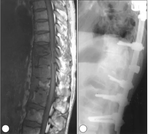
1996년 2월부터 2000년 3월까지 흉요추 결핵으로 진 단되어 본원 신경외과에서 수술적 치료를 받은 10명의 척 추결핵 환자 대하여 12개월부터 36개월까지 추적 조사하 여 임상증상, 방사선학적 소견, 수술 방법, 수술 후 합병증 및 예후에 대하여 분석하였다. 연령별로는 30세이상 39세 미만이 4명(40%)으로 가장 많았고 40세이상 49세미만, 60세이상 69세미만이 각각 2명이였으며 남자는 4명, 여자 는 6명이었다(Table 1). 증상이 발생하여 입원까지 기간은 최저 1개월에서 최고 2년으로 평균은 6.8개월 이였으며, 입 원당시 폐결핵이 동반된 경우는 3례, 신장 결핵이 동반된 경우가 1례였으며, 병소가 척추에만 국한되고 타장기에서 병 변이 발견되지 않는 경우가 6례였다. 환자의 주 증상은 요 통 10례, 하지 방사통 8례, 하지 마비 5례 그리고 하지 감 각 저하 5례였다. 병소 부위별 발생빈도는 요추 5례(50%), 흉추 4례(40%), 흉요추 1례(10%)였다. 수술적 방법으로 5례는 Hong Kong 술식과 전방척추고정술(Fig. 1)을 병행 하였으며, 2례는 척추체 병소 절제술 및 골 이식을 통한 전 방골융합술(Hong Kong 술식)을 시행하였고(Fig. 2), 1례 는 Hong Kong 술식과 후방척추고정술(Fig. 3)을 병행하 였으며 2례는 후방감압술 및 후방척추고정술(Fig. 4)을 시
Table 1. Age and sex distribution Sex
Age Male Female Total
20- 29 1 - 1
30-39 2 2 4 40-49 1 1 2 50-59 1 - 1
60-69 - 2 2
Total 5 5 10
Fig. 1. Gd-DTPA enhanced sagit- tal T1W(A) of T spine MRI of a 39- year-old woman who had tuber- culosis in T8-T9 with severe bony destruction. Postoperative anter- oposterior radiograph(B)showing anterior interbody fusion and an- terior intsrumentation from T7 to T10.
AAA
A BBBB
결핵성 척추염 10례의 수술적 치료에 관한 임상적 고찰
행하였다(Table 2). 사용된 금속 내고정물은 Z-plate가 3 례, titanium mesh를 이용한 자가골융합술이 6례였다. 이 식골은 자가 장골 3례, 자가 늑골 5례, 동종이식(allograft) 1례 등이 사용되었으며 이들 이식골의 유합 여부는 측면 방사선 골 소주의 연결로 평가하였다. 그리고, 척추 후만각 변화는 수술 전후에 측면사진을 촬영하여 Cobb방법으로 측 정하였다.
결 과
남녀 성별비에서는 뚜렷한 차이는 없었으며 30대에서 많 이 발생하였다. 모든 환자에서 수술 전에 호소하였던 요통 과 신경근병증은 호전을 보였으며, 특히 수술 전 5명의 환 자에서 흉추 2례 및 흉요추 1례에서 하지운동장애(Grade
Fig. 2. Gd-DTPA enhanced sagit- tal T1W(A) image shows rim enh- ancement of L2-L3 vertebral bo- dies, and involvement of cortical end plate and intervertebral disc.
Postoperative lateral spinal radi- ograph(B) of a 38-year-old wo- man who had tuberculosis in L2- L3 interspace showed anterior decompression and interbody fusion with rib graft(Hong Kong procedure).
Fig. 3. Gd-DTPA enhanced sagit- tal T1W(A) of thoracic spine MRI of a 43-year-old man who had tuberculosis in T10-12 showed destruction of T10-T12 vertebral bodies with loss of intervening disc height. Postoperative lateral spinal radiograph(B) of a 43-year-old man who had tuberculosis in T10-T12 interspace shows anterior decompression and interbody fusion with rib graft using posterior instrumentation.
A AA
A BB BB
AAA
A BBBB
Ⅲ-Ⅳ), 요추 2례에서 발목 및 무릅의 신전 약화 소견을 보였으나 수술 후 완전히 회복되었다. 10명의 환자에서 결 핵에 감염된 척추체는 모두 28개로 환자 1인당 평균 2.8 개였고, 2개의 요추부 척추체 감염된 것이 5례로 가장 많
았다. 결핵성 척추염의 침범부위에 따른 방사선학적 분류 로는 추간판 주위성(peridiscal) 8례, 중심성(central) 1례, 전방성(anterior) 1례였다(Table 3). 술전 평균 후만각은 흉추 24.5도, 흉요추 25도, 요추 -1도, 수술 직후에 각각 21.3도, 20도, -3도로 흉추에서 3.2도, 흉요추에서 5도, 요 추에서 -2도의 평균 교정각을 얻었다. 최종 추시시의 평균 후만각은 흉추 20.3도, 흉요추 18도, 요추부 -6.4도였으 며 각각 1도, 2도, 3.4도의 교정 후만각의 소실을 보였다 (Table 4). 골융합술을 시행한 9례의 이식골의 융합은 술 후 평균 3.6개월부터 이루어졌다. 골융합술에 따른 이식골 의 흡수, 골절 또는 탈출은 없었으며 전례에서 골융합을 얻 었다. 특히, 추간판 주위성(peridiscal) 형태는 흉추에 3례, 요추에 5례의 분포를 보였으나 본 연구에서는 감염된 척추 체의 소파술 및 척추 고정 기구를 사용한 최소 운동 분절의 고정만으로도 치유가 가능하였으며 병소 부위에 금속 내고
Table 2. Surgical treatments
Surgical modality T T-L L Total Hong Kong procedure(H-K) - - 2 2
H-K + AIF 2 - 3 5
Laminectomy + PIF 1 1 - 2
H-K + PIF 1 - - 1
Total 4 1 5 10
Hong Kong procedure(Anterior debridement and fusion with bone graft)
AIF(Anterior instrument fixation) PIF(Posterior instrument fixation)
Table 3. Types of tuberculous spondylitis
Types T T-L L Total
Peridiscal 3 - 5 8
Central 1 - - 1
Anterior - 1 - 1
Posterior - - - -
Total 4 1 5 10
Table 4. Angle of correction and loss of correction
Angle of correction Loss of correction
Thoracic 3 .2 1
Thoracolumbar 5 2
Lumbar -2 3 .4
Fig. 4. Gd-DTPA enhanced sagit- tal T1W(A) image showing heter- ogeneous enhancement of T9- L2 vertebral bodies with destru- ction of T9 vertebral body. Posto- perative lateral spinal radiography (B) of a 50-year-old woman who had tuberculosis in T9-L2 interface demonstrated posterior decom- pression and posterior instrument- ation.
A AA
A BBBB
결핵성 척추염 10례의 수술적 치료에 관한 임상적 고찰
정물 및 척추 고정술을 시행한 이후에 추적 관찰시 감염의 지속 및 재발 소견은 없었다. 수술 후 합병증은 수술 부위 누공 1례가 있었으나 보존적 치료를 하여 치유되었다.
고 찰
결핵은 후진국에서 선진국으로 갈수록 그 빈도가 감소하 는 경향이 있으나, 우리 나라에서는 활동성 폐결핵의 유병 율은 10만 명당 10.1명으로 보고되고 있고 또한 폐결핵의 신환 발생률은 20~30대 젊은 연령층에서 아직도 높게 나 타나는 실정이다. 결핵성 척추염은 결핵성 질환의 2~3%를
차지하며2)19) 이중에서 척추 결핵은 골 관절 결핵의 50%를
차지한다고 알려져 있다5). 대부분 흉추 하부 및 상부 요추 에 발생하고14)20), 약 3~5%에서 경추에 발생하는 것으로 보고되고 있다4)9). 국내에서는 한과 석 등 및 김 등이 각각 45%와 39%로 요추가 가장 흔한 침범부위로 보고하였
다6)11). 본 연구는 요추가 5례(50%)로 가장 많았으며 흉추
4례, 흉요추 1례이었다. 그리고, 임상증상, 방사선학적 소 견, 조직생검 및 병리학적으로 진단된 척추결핵 치료의 목 표는 감염의 박멸과, 신경증상 및 척추 변형의 예방 또는 치료에 있으며, 수술적 치료의 적응증은 추체 전방의 농양 이 주위 연부 조직을 눌러 증상을 유발할 때, 불안정성 척 추가 초래되는 경우, 중증의 신경학적 장애가 유발된 경우, 경미한 신경학적 장애가 항결핵제를 투여한 6주 후에도 호 전이 없는 경우, 약물 치료가 실패한 경우 등에 시행하게 된
다12)20-22). 수술적 치료로는 1779년 Percival Pott가 수술
적 배농술을 기술한 이래 후궁절제술, 늑척추 횡돌기 절제 술, Albee(1911)와 Hibbs(1912)에 의해 후방융합술, Wi- lkinson(1950)에 의해 전측방융합술, 전방융합술 등으로 결과를 향상시켜왔으며 전방융합술은 1934년 Ito 등에 의 하여 처음 도입된 후, 1956년 Hodgson과 Stock 등이 척 추 전방 도달법에 의한 척추체의 근본적인 병소제거, 감압 및 전방 추체간 융합술(Hong Kong 술식)을 발표한 이래 이 방법이 척추 결핵의 수술적 치료법의 표준 치료법으로 보편화되었다3)8)10)15). 전방접근술은 병변부위를 육안으로 직접 확인 후 가능한 한 모든 병소를 제거하고 척추체간을 신전시킨 후 골이식을 시행하므로 조속한 골융합을 얻을 수 있고 내부고정기기의 사용으로 이식골의 탈출방지 및 환자 의 재활을 촉진하고 재발의 빈도를 감소시킬 수 있는 장점 이 있다. 전방융합술 시행후 후만각의 증가가 초래될 수 있 는데23), 그 원인으로는 이식골의 전위, 수술전 후만각의 정 도, 이환 부위, 이식골의 종류 및 골절 여부, 이식골의 불충 분 및 흡수 여부, 지속적인 결핵 감염, 수술 후 고정방법 및
기간, 골조소증의 정도, 그리고 완치기간 등이 영향을 미친 다고 알려져왔다. Rajasekaran과 Soundarapandian은 수 술후 후만각 증가를 방지하기 위해 1차 수술 시행후 6~12 주경 2차 후방융합술을 시행할 것을 제안하였다19). 후방융 합술은 시술이 간편한 점이었고 병변이 없는 부위에 골 이 식을 하므로 골융합술이 좋은 장점은 있으나, 척수압박 요 인의 제거가 불가능하고 정확한 진단이 어려운 점이 있다 고 알려져 있다. 척추기기고정술이 보편화됨에 따라 병소 부 위에 금속 내고정물을 장착함으로서 결핵의 지속 또는 재 발이 우려되고 있으나, Oga 등이 금속 내고정물이 척추 결 핵의 지속 및 재발에 대해 미치는 영향에 대해서 결핵균이 다른 일반세균 감염에 비해 집락 형성 능력이 낮고 금속에 형성한 biofilm이 얇고 접착력이 약하여 면역 기전에 대한 저항 능력과 항생제에 대한 저항력이 약하기 때문에 척추 결 핵의 치료에 있어서 철저하게 병소를 제거하고 후방 고정술 을 시행한 후 항결핵제를 투여하면 성공적인 치료 결과를 얻 을 수 있다고 보고하였다16). 본 연구에서는 전방융합술의 단점을 보완하기 위하여 시행한 골융합술과 척추기기고정술 은 조기보행, 입원기간 단축 및 운동분절(motion segment) 의 고정을 최소화는 장점을 가지고 있어 유용하였다.
결 론
결핵성 척추염의 수술적 치료에 있어서 감염된 척추체의 소파술 및 척추 고정 기구를 사용한 최소 운동 분절의 고정 만으로도 치유가 가능하였으며 고정기구에 따른 감염의 지 속이나 악화는 발생하지 않았다. 그러나, 장기적 결과를 위 해서는 보다 많은 증례와 장기적 추적관찰이 필요하다.
•논문접수일:2001년 7월 1일
•심사완료일:2001년 9월 5일
•책임저자:어 환
135-710 서울 강남구 일원동 50
성균관대학교 의과대학 삼성의료원 신경외과학교실 전화:02) 3410-3491, 전송:02) 3410-0048 E-mail:[email protected]
References
1) Albee FH:Transplantation of a portion of the Tibia into the spine for Pott’s disease. J Am Med Asso 57:885, 1911 2) Alvarez S, McCabe WR:Extrapulmonary tuberculosis revi-
sited:A review of experience at Boston city and other hospitals.
Medicine 6:25-55, 1984
3) Bailey HL, Mary G, Hodgson AR, Shin JS:Tuberculosis of the spine. operative findings and results in one hundred conescutive
patients treated by removal of the lesion anterior grafting. J Bone and Joint Surg 54-A:1633-1657, 1972
4) Dobson J:Tuberculosis of the spine:An analysis of the results of conservative treatment and of the factors influencing the pro- gnosis. J Bone and Joint Surg[Br] 33:517-531, 1951 5) Grenshaw:Campbells operative orthop. 8th Ed., Saint Louis:
Mosby, 1992, pp3802-3823
6) Hahn MS, Lee HK, Lee DY, Suk SI:Part Ⅱ:Long term follow-up of anterior fusion in spinal tuberculosis. J Korean Orthop Assoc 19:75-85, 1984
7) Hibbs RA:An operation for Pott’s disease of the spine. J Am Med Asso 59:433, 1912
8) Hodgson AR, Stock FE:Anterior spine fusion for the treat- ment of the spine. The operative findings and results of treat- ment in the first one hundred cases. J Bone and joint Surg 42- A:295-310, 1960
9) Hsu LC, Leong JC:Tuberculosis of the lower cervical spine (C2 to C7). J Bone and Joint Surg 66:1-5, 1984
10) Kemp HBS, Jackson JW, Blesovsky A:Anterior fusion of the spine for infective lesions in adults. J Bone and Joint Surg 55-B:715- 735, 1973
11) Kim BJ, Ko HS, Lim Y, Seo JK:Surgical treatment of para- plegia in spinal tuberculosis. J Korean Orthop Assoc 28: 1595-1602, 1993
12) Lifeso RM, Weaver P, Harder EH:Tuberculosis spondylitis in adults. J Bone and Joint Surg 67:1405-1413, 1985
13) Medical Research Council Working Party on Tuberculosis of the Spine:A controlled trial of ambulant out-patient treatment and in-patient rest in bed in the management of tuberculosis of the spine in young korean patients on standard chemotherapy.
A study in Masan, Korea. J Bone and Joint Surg 55-B:678- 697, 1973
14) Medical Research Council Working Party on Tuberculosis of the Spine:Five-year assessment of controlled trials of ambu- latory treatment. Debridement and anterior spinal fusion in the management of the tuberculosis of the spine. Studies in Bul-
awayo(Rhodesia) and in Hong Kong. J Bone and Joint Surg 60- B:163-177, 1978
15) Medical Research Council Working Party on Tuberculosis of the Spine:A 10-year assessment of a controlled trial comparing debridement and anterior spinal fusion in the management of the tuberculosis of the spine in patients on standard chem.- otherapy in Hong Kong. J Bone and Joint Surg 64-B:393- 398, 1982
16) Oga M, Arizono T, Takasita M, Sugioka Y:Evaluation of the risk of instrumentation as a foreign body in spinal tuberculosis.
clinical and biological study. Spine 18:1890-1894, 1993 17) Pott P:Remarks on that kind of palsy of the lower limb, which
is frequently found to accompany a curvature of the spine, and is supposed to be caused by it. Together with its method of cure.
Medical Classics:281-297, 1936
18) Rajasecaran S, Shanmugasundaram TK:Prediction of the angle of gibbus deformity in tuberculosis of the spine. J Bone and Joint Surg 69-A:503-509, 1987
19) Rajasekaran S, Soundarapandian S:Progression of the kyphosis in tuberculosis of the spine treated by anterior arthrodesis. J Bone and Joint Surg 71-A:1314-1323, 1989
20) Rebecca W, Zafar Q, David S, Langer B:Cervical tuberculosis vertebral osteomyelitis:Case report and discussion of the li- terature. Clin Infect Dis 16:806-808, 1993
21) Roy TM, Giles C, Mendieta J, Ossorio MA:Pott’s disease in Kentucky:Diagnosis and treatment. J Ky Med Assoc 86:499- 502, 1988
22) Slater RR, Beale RW, Bullitt E:Pott’s disease of the cervical spine. South Med J 84:521-523, 1991
23) Upadhyay SS, Saji MJ, Sell P, Sell B, HSL LCS:Spinal de- formity after childhood surgery for tuberculosis of the spine. A cpmparision of radical surgery and debridement. J Bone Joint Surg 76-B:91-98, 1994
24) Wilkinson MC:Curettage of tuberculosis vertebral disease in the treatment of spinal caries. Proc Roy Soc Med 43:114- 115, 1950