Original Article: Bioactive Materials
Antiulcerogenic and Anticancer Activities of Korean Red Ginseng Extracts Bio-transformed by Paecilomyces tenuipes
Young-Man Kim · Won-Sik Choi · Hye Jin Kim · Eun-Woo Lee · Byeoung-Soo Park · Hoi-Seon Lee · Jong Hwa Yum*
Received: 30 August 2013 / Accepted: 7 September 2013 / Published Online: 31 March 2014
© The Korean Society for Applied Biological Chemistry 2014
Abstract In the present study, red ginseng extracts were fermented by Paecilomyces tenuipes and the protopanaxdiol-type ginsenosides in the extracts were bio-transformed to F2, Rg3, Rg5, Rk1, Rh2, and CK determined by a high-pressure liquid chromatography analysis. It indicates that P. tenuipes is a microorganism to biotransform protopanaxdiol-type ginsenosides to their less glucosidic metabolites. Other biotransformed metabolites during fermentation were also analyzed using a GC-MS and identified as 2-methyl-benzaldehyde, 4-vinyl-2-methylphenol, palmitic acid, and linoleic acid. Antiulcerogenic activity of the fermented red ginseng extract (FRGE) on gastric mucosal damage induced by 0.15 M HCl in ethanol in rats was evaluated. FRGE was shown to have a potent protective effect on gastritis with
60.5% of inhibition rate at the dose of 40 mg/kg when compared to 54.5% of the inhibition rate at the same dose for stillen, the currently used medicine for treating gastritis. Linoleic acid showed a strong inhibition on gastritis with 79.3% of inhibition rate at the dose of 40.0 mg/kg. FRGE exhibited a distinct anticancer activity including growth inhibition of the two human colon cancer cells HT29 and HCT116. HT29 cells were less susceptible to FRGE in comparison with HCT116 cells. Taken together, fungal fermentation of the red ginseng extract induced hydrolysis of some ginsenosides and FRGE exhibited potent antiulcerogenic and anticancer activities. These results refer to use FRGE as a new source for treating human diseases.
Keywords fungal fermentation · gastritis, rat · human colon cancer cells · Paecilomyces tenuipes · red ginseng extract
Introduction
Korean ginseng (Panax ginseng C. A. Myer) has been known to possess heterogeneous pharmacological activities including prevention of obesity in high fat diet-induced obese C57BL/6J mice (Lee et al., 2012a), antidiabetic effects on type 2 diabetic rats induced by high-fat and streptozotocin (Liu et al., 2013), restoring antioxidant capacity in aged rats (Ramesh et al., 2012), antiapoptotic activity in rat pancreatic b-cells (Che et al., 2012) and anticancer activity in breast cancer cells (Kang et al., 2011).
However, there are two major problems that should be blended before use of ginseng extracts. One is safety and tolerability of ginseng extracts. The other one is bioavailability of bioactive compounds in ginseng extracts. Recently, a clinical trial report reveals that Panax ginseng root extract has been shown to be safe, tolerable, and free of any untoward toxic effect in Korean healthy male and females (Lee et al., 2012b). Bioavailability is serious bottleneck of intake of bioactive ginsenosides from the ginseng extract (Xu et al., 2003). The authors demonstrated that only Y.-M. Kim and W.-S. Choi contributed equally.
Y.-M. Kim
Department of Food Science and Nutrition, Dong-eui University, Busan 614-714, Republic of Korea
W.-S. Choi
Department of Biotechnology, Soonchunhyang University, Asan 336-745, Republic of Korea
H. J. Kim
Department of Dental Hygiene, Dong-eui University, Busan 614-714, Republic of Korea
E.-W. Lee
Department of Life Science and Biotechnology, Dong-eui University, Busan 614-714, Republic of Korea
B.-S. Park
Research Station, Nanotoxtech Inc., Ansan 425-702, Republic of Korea H.-S. Lee
Faculty of Biotechnology, Chonbuk National University, Chonju 561-756, Republic of Korea
J. H. Yum
Department of Clinical Laboratory Science, Dong-eui University, Busan 614-714, Republic of Korea
*Corresponding author (J. H. Yum: [email protected] )
4.35% of Rb1 and 18.40% of Rg1 were available and absorbed through the digestive tract after oral administration in rats (Xu et al., 2003). In relation to increase bioavailability of bioactive ginsenosides, many researchers have introduced fermentation method to metabolize ginsenosides to be less glucosidic compounds using Lactobacillus plantarum (Kim et al., 2011), Phellinus linteus (Lee et al., 2009), and Lentus edodes and Inonotus obliquus (Bae et al., 2011). The biotransformation of ginsenosides by living organisms successfully increases bioavailability (Ryu et al., 2013).
Paecilomyces tenuipes originally known as entomopathogenic fungus is a mushroom showing anticancer and immune activities with other medicinal interest (Han et al., 2004; Chen et al., 2008).
Even if P. tenuipes is a carnivorous fungus, it can survive without supply of minute insects as nutrients. Recently, P. bainier sp. 229 has been isolated for ginsenoside hydrolysis as it mediates the biotransformation of Rb1 and Rb1 to Rd and CK, respectively (Yan et al., 2008; Ye et al., 2010).
The purpose of this study was to enhance bioavailability of red ginseng extracts by a biotransformation process of ginsenosides using P. tenuipes and find some pharmacological activities. In the present study, biological activities of fermented red ginseng extract (FRGE) were determined in comparison to the non- fermented ginseng extracts (NFRGE).
Materials And Methods
Materials. Human colorectal cancer cell lines HCT-116 and HT- 29 cells were purchased from Korean Cell Line Bank (Korea).
Paecilomyes tenuipes mycelia were kindly presented by Dr. Shin (Bion Co., Korea). Red ginseng fermented with P. tenuipes (FRGE) was manufactured by Nanotoxtech Inc. (Korea). Red ginseng extract was purchased from Nonghyub Company (Korea). RPMI1640 and DMEM culture media were obtained by Invitrogen Co. (USA). Standard ginsenosides Rg1, Re, Rf, Rh1, Rg2(s), Rg2(r), Rb1, Rc, Rb2, Rd, F2, Rg3(s), Rg3(r), CK, Rg5, Rk1, Rh2(s), and Rh2(r) were supplied by Prof. Moon (Hankyong National University, Korea). Other reagents and antibiotics were purchased from Sigma Chemical Co. (USA) and were used as received.
Biological materials. Male Sprague-Dawley rats (180–210 g) were used in the experiments. All animals were housed in a temperature-controlled room with a 12 h light period. They were fed commercial solid food (Samyang Yuji Co. Ltd., Korea) and tap water ad libitum. The test materials were suspended in 2%
carboxymethylcellulose solution and given in a volume of 0.2 mL/
100 g body weight. The doses of the test materials were chosen based on the yields obtained from the original extract or fractions.
The room temperature was maintained at 25oC.
Microorganisms and fermentation. The strain of P. tenuipes maintained on potato dextrose agar slant was produced as previously described by fermentation of P. tenuipes mycelia with the following modification (Lee et al., 2009). A potato dextrose
broth or potato dextrose agar was used as a medium which seed cultivations were carried out for 10 days at 24.5 and 145 rpm. Ten percent red ginseng extract (2 L) was prepared as a media for the fermentation and two hundreds milliliters of the seed culture broth were then added to initiate the fermentation. After 10 days of cultivation, mycelia and culture media were harvested by filtration of culture broths and were filtered for further high-pressure liquid chromatography (HPLC) analysis. A filtered sample was ready to be analyzed by HPLC.
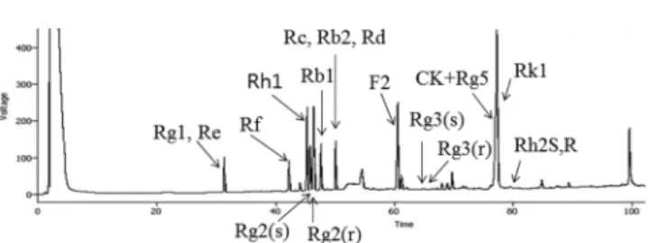
Determination of ginsenosides using HPLC. The levels of major 11 ginsenosides were determined by using a method previously reported (Ryu et al., 2013). A HPLC system as an Agilent 1100 (Agilent Technologies, USA) was equipped with a quaternary solvent delivery system and a column of Imtakt Cadenza CD-C18 (4.6×75 mm; Imtakt Co., Japan). Gradient elution was employed, using 10% acetonitrile and 90% acetonitrile at 35oC. The flow rate was at 1.0 mL/min, and the volume of the injected sample was 2µL. A typical HPLC chromatograme of 18 ginsenoside standards is shown in Fig. 1.
Determination of fatty acids and polyacetylenes using GC- MS. After a 10-day fermentation, the culture medium was filtered through glass wool and centrifuged (6,000×g, 10 min). The supernatant was extracted with ethyl acetate (3×500 mL). The extracts were combined and some metabolites were analyzed on a gas chromatograph-mass spectrometer (GC-MS). GC-MS analyses were performed using a GC-MS-QP 2010 spectrometer (Shimadzu, Japan), equipped with a 30 mm×0.25 mm (i.d.), 0.25µm DM-5 capillary GC column (Dikma Technologies, China). The carrier gas was helium at a rate of 2 mL/min. The column temperature started from 120oC (2 min), increased to 280oC at a rate of 2oC min−1, and was held at 280oC for 10 min. Injector and analyzer temperatures were 270 and 280oC, respectively. The mass spectrometer was operated in EI mode. Initially, detection of the two compounds was performed using the full-scan mode in the range m/z 40–380. Selected ion monitoring (SIM) GC-MS analysis was applied to identify polyacetylenes in ginseng and SIM mode with scanning at 159 and m/z 121 for the detection of panaxynol and panaxydol, respectively, was carried out for the identification of the polyacetylenes.
Cell culture and cell viability assay. HCT-116 and HT-29 cells were kept in McCoy’s 5A medium supplemented with 10% fetal bovine serum and penicillin-streptomycin (50 units/mL). Cells were grown and maintained in a tissue culture dish (100 mm i.d.) and kept in a humidified incubator (5% CO2 in air at 37oC) with a medium change every 2–3 days. Cell viability was measured by a method using 3-[4,5-dimethylthiazol-zyl]-2,5-diphenyltetrazolium bromide (MTT) (Duan et al., 2005). In a reaction mixture MTT at a concentration of 0.5 mg/mL was added to every well and incubated for 4 h, after which the media was removed and replaced with dimethyl sulphoxide (DMSO). Level of optical density was measured by Spectra MAX 190 Reader (MDS Inc., USA) at 570 nm after reduced MTT was dissolved in DMSO for 30 min.
Ethanol/HCl-induced gastric lesion in rats. The gastroprotective
activity of the samples was assessed in the ethanol-HCl induced lesion model as previously described.7 Rats were randomly allotted into groups of eight animals each and fasted for 24 h with free access to water prior to the experiment. Fifty minutes after oral administration of samples and the currently used medicines, all groups were orally treated with 0.2 mL of a solution containing 60% ethanol-0.15 M HCl (ethanol-HCl) for gastric lesion induction.
Animals were sacrificed by an overdose of ether 1 h after the administration of ethanol-HCl, and the stomachs were excised and inflated by injection of saline (1 mL). The ulcerated stomachs were fixed in 5% formalin for 30 min and opened along the greater curvature. Gastric damage visible to the naked eye was observed in the gastric mucosa as elongated black-red lines, parallel to the long axis of the stomach similar to the ethanol-HCl- induced lesions in rats. The length (mm) of each lesion was measured and the lesion index was expressed as the sum of the length of all lesions. The gastritis pictures were obtained at this stage.
Statistical analysis. The results were expressed as the mean (±SEM) of the indicated number of experiments. The statistical significance was estimated using a student’s t-test. Results with a p <0.05 were considered statistically significant.
Results and Discussion
A representative HPLC chromatogram of the 18 standard for ginsenosides is shown in Fig. 1. Changes of ginsenoside constitution in FRGE are disclosed in Table 1 and Fig. 2. The total ginsenoside amounts in FRGE and NFRGE was 26.5 mg/g and 30.0 mg/g, respectively. In addition to the total amount of ginsenosides, the sum of two major constituents Rg1 and Rb1 was different in two samples as 10.9 and 1.66 mg/g in NFRGE and FRGE, respectively.
The level of each metabolite in the two samples was determined and they were significantly different. The protopanaxdiol metabolites F2, Rg3, Rg5, Rk1, Rh2, and CK increased significantly in FRGE when compared to those of NFRGE (Table 1 and Fig. 2). A probable protopanaxtriol metabolite was not determined in FRGE, even NFRGE possessed high concentrations of protopanaxtriols and P. tenuipes decreased their levels during fermentation. It is likely that protopanaxtriol metabolites might be undertaken additional structural changes in the fermentation period. Our results are very similar to the results previously reported (Ryu et al., 2013).
Results obtained by a previous study (Ryu et al., 2013) demonstrated that the ginsenoside composition of the fermented red ginseng extract was significantly different and the protopanaxdiol metabolites increased dramatically. However, they found that the protopanaxtriol metabolites Rh1, Rg2(s), Rg2(r), and Rd increased in the fermentation with P. linteus. The dissimilarity between our results and the results supplied by the scientists (Ryu et al., 2013) has been caused by two factors as fungi difference and fermentation period. The study (Ryu et al., 2013) used P. linteus and 5 days fermentation instead of P. tenuipes and 10 days fermentation. With the analysis of the composition of ginsenoside metabolites in
FRGE, it indirectly proves to increase the bioavailability of FRGE when compared to NFRGE as reported (Ryu et al., 2013).
Additionally, other products in FRGE during fermentation using a GC-MS analysis have been identified as 2-methyl-benzaldehyde, 4-vinyl-2-methoxyphenol, palmitic acid, and linoleic acid as shown in Fig. 3. Two increased fatty acids might be hydrolyzed from triglycerols by a lipase excreted from the fungi.
FRGE has showed its protection activity with 60.5% of inhibition rate at 40 mg/kg dose, while stillen showed its protection activity with 54.5% of inhibition rate at 40 mg/kg dose against gastritis (Table 2). In our findings, FRGE potentially inhibited HCl- ethanol-induced gastric mucosal damage in rats. The inhibitory effect on gastritis was stronger than that of stillen, which is the currently used medicine for treating gastritis. In respect to the result, linoleic acid was also used to treat gastritis in this study because it was one of major biotransformed products by the fermentation. Linoleic acid showed its protection activity with 79.3% of inhibition rate at 40 mg/kg dose. Therefore, linoleic acid is one of the biologically active compounds to suppress gastritis in rats. Figure 4 also shows images of severe gastritis after HCl- ethanol treatment without any drug as a control. However, the currently used medicine, stillen controlled gastritis in rats. When compared to these findings, linoleic acid had a potent protective Fig. 1 Chromatogram of standard ginsenosides by HPLC assay. Gradient elution was employed using 10% acetonitrile and 90% acetonitrile at 35oC.
Table 1 Changes of ginsenosides content in fermented red ginseng extract by Paecilomyces tenuipes
Ginsenoside NFRGE1 (mg/g)
FRGE2 (mg/g)
Ratio of FRGE to NFRGE
Rg1 2.50±0.07 0.21±0.02a) 0.08
Rf tr Tr -
Rh1 2.73±0.13 0.43±0.07a) 0.16
Rb1 8.41±0.35 4.45±0.18a) 0.53
Rc, Rb2, Rd 1.63±0.28 0.72±0.04a) 0.44
F2 1.32±0.14 2.56±0.13a) 1.93
Rg3 2.18±0.31 5.22±0.37a) 2.39
CK+Rg5 3.11±0.12 5.64±0.85a) 1.81
RK1 4.34±0.27 9.85±0.56a) 2.27
Rh2 ND tr -
total 30.0 26.5 0.88
Rg1+Rb1 10.9 1.66 0.15
1NFRGE; Non-fermented red ginseng extract.
2FRGE; Fermented red ginseng extract by Paecilomyces tenuipes.
a)The fermented red ginseng extract shows a significant difference when compared to the non-fermented red ginseng extract (p <0.05).
effect on the gastritis induced by HCl-ethanol treatment (Fig. 4).
Recently, many efforts in Korea have concentrated to discover new gastroprotective drugs from natural products such as araloside A from Aralia elata, scoparone from an ethanol extract from Hericium erinaceus cultivated with Artemisia iwayomogi, and DA-9601 obtained from Artemisia asiatica (Huh et al., 2003; Lee et al., 2005). Occurrence of gastric cancer with high consumption of alcohol, Helicobacter pylori infection, and administration of non-steroidal anti-inflammatory drugs has been recognized as the first leading cause of cancer death in Korea. Therefore, it is important to express that FRGE may be applied to treat or prevent gastritis occurred by high consumption of alcohol.
Anticancer properties of ginsenosides have been well documented and compound K (CK) has been known to induce apoptosis in HT-29 colon cancer cells regulated by CAMK-IV/AMPK (Kang et al., 2011; Kim et al., 2012). Similarly, anticancer activities of ginseng extract fermented with P. linteus have been reported and ginsenosides Rg3, Rh1 and Rh2 are important mediators to
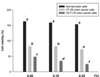
control angiogenesis on A549 cells among five different cell lines as A549, L1210, NIH3T3, B16F10 and Raw 264.7 cells (Lee et al., 2009). In the present study, we used two human colon cancer cells as HT-29 and HCT116 for elucidating anticancer activity of FRGE by P. tenuipes. Anti-proliferation effects of FRGE on two colon cancer cells are shown in Fig. 5. HCT116 colon cancer cells were more susceptible to the FRGE when compared to HT-29 colon cancer cells, while normal colon cells increased until the level of the treated concentrations of FRGE was reached to 0.25%. After 0.25% addition of FRGE, the viability of normal colon cells decreased dramatically as below 50 at 0.50% addition of FRGE. FRGE inhibited HCT116 cell proliferation in a dose- dependence manner as shown in Fig. 5. These results indicate that the biotransformed ginsenosides in FRGE increase normal colon cell growth and inhibit the proliferation of human colon cancer cells. These results are similar to the results previously reported (Seo et al., 2011). The authors indicated that the red ginseng extract possessed the inhibitory activity on the proliferation of SW480 human colon cancer cells induced by phorbol-12 myristate 13-acetate. Therefore, it is important to express that FRGE may be used to treat human colon cancer.
In conclusion, fungal biotransformation on red ginseng extract produced the protopanaxdiol-type metabolites and they referred antiulcerogenic activity on HCl-ethanol induced gastric mucosa damage in rats and anticancer activity on two human colon cancer cells HT-26 and HCT116. Therefore, fungal fermentation by P.
Fig. 4 Pictures of gastritis induced by ethanol-HCl solution in rat stomach. (a), control without any medicine on gastritis; (b), stomachs treated with 40 mg/kg of fermented red ginseng extract; (c), stomachs treated with 40 mg/kg of stillen as positive control.
Fig. 2 A chromatogram of (A) non-fermented red ginseng extract and (B) fermented red ginseng extract fermented by Paecilomyces tenuipes using a HPLC assay. (1) Rg1; (2) Rf; (3) Rh1; (4) unknown; (5) Rb1; (6) Rb2, Rc, Rd; (7) F2; (8) Rg3; (9) CK & Rg5; (10) Rk1; (11) Rh2.
Fig. 3 A GC-MS chromatogram of (A) non-fermented red ginseng extract and (B) fermented red ginseng extract fermented by Paecilomyces tenuipes. (1) maltol, (2) 2-methyl-benzaldehyde, (3) 4-vinyl-2- methoxyphenol, (4) palmitic acid, (5) panaxynol (falcarinol), (6) linoleic acid, (7) panaxydol, (8) a non-matched compound.
Table 2 Gastroprotective activities of fermented red ginseng extract (FRGE) and linoleic acid, one of major biotransformed components by Paecilomyces tenuipes using ethanol/HCl-induced gastric lesion in rats
Group Dose
(mg/kg)
Number of rats
Gastric Lesion rate (Mean±SD, %)
Inhibition rate (%)
Control - 8 41.8±4.7 -
Stillen 40 8 19.0±4.9* 54.5
Linoleic acid 40 8 8.7±3.9*,** 79.3
FRGE 40 8 16.5±5.4* 60.5
1Asterisk (*) are statistically different from the control group at *p
<0.05 and **p <0.01.
tenuipes can contribute to enhance bioavailability to humans and the fermented extracts increase the value of red ginseng extracts for using it as a new resource for medicinal purposes.
Acknowledgment This work was supported by Dong-eui University Grant (Grant No. 2012AA132).
References
Bae SH, Lee HS, Kim MR, Kim SY, Kim JM, and Suh HJ (2011) Changes of ginsenoside content by mushroom mycelial fermentation in red ginseng extract. J Ginseng Res 35, 235–42.
Che F, Chen Y, Kang X, Zhou Z, Zhang Z, and Liu D (2012) Antiapoptotic function and mechanism of ginseng saponins in Rattus pancreatic b-cells.
Biol Pharm Bull 35, 1568–73.
Chen X, Lu J, Zhang Y, He J, Guo X, Tian G et al. (2008) Studies of macropharge immune-modulating activity of polysaccharides isolated from Paecilomyces tenuipes. Int J Biol Macromol 43, 252–6.
Duan W, Yu Y, and Zhang L (2005) Antiatherogenic effects of Phyllanthus emblica associated with corilagin and its analogue. Yakugaku Zasshi 125, 587–91.
Han HC, Lindequist U, Hyun JW, Kim YH, An HS, Lee DH et al. (2004) Apoptosis induction by 4b-acetoxyscirpendiol from Paecilomyces
tenuipes in human leukaemia cell lines. Pharmazie 59, 42–9.
Huh K, Kwon TH, Shin US, Kim WB, Ahn BO, Oh TY et al. (2003) Inhibitory effects of DA-9601 on ethanol-induced gastrohemorrhagic lesions and gastric xanthine oxidase activity. J Ethnopharmacol 88, 269–
73.
Kang JH, Song KH, Woo JK, Park MH, Rhee MH, Choi C et al. (2011) Ginsenoside Rp1 from Panax ginseng exhibits anti-cancer activity by down-regulation of the IGF-1R/AKt pathway in breast cancer cells.
Plant Foods Hum Nutr 66, 298–305.
Kim BG, Shin KS, Yoon TJ, Yu KW, Ra KS, Kim JM et al. (2011) Fermentation of Korean red ginseng by Lactobacillus plantarum M-2 and its immunological activities. Appl Biochem Biotechnol 165, 1107–
19.
Kim DY, Park MW, Yuan HD, Lee HJ, Kim SH, and Chung SH (2012) Compound K induces apoptosis via CAMK-IV/AMPK pathways in HT- 29 colon cancer cells. J Agric Food Chem 57, 10573–8.
Lee EB, Kim OJ, Kang SS, and Jeong C (2005) Araloside A, an antiulcer constituent from the bark of Aralia elata. Biol Pharm Bull 28, 523–6.
Lee H, Park D, and Yoon M (2012a) Korean red ginseng (Panax ginseng) prevents obesity by inhibiting angiogenesis in high fat diet-induced obese C57BL/6J mice. Food Chem Toxicol 53C, 402–8.
Lee JJ, Kwon HK, Jung IH, Cho YB, Kim KJ, and Kim JL (2009) Anti- cancer activities of ginseng extract fermented with Phellinus linteus.
Mycobiology 37, 21–7.
Lee NH, Yoo SR, Kim HG, Cho JH, and Son CG (2012b) Safety and tolerability of Panax ginseng root extract: a randomized, placebo- controlled, clinical trial in healthy Korean volunteers. J Altern Complement Med 18, 1061–9.
Liu Z, Li W, Li X, Chen L, Zheng YN, Sun GZ et al. (2013) Antidiabetic effects of malonyl ginsenosides from Panax ginseng on type 2 diabetic rats induced by high-fat diet and streptozotocin. J Ethnopharmacol 145, 233–40.
Ramesh T, Kim SW, Hwang SY, Sohn SH, Yoo SK, and Kim SK (2012) Panax ginseng reduces oxidative stress and restores antioxidant capacity in aged rats. Nutr Res 32, 718–26.
Ryu JS, Lee HJ, Bae SH, Kim SY, Park Y, Suh HJ et al. (2013) The bioavailability of red ginseng extract fermented by Phellinus lintus. J Ginseng Res 37, 108–16.
Seo EY and Kim WK (2011) Red ginseng extract reduced metastasis of colon cancer cells in vitro and in vivo. J Ginseng Res 35, 315–24.
Xu QF, Fang XL, and Chen DF (2003) Pharmacokinetics and bioavailability of ginsenoside Rb1 and Rg1 from Panax notoginseng in rats. J Ethnopharmacol 84, 187–92.
Yan Q, Zhou XW, Zhou W, Li XW, Feng MQ, and Zhou P (2008) Purification and properties of a novel beta-glucosidase, hydrolyzing ginsenoside Rb1 to CK from Paecilmyces bainier. J Microbiol Biotechnol 18, 1081–9.
Ye L, Zhou CQ, Zhou W, Zhou P, Chen DF, Liu XH et al. (2010) Biotransformation of ginsenoside Rb1 to ginsenoside Rd by highly substrate-tolerant Paecilomyces bainier 229-7. Bioresour Technol 101, 7872–6.
Fig. 5 Anti-proliferation effects of non-fermented red ginseng extract and fermented red ginseng extract on two human colon cancer cells HT-29 and HCT116. Different italics indicate significantly difference between control and fermented red ginseng extract (p <0.05).