• 검색 결과가 없습니다.

후부공막염으로 인한 이차성 폐쇄각에서 레이저홍채절개술 후 발생한 장액망막박리 1례

N/A
N/A
Protected

Academic year: 2021

Share "후부공막염으로 인한 이차성 폐쇄각에서 레이저홍채절개술 후 발생한 장액망막박리 1례"

Copied!
6
0
0

로드 중.... (전체 텍스트 보기)

전체 글

(1)

© Copyright

Keimyung University School of Medicine 2017

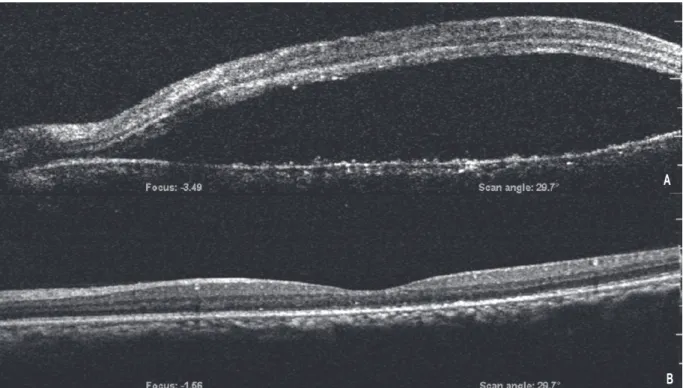
This study was aimed to report a case of serous retinal detachment following laser peripheral iridotomy (LPI) for the treatment of angle closure secondary to posterior scleritis. A 55-year-old man with bilateral ocular pain, redness, and headache was referred to Keimyung University Dongsan Medical Center. At the initial examination, his visual acuity was 1.0 in the both eyes. The intraocular pressure (IOP) was 25 mmHg in the right eye and 28 mmHg in the left eye. Slit lamp examination showed a shallow anterior chamber, which was found to be Shaffer grade I by gonioscopy. There were no specific findings in the fundus, except a slightly edematous disc margin in both eyes. LPI was performed on both eyes. Fourth day after LPI, the patient complained of a central scotoma and visual disturbance of the left eye, in which the visual acuity had decreased to 0.06. The optical coherence tomography showed serous retinal detachment at the posterior pole. Fluorescein angiography revealed a focal leakage in the superotemporal area, as well as multiple hyperfluorescence lesions.

Posterior scleritis of the left eye was diagnosed. Systemic steroid therapy was initiated and the area with the leakage was treated by focal laser photocoagulation. Two weeks later, the serous retinal detachment of the left eye resolved and visual acuity improved to 0.63. Laser peripheral iridotomy can exacerbate serous retinal detachment in patients with posterior scleritis that presented as acute angle closure.

Received: March 27, 2017 Revised: April 26, 2017 Accepted: May 31, 2017

Corresponding Author: Chong Eun Lee, M.D., Department of Ophthalmology, Keimyung University School of Medicine,

56 Dalseong-ro, Jung-gu, Daegu 41931, Korea Tel: +82-53-250-8425

E-mail: [email protected]

• The authors have no conflicts of interest to report with this submission. There was no financial support for this manuscript.

• This article was presented as a poster at the 114th Annual Meeting of the Korean Ophthalmological Society 2015

Department of Ophthalmology, Keimyung University School of Medicine, Daegu, Korea

A Young Choi, M.D., Kwang Soo Kim, M.D., Chong Eun Lee, M.D.

Serous Retinal Detachment Following Laser Peripheral Iridotomy for the Angle Closure Secondary to Posterior Scleritis

계명대학교 의과대학 안과학교실

최아영·김광수·이종은

후부공막염으로 인한 이차성 폐쇄각에서 레이저홍채절개술 후 발생한 장액망막박리 1례
(2)

Keywords: Acute angle closure, Laser peripheral iridotomy, Posterior scleritis, Serous retinal detachment

서 론

폐쇄각녹내장은 많은 경우 상대적 동공차단으로 인한 급성 발작의 형태로 나타나며 이 경우 전신적 또는 점안 약물 치료를 통해 안압을 저하시킨 후 동공차단을 제거하고 앞방각을 넓혀주기 위해 레이저 홍채절개술을 시행하는 것이 일반적인 치료 방법으로 알려져 있다[1]. 그러나 동공차단이 아닌 수정체의 섬모체소대 이완, 맥락막 삼출 등과 같은 이차적 원인에 의해 수정체가 전방으로 이동되어 발생하는 이차성 폐쇄각녹내장의 경우 임상양상과 전방각경 검사만으로는 진단이 어려우며, 동공 폐쇄에 의한 폐 쇄 각 녹 내 장 과 는 그 치 료 법 이 달 라 감 별 이 중요하다[2].

후부공막염은 주로 단안에 드물게 발생하는 만성 염증성 질환으로 이환된 눈의 통증 및 시력저하와 같은 비특이적인 초기 증상과 시신경유두부종, 삼출망막박리, 맥락막주름, 앞공막염 등 다양한 임상증상을 보여 초기진단이 어렵다[3-5]. 또한 맥락막 삼출 및 모양체 부종을 야기하여 이차성 전방각 폐쇄 및 안압상승을 유발할 수 있어 전방각 폐쇄를 첫 증상으로 나타내는 경우 상대적 동공차단에 의한 폐쇄각녹내장과 감별이 어려울 수 있다[6,7].

저자들은 첫 증상으로 좁은 전방각을 보인 후부공막염 환자에서 레이저홍채절개술을 시행한 후 발생한 장액망막박리를 경험하였기에 이를 보고하고자 한다.

증 례

55세 남자가 내원 2주 전부터 지속된 양안 충혈 및 두통을 주소로 내원하였다. 과거력상 고혈압 및 심근경색으로 관상동맥우회술을 시행받은 기왕력이 있었고, amlodipine과 atorvastatin을 복용 중이었다.

안과적 과거력이나 수술력은 없었다. 상기 증상으로

내원 1주일 전 개인 의원 방문하여 시행한 검사상 우안 안압 25 mmHg, 좌안 안압 28 mmHg 측정되어 양안에 brimonidine/timolol fixed combination제제 (Combigan

, Allergan Inc, Irvine, CA, USA)를 점안하고, 경구용 탄산 탈수효소억제제 (acetazolamide)를 처방 받 아 복 용 중 이 었 다 . 본 원 내 원 당 시 양 안 의 최대교정시력은 1.0 이었으나 안압은 우안 23 mmHg, 좌 안 2 3 m m H g 으 로 약 간 증 가 되 어 있 었 다 . 세 극 등 현 미 경 검 사 에 서 양 안 의 심 한 결 막 및 상 공 막 충 혈 이 관 찰 되 었 고 , 전 방 내 염 증 반 응 은 없었으며 중심 전방깊이는 얕아져 있었다. 전방각경 검사에서 양안 전방각이 Shaffer 분류상 grade 1로 좁아진 소견이 관찰되었고, 압박 전방각경검사 시 주변홍채앞유착은 관찰되지 않았다. 안저검사상 시신경유두함몰비는 정상이었고, 양안의 경미한 시 신 경 부 종 소 견 이 관 찰 되 었 으 나 후 극 부 의 특이소견은 관찰되지 않았다.

양안 원발폐쇄각 의심 하에 추가적으로 2% pilocarpine (Isopto carpine eyedrops 2%

, Alcon Laboratories I n c., T e x a s, U S A)을 15분 간격으로 3차례, Brimonidine (Aphagan

, Allergan Inc., Irvine, CA, USA)을 1차례 점안한 다음 우안에 아르곤 레이저와 Nd-YAG 레이저를 이용한 레이저 홍채절개술을 시행하였고, 3일 뒤 같은 방법으로 좌안에 아르곤 레이저(900 mW, 50 µm, 0.01 sec, 70회)와 Nd-YAG 레이저(8회, 4 mJ)를 이용한 레이저 홍채절개술을 시행하였다. 시술 이후 우안 안압은 19 mmHg, 좌안 안압은 19 mmHg로 감소 하였고, 국소 스테로이드 안약 0.1% Fluorometholone, (Ocumetholone

, 삼일제약, 서울, 대한민국)을 하루 4번 3일간 점안하고, brimonidine/timolol fixed combination제제 (Combigan

, Allergan Inc., Irvine, CA, USA)를 제외한 나머지 약제는 중단하였다.

좌안 레이저홍채절개술 4일 후 좌안 중심암점 및 시력

저하를 호소하며 내원하였고 내원 당시 좌안 교정시력은

0.06, 안압은 20 mmHg였다. 세극등현미경검사에서

좌안의 결막 및 상공막충혈이 있었고 경미한 전방

내세포반응이 관찰되었으며 레이져 홍채절개 부위는

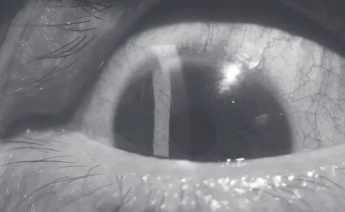
막힘없이 잘 관찰되었다(Fig. 1). 안저검사상 및

빛간섭단층촬영상 후극부에 황반부를 침범하는

(3)

장액망막박리가 넓게 관찰되었고, 형광안저촬영상 좌안 상이측에 국소 형광누출 부위와 함께 후기에 황반부 장액망막박리와 일치하는 부위에 형광 고임 소 견 이 보 였 고 망 막 전 반 에 망 막 색 소 상 피 층 의 변성으로 인한 다발성 과형광이 관찰되었다(Fig. 2).

혈액검사에서는 항핵항체 양성, 류마티스인자가 증가된 소견을 보여 류마티스내과 협진 진료상 류마티스관절염으로 진단되어, 경구 prednisolone 4 mg과 methotrexate 7.5 mg 주 1회 병합투여를 2주간 복용 후 감량하여 유지 하였다.

Fig. 1. Anterior segment photograph of the left eye taken 4 days after laser peripheral iridotomy (LPI). There were mild conjunctival and episcleral injections. A patent LPI site was observed at 11 o’clock position of peripheral iris.

Fig. 2. Fundus photographs. The finding taken 4 days after LPI in the left eye (A) shows serous retinal

detachment at posterior pole. Fluorescein angiograms early phase (B) and late phase (C) of also show diffuse

leakage of fluorescein at the posterior pole, focal leaking point at superotemporal arcade, and multiple focal

hyperfluorescence.

(4)

좌안 후부공막염 진단 하에 경구 스테로이드 치료를 지속하며, 형광안저촬영상 관찰되는 국소 누 출 부 위 에 는 직 접 국 소 레 이 저 광 응 고 술 을 시행하였다. 레이저시술 2주 후 빛간섭단층촬영상 좌안의 망막하액은 대부분 흡수되었고 교정시력은 0 . 6 3 으 로 회 복 되 었 으 며 안 압 은 1 9 m m H g 로 유지되었다(Fig. 3). 시술 1달 후 좌안 교정시력은 0.8로 향상되었고, 빛간섭단층촬영상 장액망막박리는 관찰되지 않았다.

고 찰

후부공막염은 시력저하를 유발할 수 있는 공막의 만 성 염 증 성 질 환 으 로 약 6 0 % 의 환 자 에 서 는 앞 공 막 염 이 동 반 되 나 나 머 지 의 경 우 에 는 후부공막에만 독립적으로 발생하고 장액망막박리, 시신경유두부종, 망막색소상피 의 변화 등 다양한

임상양상을 보여 초기 진단이 어려운 경우가 많다[3].

후부공막염에서 안압이 증가되는 경우는 12-46%로 보고되며[8], 그 기전으로 방수유출로의 염증, 염증세포로 인한 섬유주의 막힘 및 방수의 점도 증가, 주변 홍채 유착, 상공막정맥압의 증가 등의 다양한 원인에 의해 대부분 급성 염증기에 안압이 오르는 양상을 보인다고 보고되었다[9].

또한 공막의 염증 및 비후화는 맥락막 조직액의 공막을 통한 유출을 방해하여 섬모체 뒤쪽으로 축적되면서 맥락막 삼출 및 섬모체 부종을 야기하고, 섬모체를 앞쪽으로 회전시켜 섬모체 소대가 이완되고 수정체를 앞으로 이동시켜 동공차단 없이 이차성 폐쇄각녹내장을 유발할 수 있다[6]. 이러한 수정체-홍채 면의 앞쪽 이동은 근시를 유발할 수가 있으며, Ugurbas 등[7]은 6.0 디옵터의 근시와 폐쇄각녹내장을 보인 후부공막염 사례를 발표하였다.

이러한 후부공막염에 동반된 이차성 폐쇄각녹내장의 경우 임상증상과 전방각경 검사만으로 감별하기 어려우며,

Fig. 3. Optical coherence tomography (OCT) of the left macula taken 4 days after LPI (A) shows hyporeflectivity

due to massive subretinal fluid accumulation; 2 weeks after focal laser photocoagulation (B), subretinal fluid

collection markedly decreased.

(5)

초음파생체현미경이 진단에 효과적이다. 초음파 생체현미경 상에서 공막극 주변의 섬모체맥락막 삼출이 360도 관찰되고 그로 인한 섬모체의 앞쪽 회전 및 수정체-홍채 면의 앞쪽 이동을 보이며, 이러한 섬모체맥락막 삼출은 후부공막염 뿐만 아니라 보그트- 고야나기-하라다 증후군, 주변부 홍채섬모체염, 전안구염 등의 염증성 질환, 안내 종양, 정맥 울혈, 외상, 약물 등에 의해서도 발생할 수 있어 원인 질환의 감별이 중요하다[2,10,11].

본 증례에서는 경미한 전방 염증과 함께 심한 공막 및 결막충혈을 보여 앞공막염이 동반된 것으로 추정되며, 양안 후극부의 다발성 장액망막박리 부위에 형광안저촬영 검사에서 조약돌모양의 형광 고임 소견이 관찰되는 보그트-고야나기-하라다 증후군과는 달 리 단 안 에 하 나 의 광 범 위 한 장 액 망 막 박 리 가 관찰되었으며 보그트-고야나기-하라다 증후군에서 볼 수 있는 피부과적 증상과 뇌막자극증상, 신경과적 증상은 관찰되지 않았다.

후부공막염으로 인한 이차성 급성 폐쇄각의 치료는 일반적인 급성 폐쇄각녹내장의 치료와 달라 감별이 중요하다. 축동제는 안구내 염증을 악화시킬 수 있고, 수정체-홍채 면의 전방 이동, 섬모체 수축으로 인한 전방각 폐쇄를 더욱 악화시킬 수 있어 금기이고 조절 마비제가 증상 완화에 효과적이다[12]. 또 동공차단에 의한 폐쇄각녹내장이 아니므로 레이저홍채절개술은 효과가 적다. 또한 전신적 스테로이드 치료가 염증 조 절 을 조 절 하 여 섬 모 체 맥 락 막 삼 출 물 을 흡수시키는데 필수적이다.

Liu 등[13]은 폐쇄각녹내장의 치료로 아르곤 레이저와 Nd-YAG 레이저를 이용하여 주변부홍채절개술을 시행한 후 중심장액망막병증이 발생한 증례를 보고하였고, 레이져 시술로 인한 포도막의 과도한 염증과 정신적 스트레스로 인한 내인성 부신피질호르몬의 생성 증가가 장액망막박리를 유발시키는 것과 연관이 있다고 보고하였다. 본 증례에서도 폐쇄각녹내장의 첫 증 상 을 보 인 후 부 공 막 염 환 자 에 서 시 행 한 레이저홍채절개술이 오히려 후부 염증을 악화시키고, 그 로 인 한 정 신 적 스 트 레 스 가 장 액 망 막 박 리 가 촉발시킨 요인으로 작용했을 것으로 추정되며, 전 신 적 인 스 테 로 이 드 치 료 로 염 증 이 감 소 되 고

섬모체맥락막 삼출이 흡수되며 전방이 깊어지며 안압이 정상화되었다.

국소 레이저광응고술은 주로 중심장액맥락망막병증 에서 형광안저조영소견에서 관찰되는 누출점을 응고하여 더 이상의 누출을 막고, 망막하삼출액의 빠른 흡수를 통해 병의 경과를 단축시킬 수 있는 방법으로 널리 사용되는 치료법으로[14] 본 증례에서는 후부 공막염에서 발생한 장액망막박리의 국소 누출부위에 국소 레이저광응고술을 시 행 하 여 망 막 하 액 이 흡 수 되 는 효 과 를 보 여 중심소와에서 벗어난 부위에 누출점이 있는 경우 전신 스테로이드 치료에 효과적인 병합 치료법이 될 수 있을 것으로 생각된다.

후 부 공 막 염 은 류 마 티 스 성 관 절 염 , 웨 게 너 육아종증, 전신성 혈관염 등의 다양한 면역관련질환과 매독, 결핵 등의 감염성 질환과 동반되어 나타날 수 있으며, 본 증례에서와 같이 류마티스 관절염이 가장 흔하게 동반 되는 질환이다[15]. 공막염으로 진단받은 환자 중 33%가 류마티스관절염과 연관된 공막염으로 보고되며, 이러한 경우 장기적 환자관리를 위해 류 마 티 스 내 과 의 긴 밀 한 협 진 이 필 수 적 이 며 장기적으로 전신 스테로이드 를 사용해야하는 경우 methotrexate와 같은 면역억제제의 병합투여를 고려할 수 있다.

후부공막염에 의한 이차성 폐쇄각녹내장은 동공 차단에 의해 발생하는 급성 폐쇄각녹내장과 그 치료법에 차이가 있으므로 감별하는 것이 중요하며, 레이저홍채절개술은 장액망막박리를 악화시킬 수 있어 주의가 필요하다. 전신적 스테로이드요법과 함께 시행한 국소 레이저광응고술은 국소 누출부위가 있는 경우 효과적인 치료법이 될 수 있다.

참 고 문 헌

1. Robin AL, Pollack IP. Argon laser peripheral iridotomies in the treatment of primary angle closure glaucoma. Long-term follow-up. Arch Ophthalmol 1982;100:919-23.

2. Ikeda N, Ikeda T, Nomura C, Mimura O. Ciliochoroidal

effusion syndrome associated with posterior scleritis.

(6)

Jpn J Ophthalmol 2007;51:49-52.

3. McCluskey PJ, Watson PG, Lightman S, Haybittle J, Restori M, Branley M. Posterior scleritis: clinical features, systemic associations, and outcome in a large series of patients. Ophthalmology 1999;106:2380-6.

4. Lavric A, Gonzalez-Lopez JJ, Majumder PD, Bansal N, Biswas J, Pavesio C, et al. Posterior scleritis: analysis of epidemiology, clinical factors, and risk of recurrence in a Cohort of 114 Patients. Ocul Immunol Inflamm 2015:1-10.

5. Gonzalez-Gonzalez LA, Molina-Prat N, Doctor P, Tauber J, Sainz de la Maza M, Foster CS. Clinical features and presentation of posterior scleritis: a report of 31 cases. Ocul Immunol Inflamm 2014;22:203-7.

6. Jain SS, Rao P, Kothari K, Bhatt D, Jain S. Posterior scleritis presenting as unilateral secondary angle- closure glaucoma. Indian J Ophthalmol 2004;52:241-4.

7. Ugurbas SH, Alpay A, Ugurbas SC. Posterior scleritis presenting with angle closure glaucoma. Ocul Immunol Inflamm 2012;20:218-20.

8. Watson PG, Hayreh SS. Scleritis and episcleritis. Br J Ophthalmol 1976;60:163-91.

9. Heinz C, Bograd N, Koch J, Heiligenhaus A. Ocular hyper-

tension and glaucoma incidence in patients with scleritis.

Graefes Arch Clin Exp Ophthalmol 2013;251:139-42.

10. Maruyama I, Ohguro H, Nakazawa M. A case of acute angle-closure glaucoma secondary to posterior scleritis in patient with Sturge-Weber syndrome. Jpn J Ophthalmol 2002;46:74-7.

11. Ikeda N, Ikeda T, Nagata M, Mimura O.

Ciliochoroidal effusion syndrome induced by sulfa derivatives. Arch Ophthalmol 2002;120:1775.

12. Poinoosawmy D, Nagasubramanian S, Brown NA.

Effect of pilocarpine on visual acuity and on the dimensions of the cornea and anterior chamber. Br J Ophthalmol 1976;60:676-9.

13. Liu DT, Fan AH, Li CL, Lee VY, Lam PT, Lam DS.

Central serous chorioretinopathy after sequential argon-neodymium: YAG laser iridotomies.

Ophthalmic Surg Lasers Imaging 2009;40:177-9.

14. Burumcek E, Mudun A, Karacorlu S, Arslan MO.

Laser photocoagulation for persistent central serous retinopathy: results of long-term follow-up.

Ophthalmology 1997;104:616-22.

15. Benson WE. Posterior scleritis. Surv Ophthalmol

1988;32:297-316.

수치

Fig. 1. Anterior segment photograph of the left eye taken 4 days after laser peripheral iridotomy (LPI)
Fig. 3. Optical coherence tomography (OCT) of the left macula taken 4 days after LPI (A) shows hyporeflectivity  due to massive subretinal fluid accumulation; 2 weeks after focal laser photocoagulation (B), subretinal fluid  collection markedly decreased.

참조

관련 문서

The purpose of this study was to examine the effects of football on PAPS in sports club activity for 12 weeks in middle school boys.. The following

Purpose: This study was aimed to compare the functional outcomes of the continuous and discontinuous fractures, retrospectively, in multiple thoracolumbar

Purpose: The purpose of this study is to report the safety and effectiveness of arthroscopic-assisted implantation of Cartistem®, an articular cartilage

Results : The expression of p21 was increased in boderline serous tumor and serous cystadenocarcinoma in contrast to benign serous tumors. The expression of

The purpose of this study was to provide primary material in discussing methods to activate the treatment effects of diabetes and reduce the rate of

The purpose of this study was to evaluate the curvature of Vertucci's type II mesial canals of mandibular molar using new method; The radius and angle

The purpose of this study was to investigate the leisure activities of secondary school students with mental retardation during the summer break and

This study aims to suggest a basic data for the activation of aromatherapy pimple treatment program with the pimple skin care and treatment by testing the actual