MG63 Cell Attachments on the Titanium Disks after Micro-Arc Oxidation
Jeong-Won Choi, D.D.S., M.S.D., Seong-Joo Heo, D.D.S., Ph.D.,
Ik-Tae Chang, D.D.S., Ph.D., Jai-Young Koak, D.D.S., Ph.D., Jai-Bong Lee, D.D.S., Ph.D., Soon-Ho Yim * , D.D.S., Ph.D.
Department of Prosthodontics, Graduate School, Seoul National University
* Sungkyunkwan University, School of Medicine
타이타늄 임플랜트의 양극산화 표면처리에 따른 세포부착 특성에 관한 연구
서울대학교 치과대학 보철학 교실, * 성균관대학교 의과대학
최정원 허성주 장익태 곽재영 이재봉 임순호 ․ ․ ․ ․ ․ *
이번 연구의 목적은 순수 타이타늄의 표면을 양극산화법으로 표면처리하여 표면의 특성변화를 연구
하고 이에 따른 세포부착 특성의 차이를 연구하는 것이다 , .
원반 모양의 타이타늄을 전해질용액에서 300V - 550V 의 전압을 주어 양극산화 하고 표면특성을 관찰
한 결과 전압이 높아짐에 따라 표면의 분화구 크기가 커지는 양상을 보였으며 아울러 표면 거칠기도 ,
증가되었다.
세포 부착 실험결과 전압이 증가함에 따라 세포부착 및 증식세포수는 감소하였다.
이상의 양극산화 전압은 표면의 거칠기는 증가시키지만 세포증식은 오히려 억제되는 것이 관찰 300V
되었다.
주용어 ; 세포부착 양극산화 전압 타이타늄 임플랜트 , , ,
Ⅰ . INTRODUCTION
Pure titanium (Ti) and titanium alloys are frequently used as dental and orthopedic implant materials because of their excellent mechanical strength, chemical stability, and biocompatibility 1) . The biocompatibility of titanium is closely related to the properties of the surface oxide layer, in terms of its structure, morphology and composition. Various physical and chemical treatments of the Ti surface have been proposed with a view to obtaining the most biocompatible implant surface. Among the techniques, which have been found to be beneficial to the biological performance of the implants, are increasing the surface roughness, the oxidation of Ti to form a TiO2 layer on the surface, and the incorporation of Ca or P ions into the surface layer, and the validity of these results has been confirmed by several different researchers 2,3) .
Recently, an electrochemical procedure for modifying the Ti surface was proposed, which has since attracted much attention. By applying a positive voltage to a Ti specimen immersed in an electrolyte, anodic oxidation (or anodizing) of Ti occurs to form a TiO 2 layer on the surface. When the applied voltage is increased to a certain point, a micro-arc occurs as a result of the dielectric breakdown of the TiO 2 layer.
At the moment that the dielectric breakdown occurs, Ti ions in the implant and OH ions in the electrolyte move in opposite directions very quickly to form TiO 2
again. This process is generally referred to as micro-arc oxidation (MAO) or plasma electrolysis 4,5) . The newly formed TiO 2 layer is both porous and firmly adhered to the substrate, which is beneficial for the biological performance of the implants. Another advantage of this MAO process is the possibility of incorporating Ca and P ions into the surface layer, by controlling the composition and concentration of the electrolyte 5,6) . The incorporated Ca and P ions were even precipitated into hydroxyapatite crystals by a hydrothermal treatment 7,8) . Recent studies on the biological response of Ti implants demonstrated that the MAO process constitutes one of the best methods of modifying the implant surface 9-11) . However, further research is necessary for the complete characterization of the oxide layer and also for the identification of the optimum conditions for the MAO process.
In vitro cell studies showed that cell proliferation level, and cell morphology and arrangement varied with surface roughness of the discs 12) .
In this study, we formed TiO2 layers with different thicknesses and roughnesses on the Ti surface, by controlling the applied voltage used in the MAO process. The phase, composition and morphology of
MG63 Cell Attachments on the Titanium Disks after Micro-arc Oxidation
Jeong-Won Choi, D.D.S., M.S.D., Seong-Joo Heo, D.D.S., Ph.D.,
Ik-Tae Chang, D.D.S., Ph.D., Jai-Young Koak, D.D.S., Ph.D., Jai-Bong Lee, D.D.S., Ph.D., Soon-Ho Yim * , D.D.S., Ph.D.
Department of Prosthodontics, Graduate School, Seoul National University
* Sungkyunkwan University, School of Medicine
were evaluated by in-vitro tests, in terms of the proliferation and differentiation of certain cell lines
Ⅱ . MATERIALS AND METHODS 1. Micro-arc Oxidation (MAO)
Commercially available pure Ti (CP-Ti, Grade 2, Ka-Hee Metal Industry Co., Seoul, Korea), machined into disks with dimensions of 12 mm (diameter) × 1 mm (thickness), was used as the substrate. These disks were ground using 400-grit SiC sandpaper and cleaned ultrasonically in acetone, ethanol and de-ionized water.
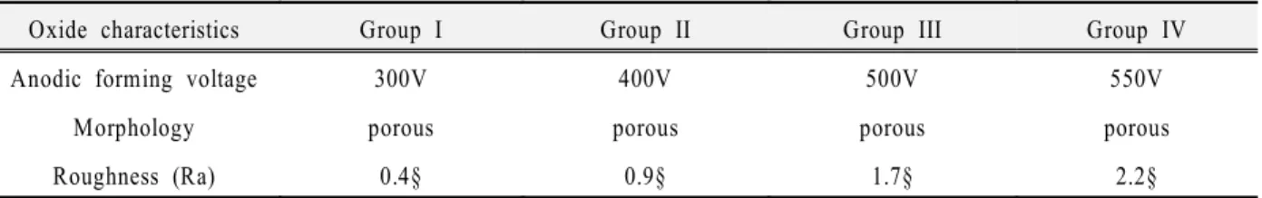
Micro-arc oxidation (MAO) of the specimen was carried out in an aqueous electrolyte, by applying a pulsed DC field to the specimen. The frequency and duty of the pulsed DC power were 660 Hz and 10 %, respectively. The electrolyte was prepared by dissolving 0.15 mol calcium acetate monohydrate {Ca(CH 3 COO) 2 ․ H 2 O} and 0.02 mol calcium glycerophosphate (CaC 3 H 7 O 6 P) in de-ionized water [10]. To obtain oxide layers with different degrees of roughness and thickness, a wide range of DC fields (300-550 V) were applied to the specimens, with each treatment lasting 3 min. All of the MAO processing was carried out in a water-cooled bath made of stainless steel, and a stainless steel plate (100 × 60 × 1 mm) was used as the counter electrode. The titanium disks which were anodized at 6 different voltage were examined.
2. Biological properties
The biological properties of the specimens were evaluated by in-vitro cell tests. For the in-vitro tests, the MG63 and human osteosarcoma (HOS) cell lines were used to characterize the proliferation and differentiation behaviors of the cells, respectively. The pre-incubated cell lines were plated onto specimens with a cell density of 1.5 × 10⁴ cells/cm 2 for the
CO 2 at 37 ℃ . Dulbecco ꡑ s modified Eagle’s medium (DMEM, Life Technologies, Inc., USA) supplemented with 10% fetal bovine serum (FBS, Life Technologies, Inc., USA) was used as the culturing medium.
The proliferation behavior was determined by counting the number of cells after culturing them for 7 days. The cells were detached from the specimens with 0.05% trypsin-EDTA and counted using a hemocytometer (Superior Co., Germany). The differentiation behavior was estimated by measuring the alkaline phosphatase (ALP) activity of the HOS cells after culturing them for 10 days [20]. The cell layers were washed with Hank ꡑ s balanced salt solution (HBSS) and detached using trypsin-EDTA solution. After centrifugation at 1200 rpm for 7 min, the cell pellets were washed once with PBS and resuspended by vortexing them in 200 ㎕ of 0.1 % Triton X-100. The pellets were disrupted by 4 cycles of successive freezing and thawing. After centrifugation, the cell lysates were assayed colorimetrically in order to measure their ALP activity using p-nitrophenyl phosphate as the substrate (Sigma Kit, as described fully in Procedure No. 104). The reaction lasted for 60 min at 37 ℃ , and was then stopped by quenching on ice. The quantity of p-nitrophenol produced was measured at 410 nm using a spectrophotometer (Shimadzu, Japan). The morphology of the proliferated cells was observed by means of SEM after fixation with 2.5 % glutaraldehyde, dehydration with graded ethanols (70, 90 and 100 %), and critical point drying using CO 2 . Each set of tests was performed in triplicate, and the data was normalized by taking the surface area into consideration.
Ⅲ . RESULTS 1. Morphology of oxide layer
Before the oxidation treatment, only the machining
Fig. 2. Number of MG63 cells after proliferation for 7 days. Each set of tests was performed in triplicate and error bars stand for ± 1 standard deviations.
0 200 400 600
0 2 4 6