• 검색 결과가 없습니다.

Mechanisms of tissue factor induction by Porphyromonas gingivalis in human endothelial cells

N/A
N/A
Protected

Academic year: 2021

Share "Mechanisms of tissue factor induction by Porphyromonas gingivalis in human endothelial cells"

Copied!
6
0
0

로드 중.... (전체 텍스트 보기)

전체 글

(1)

Introduction Introduction

Periodontal diseases are among the most widespread in- fectious diseases in humans, and they are characterized by plaque-induced inflammation in the supporting tissues of the teeth [1,2]. A relationship between periodontal diseases and systemic diseases has been increasingly recognized over the past decades. The systemic diseases involved include cardio- vascular disease, gastrointestinal and colorectal cancer, diabe- tes and insulin resistance, and Alzheimer’s disease, as well as respiratory tract infection and adverse pregnancy outcomes [3].

The presence of periodontal pathogens and their metabolic by- products in the mouth may modulate the immune response beyond the oral cavity, thus promoting the development of

systemic conditions [4].

Porphyromonas gingivalis is a well-established pathogen in severe forms of adult periodontal diseases. P. gingivalis is a gram-negative black pigmented anaerobe that colonizes in periodontal pockets and spreads into deeper tissues [5,6]. It has been detected within atheromatous plaques and been shown to induce inflammatory, immune, and procoagulant responses [7,8]. P. gingivalis invades endothelial cells, and it has been proposed that endothelial cell dysfunction is an early manifestation of atherosclerotic vascular disease [9]. There is evidence that P. gingivalis is able to induce and maintain a chronic state of inflammation at distant sites, including athero- matous plaques, and this species has been shown to acceler- ate atherosclerosis in animal models [10,11].

Int J Oral Biol 46:99-104, 2021 pISSN: 1226-7155 • eISSN: 2287-6618 https://doi.org/10.11620/IJOB.2021.46.3.99

Mechanisms of tissue factor induction by

Porphyromonas gingivalis in human endothelial cells

So-Hee Kim, Ji-Yeon Jung, Won-Jae Kim, Ok-Joon Kim, Young Kim, and In-Chol Kang*

Dental Science Research Institute, Chonnam National University, Gwangju 61186, Republic of Korea

Associations between periodontal infection and cardiovascular disease have been documented. Porphyromonas gingivalis is a well-established periodontal pathogen, and tissue factor (TF) is a key initiator of the coagulation cascade. In this context, P. gingivalis has been reported to enhance TF expression in human endothelial cells.

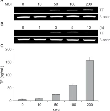
The present study investigated the underlying mechanisms of TF induction by P. gingivalis in human umbilical vein endothelial cells. P. gingivalis increased TF expression in a dose- and time-dependent manner. Not only live bacteria but also glutaraldehyde-fixed bacteria increased TF expression to the same extent. However, sonicates of P. gingivalis did not induce TF expression. Cytochalasin D and SMIFH2, which are inhibitors of actin polymerization and actin nucleation, respectively, inhibited the TF expression induced by P. gingivalis. Finally, TF production was decreased or increased in the presence of various signaling inhibitors, including mitogen-activated protein kinases.

These results suggest that P. gingivalis induces endothelial TF expression by a bacterial internalization-dependent mechanism and through diverse signal transduction mechanisms.

Keywords: Porphyromonas gingivalis, Tissue factor, Endothelial cells

Received June 26, 2021; Revised August 19, 2021; Accepted August 24, 2021

*Correspondence to: In-Chol Kang, E-mail: ickang@jnu.ac.kr https://orcid.org/0000-0002-5993-8728 Copyright © The Korean Academy of Oral Biology

CC

This is an open-access article distributed under the terms of the Creative Commons Attribution Non-Commercial License (http://creativecommons.org/licenses/by- nc/4.0/), which permits unrestricted non-commercial use, distribution, and reproduction in any medium, provided the original work is properly cited.

Original Article IJOB

(2)

Tissue factor (TF) is a key initiator of the coagulation cascade.

It is a 47-kD membrane-bound glycoprotein expressed in both vascular and nonvascular cells. TF is constitutively expressed in subendothelial cells such as vascular smooth muscle cells leading to rapid initiation of coagulation when the vessel is damaged. Endothelial cells do not express TF under physi- ological conditions. In response to various stimuli, however, TF expression and activity can be induced in endothelial cells. TF binds factor VIIa resulting in activation of factor IX and factor X, leading to fibrin formation [12-14].

Previous studies demonstrated that P. gingivalis induces procoagulant effects in human aortic endothelial cells and smooth muscle cells [15,16]. They revealed that P. gingivalis enhanced TF expression in endothelial cells, but not in vascular smooth muscle cells. However, they did not report regarding the mechanisms of TF induction by P. gingivalis. The purpose of the present study was to investigate the mechanisms re- sponsible for TF induction in endothelial cells.

Materials and Methods Materials and Methods

1. Reagents

PD98059, SB203580, SP600125, GF109203X, and U73122 were purchased from Calbiochem (San Diego, CA, USA).

Wortmannin, genistein, SC-514, cytochalasin D (CD), SMIFH2 were purchased from Sigma (St. Louis, MO, USA).

2. Bacterial culture

P. gingivalis 381 was grown in Trypticase soy broth supple- mented with yeast extract (1 mg/mL), hemin (5 μg/mL), and menadione (1 μg/mL). The bacteria were incubated anaerobi- cally (85% N

2

, 10% H

2

, and 5% CO

2

) at 37℃. For experiments, bacterial concentrations were determined by measuring optical density at 600 nm using a spectrophotometer.

3. Cell culture

Human umbilical vein endothelial cells (HUVEC) were pur- chased from Cascade Biologics (Portland, OR, USA) and cul- tured in Medium 200 supplemented with 10% fetal bovine serum and 50 μg/mL gentamicin at 37℃ in 5% CO

2

. For glu- taraldehyde (GA) fixation, P. gingivalis was washed twice in phosphate-buffered saline (PBS) and then incubated with 2.5%

GA in PBS for 2 hours at room temperature. After fixation, the

bacteria were washed twice in PBS and then resuspended in a small volume of PBS. For sonication, P. gingivalis was washed twice in PBS and the bacterial cells were disrupted in iced bath using a Vibra-Cell VCX-600 ultrasonic processor (Sonics

& Materials, Newtown, CT, USA) with 13 mm diameter horn.

Amplitude of the ultrasonic device is maintained at 20% for 20 minutes.

4. Reverse transcription-polymerase chain reaction (RT-PCR)

Total RNA was prepared with Trizol reagent (Invitrogen, Carlsbad, CA, USA) as specified by the manufacturer and was quantified spectrophotometrically. First-strand cDNA was synthesized from 1 μg of RNA using random primers (Pro- mega, Madison, WI, USA) and Molony murine leukemia virus reverse transcriptase (Promega). Two μL of cDNA products were amplified in 25 μL volumes under a layer of mineral oil using a GeneAmp 2700 thermal cycler (Applied Biosystems, Foster City, CA, USA). Each PCR reaction mixture contained 50 mM KCl, 10 mM Tris-HCl, 1.5 mM MgCl

2

, 0.2 μM each dNTP, 1 U Taq DNA polymerase, and 0.5 μM of each primer.

Each cycle consisted of denaturation at 94℃ (30 s), annealing at 57℃ (30 s), and extension at 72℃ (60 s). The sequences of primers were 5′-TGAAGGATGTGAAGCAGACG-3′, 5′-GC- CAGGATGATGACAAGGAT-3′ for TF (525 bp); 5′-CAGCCA- GATGCAATCAATGC-3′, 5′-GTGGTCCATGGAATCCTGAA-3′

for monocyte chemoattractant protein (MCP-1) (198 bp); and 5′-AGCGGGAAATCGTGCGTG-3′, 5′-CAGGGTACATGGTGGT- GCC-3′ for β-actin (300 bp). The PCR products of 10 μL were fractionated on 1.2% (w/v) agarose gels containing RedSafe (iNtRON Biotechnology, Seongnam, Korea), visualized by UV transillumination, and photographed.

5. Enzyme-linked immunosorbent assay (ELISA)

After the cell culture supernatant was removed, cells were lysed by Cell Lysis Butter 1 (R&D Systems, Minneapolis, MN, USA). After cell debris was removed by centrifugation, the levels of TF were quantified using commercial ELISA kits (R&D Systems) according to the manufacturer’s directions.

6. Statistical analysis

Our experiments were conducted in three independent ex-

periments to confirm the reproducibility of the results. The data

(3)

are presented as means with standard deviations. Statistical analysis of one-way analysis of variance with Tukey–Kramer multiple comparisons test was performed using GraphPad In- Stat (GraphPad Software, La Jolla, CA, USA). A p-value < 0.05 was considered statistically significant.

Results Results

1. Dose- and time-dependent TF induction by P. gingivalis

We first determined the dose-dependency of P. gingivalis- stimulated TF expression. Various doses of P. gingivalis were added to HUVEC cultures and the TF expression was deter- mined by RT-PCR. TF expression was increased dose-de- pendently by P. gingivalis. The induction of TF expression was evident from the multiplicity of infection (MOI) of 50 (Fig. 1A).

Time course experiments showed that the mRNA expression was induced around 3 hours and maintained around 5 hours after P. gingivalis stimulation. The TF mRNA level declined nearly to the resting level around 10 hours (Fig. 1B). Levels of TF protein in lysed cell samples was determined by ELISA.

The ELISA results showed that P. gingivalis dose-dependently increased the production of TF protein by HUVEC (Fig. 1C).

These results demonstrated that P. gingivalis stimulates HU- VEC to express TF in dose- and time-dependent manners.

2. Comparison between live, killed, and sonicated P. gingivalis

Next, we determined if there is difference between live and killed P. gingivalis in stimulating HUVEC to induce TF expres- sion. In parallel, P. gingivalis sonicates were also compared.

HUVEC cultures were incubated with the same number of live, GA-fixed, or sonicated P. gingivalis (MOI = 1:200) and the TF expression was measured by RT-PCR and ELISA. Not only live, but also killed P. gingivalis strongly stimulated HUVEC to express TF. However, P. gingivalis sonicates did not increase the TF expression at all (Fig. 2).

0 200

150

100

50

TF (pg/mL)

MOI 0

10 50 100 200

TF

-actin

TF

-actin

MOI 0 10 50 100 200

0 1 3 5 10 (h)

A

B

C

Fig. 1. Dose- and time-dependent tissue factor (TF) expression by Por- phyromonas gingivalis. (A) Human umbilical vein endothelial cells (HUVEC, 2 × 10

4

in 12-well plates) were infected with increasing multiplicity of infec- tions (MOIs) of P. gingivalis for 5 hours. (B) HUVEC were infected with P. gingivalis (MOI = 1:200) for various times. Total RNA was isolated and levels of TF mRNA were determined by reverse transcription-polymerase chain reaction. Shown are photographs of agarose gels from one experi- ment, representative of three. (C) HUVEC were infected with increasing MOIs of P. gingivalis for 18 hours. Levels of TF protein in cell lysates were measured by ELISA. Data are the mean ± standard deviation of a repre- sentative experiment performed in triplicate. Similar results were obtained in two other experiments.

None 150

100

TF (pg/mL) 50

0

Live GA-fixed Sonicates TF

-actin

A

B

None Live GA-fixed Sonicates

* *

Fig. 2. Comparison between live, killed, and sonicated Porphyromonas gingivalis in their ability to induce tissue factor (TF) expression. (A) Human umbilical vein endothelial cells (HUVEC) cultures were incubated with the same amount (multiplicity of infection [MOI] = 1:200) of live, glutaraldehyde (GA)-fixed, or sonicated P. gingivalis for 5 hours and the TF expression was measured by reverse transcription-polymerase chain reaction. Shown are a photograph of agarose gels from one experiment, representative of three. (B) HUVEC were incubated with live, killed, sonicated P. gingivalis for 18 hours. Levels of TF protein in cell lysates were measured by ELISA.

Data are the mean ± standard deviation of a representative experiment performed in triplicate. The asterisks indicate significant differences (p <

0.05). Similar results were obtained in two other experiments.

(4)

3. Effects of CD and SMIFH2 on the induction of TF and MCP-1 by P. gingivalis

As bacterial internalization has been reported to play im- portant roles in various infections [17-19], we examined the effects of inhibitors of actin polymerization on TF induction by P. gingivalis. CD prevents actin polymerization and SMIFH2 in- hibits actin nucleation. HUVEC cultures were incubated with P.

gingivalis in the presence or absence of the inhibitors and the mRNA levels of TF were examined by RT-PCR. We observed the mRNA expression of MCP-1 together because MCP-1 in- duction by P. gingivalis is dependent on bacterial internalization [18]. As shown in Fig. 3, both inhibitors inhibited P. gingivalis- stimulated TF expression as well as MCP-1 expression.

4. Effects of various signaling inhibitors on the production of TF by P. gingivalis

In order to evaluate the relative importance of various signal- ing pathways in P. gingivalis-induced TF production in HUVEC, specific pharmacological inhibitors were used. HUVEC cultures were pretreated with GF109203X (protein kinase C), wortman- nin (phosphatidylinositol 3-kinase), U73122 (phospholipase C), genistein (protein tyrosine kinase), PD98059 (extracel- lular signal-regulated kinase, ERK), SB203580 (p38 mitogen- activated protein kinase), SP600125 (c-Jun N-terminal ki- nase), or SC-514 (nuclear factor kappa B, NF-κB) for 1 hour,

and then the cells were incubated with P. gingivalis for 18 hours. TF concentrations of the cell lysates were measured by ELISA. Among the inhibitors, SB203580, PD98059, genistein, GF109203X, and SC-514 inhibited TF production stimulated by P. gingivalis. In contrast, TF production was significantly el- evated in the presence of U73122 and wortmannin (Fig. 4).

Discussion Discussion

In the present study, we investigated the mechanisms of TF induction by P. gingivalis in endothelial cells. Increased TF ex- pression was observed at the levels of both mRNA and protein.

MOI as low as 1:50 evidently stimulated HUVEC to express TF and higher MOIs resulted in higher levels of TF expression.

Time-course monitoring of TF mRNA expression showed that the mRNA level peaked at 3–5 hours after P. gingivalis stimu- lation and declined thereafter.

We prepared killed P. gingivalis by GA fixation. GA has been widely used to kill and fix bacteria [20]. There was no differ- ence between live and GA-killed P. gingivalis in the ability to stimulate HUVEC to express TF. This means that activities of viable P. gingivalis like enzyme secretion are not required for the TF induction. However, sonicated P. gingivalis could not induce the TF expression. Previous studies also reported that sonicated bacteria were less stimulatory for cytokine induction A None CD (0.5) CD (1) - CD (0.5) CD (1)

P. gingivalis

B None

TF

-actin MCP-1

TF

-actin MCP-1 SMI (5) SMI (10) - SMI (5) SMI (10)

P. gingivalis

Fig. 3. Effect of cytochalasin D (CD) (A) and SMIFH2 (SMI) (B) on the in- duction of tissue factor (TF) and monocyte chemoattractant protein (MCP-1) by Porphyromonas gingivalis. Human umbilical vein endothelial cells were pretreated with CD (0.5–1 μg/mL) or SMIFH2 (5–10 μM) and then infected with P. gingivalis (multiplicity of infection [MOI] = 1:200) for 3 hours. Total RNA was isolated and levels of TF mRNA were determined by reverse transcription-polymerase chain reaction. Shown are photographs of aga- rose gels from one experiment, representative of three.

None 350

300

250

200

150

100

50

TF (pg/mL)

0

Pg Geni GF PD SB SP U73 Wort SC

*

* *

*

*

*

*

Fig. 4. Effects of various signaling inhibitors on the production of tissue factor (TF) by Porphyromonas gingivalis. Human umbilical vein endothelial cells were pretreated for 1 hour with GF109203X (GF, 1 μM), wortmannin (Wort, 100 nM), U73122 (U73, 10 μM), genistein (Geni, 50 μM), PD98059 (PD, 50 μM), SB203580 (SB, 10 μM), SP600125 (SP, 10 μM), or SC-514 (SC, 30 μM) and then infected with P. gingivalis (Pg, multiplicity of infection [MOI] = 1:200) for 18 hours. Levels of TF protein in the cell lysates were measured by ELISA. Data are the mean ± standard deviation of a repre- sentative experiment performed in triplicate. The asterisks indicate signifi- cant differences compared to P. gingivalis stimulation without inhibitors (p

< 0.05).

(5)

than intact bacteria [21,22].

Bacterial internalization has been reported to play important roles in stimulating host cells. Also, P. gingivalis internaliza- tion into endothelial cells has been proposed as an important mechanism of pathogenesis in periodontal and cardiovascular diseases [23,24]. As actin polymerization is absolute require- ment for bacterial internalization, we used CD, an inhibitor of actin polymerization, to block bacterial internalization. We also examined the effect of SMIFH2, a formin inhibitor on the P.

gingivalis-stimulated TF expression. Formin proteins are actin nucleators that bind monomeric actin and polymerizes it into filamentous actin. SMIFH2 is a cell-permeable compound that inhibits formin-dependent actin polymerization [25]. Our re- sults showed that CD treatment prevented the TF induction.

SMIFH2 treatment resulted in decreased expression of TF.

These results suggest that P. gingivalis induce TF expression through a bacterial internalization-dependent mechanism.

Finally, we evaluated the relative importance of various sig- naling pathways in P. gingivalis-induced TF expression in HU- VEC. Pharmacological inhibition of protein kinase C, p38, ERK,

protein tyrosine kinases, NF- κB inhibited the TF production.

In contrast, the TF production was elevated with inhibition of phospholipase C and phosphatidylinositol 3-kinase. It is yet to be determined if the signaling pathways modulate the TF expression at the transcriptional level or posttranscriptional level. The signaling pathways may also indirectly involve the TF induction by affecting the P. gingivalis internalization. Further studies are needed to obtain more detailed information about involvement of each signaling pathway in the P. gingivalis- induced TF expression in endothelial cells.

Acknowledgements Acknowledgements

This study was financially supported by Chonnam National University (Grant number: 2018-3478).

Conflicts of Interest Conflicts of Interest

No potential conflict of interest relevant to this article was reported.

References References

1. Kinane DF, Stathopoulou PG, Papapanou PN. Periodontal dis- eases. Nat Rev Dis Primers 2017;3:17038. doi: 10.1038/nrdp.

2017.38.

2. K önönen E, Gursoy M, Gursoy UK. Periodontitis: a multifac- eted disease of tooth-supporting tissues. J Clin Med 2019;8:

1135. doi: 10.3390/jcm8081135.

3. Bui FQ, Almeida-da-Silva CLC, Huynh B, Trinh A, Liu J, Woodward J, Asadi H, Ojcius DM. Association between peri- odontal pathogens and systemic disease. Biomed J 2019;42:

27-35. doi: 10.1016/j.bj.2018.12.001.

4. Hasturk H, Kantarci A. Activation and resolution of periodontal inflammation and its systemic impact. Periodontol 2000 2015;

69:255-73. doi: 10.1111/prd.12105.

5. Mulhall H, Huck O, Amar S. Porphyromonas gingivalis, a long- range pathogen: systemic impact and therapeutic implica- tions. Microorganisms 2020;8:869. doi: 10.3390/microorgan- isms8060869.

6. Xu W, Zhou W, Wang H, Liang S. Roles of Porphyromonas gingivalis and its virulence factors in periodontitis. Adv Protein Chem Struct Biol 2020;120:45-84. doi: 10.1016/bs.apcsb.

2019.12.001.

7. Haraszthy VI, Zambon JJ, Trevisan M, Zeid M, Genco RJ.

Identification of periodontal pathogens in atheromatous

plaques. J Periodontol 2000;71:1554-60. doi: 10.1902/jop.

2000.71.10.1554.

8. Kozarov EV, Dorn BR, Shelburne CE, Dunn WA Jr, Progulske- Fox A. Human atherosclerotic plaque contains viable invasive Actinobacillus actinomycetemcomitans and Porphyromonas gingivalis. Arterioscler Thromb Vasc Biol 2005;25:e17-8. doi:

10.1161/01.ATV.0000155018.67835.1a.

9. Olsen I, Progulske-Fox A. Invasion of Porphyromonas gin- givalis strains into vascular cells and tissue. J Oral Microbiol 2015;7:28788. doi: 10.3402/jom.v7.28788.

10. Lalla E, Lamster IB, Hofmann MA, Bucciarelli L, Jerud AP, Tucker S, Lu Y, Papapanou PN, Schmidt AM. Oral infection with a periodontal pathogen accelerates early atherosclerosis in apolipoprotein E-null mice. Arterioscler Thromb Vasc Biol 2003;23:1405-11. doi: 10.1161/01.ATV.0000082462.26258.FE.

11. Pussinen PJ, Mattila K. Periodontal infections and atheroscle- rosis: mere associations? Curr Opin Lipidol 2004;15:583-8.

doi: 10.1097/00041433-200410000-00013.

12. Grover SP, Mackman N. Tissue factor: an essential mediator of hemostasis and trigger of thrombosis. Arterioscler Thromb Vasc Biol 2018;38:709-25. doi: 10.1161/ATVBAHA.117.

309846.

13. Steffel J, L üscher TF, Tanner FC. Tissue factor in cardiovas-

(6)

cular diseases: molecular mechanisms and clinical implica- tions. Circulation 2006;113:722-31. doi: 10.1161/CIRCULA- TIONAHA.105.567297.

14. Witkowski M, Landmesser U, Rauch U. Tissue factor as a link between inflammation and coagulation. Trends Cardiovasc Med 2016;26:297-303. doi: 10.1016/j.tcm.2015.12.001.

15. Roth GA, Aumayr K, Giacona MB, Papapanou PN, Schmidt AM, Lalla E. Porphyromonas gingivalis infection and prothrom- botic effects in human aortic smooth muscle cells. Thromb Res 2009;123:780-4. doi: 10.1016/j.thromres.2008.07.008.

16. Roth GA, Moser B, Huang SJ, Brandt JS, Huang Y, Papapanou PN, Schmidt AM, Lalla E. Infection with a periodontal patho- gen induces procoagulant effects in human aortic endothelial cells. J Thromb Haemost 2006;4:2256-61. doi: 10.1111/j.

1538-7836.2006.02128.x.

17. Gavrilin MA, Bouakl IJ, Knatz NL, Duncan MD, Hall MW, Gunn JS, Wewers MD. Internalization and phagosome escape re- quired for Francisella to induce human monocyte IL-1beta processing and release. Proc Natl Acad Sci U S A 2006;103:

141-6. doi: 10.1073/pnas.0504271103.

18. Kang IC, Kuramitsu HK. Induction of monocyte chemoattrac- tant protein-1 by Porphyromonas gingivalis in human endo- thelial cells. FEMS Immunol Med Microbiol 2002;34:311-7.

doi: 10.1111/j.1574-695X.2002.tb00639.x.

19. Shin OS, Isberg RR, Akira S, Uematsu S, Behera AK, Hu LT.

Distinct roles for MyD88 and Toll-like receptors 2, 5, and 9 in phagocytosis of Borrelia burgdorferi and cytokine induction.

Infect Immun 2008;76:2341-51. doi: 10.1128/IAI.01600-07.

20. Markowicz C, Kubiak P, Grajek W, Schmidt MT. Inactivation of Lactobacillus rhamnosus GG by fixation modifies its probiotic properties. Can J Microbiol 2016;62:72-82. doi: 10.1139/cjm- 2015-0249.

21. Barkman C, Martner A, Hessle C, Wold AE. Soluble bacterial constituents down-regulate secretion of IL-12 in response to intact Gram-positive bacteria. Microbes Infect 2008;10:1484- 93. doi: 10.1016/j.micinf.2008.08.011.

22. Jagger MP, Huo Z, Riches PG. Inflammatory cytokine (inter- leukin 6 and tumour necrosis factor alpha) release in a human whole blood system in response to Streptococcus pneu- moniae serotype 14 and its capsular polysaccharide. Clin Exp Immunol 2002;130:467-74.

23. Amar S, Wu SC, Madan M. Is Porphyromonas gingivalis cell invasion required for atherogenesis? Pharmacotherapeutic implications. J Immunol 2009;182:1584-92. doi: 10.4049/

jimmunol.182.3.1584.

24. Choi EK, Park SA, Oh WM, Kang HC, Kuramitsu HK, Kim BG, Kang IC. Mechanisms of Porphyromonas gingivalis-induced monocyte chemoattractant protein-1 expression in endothe- lial cells. FEMS Immunol Med Microbiol 2005;44:51-8. doi:

10.1016/j.femsim.2004.12.003.

25. Rengarajan M, Hayer A, Theriot JA. Endothelial cells use a formin-dependent phagocytosis-like process to internalize the bacterium Listeria monocytogenes. PLoS Pathog 2016;12:

e1005603. doi: 10.1371/journal.ppat.1005603.

수치

Fig. 1. Dose- and time-dependent tissue factor (TF) expression by Por- Por-phyromonas gingivalis
Fig. 4. Effects of various signaling inhibitors on the production of tissue  factor (TF) by Porphyromonas gingivalis

참조

관련 문서

Caffeic acid phenethyl ester induces E2F-1-mediated growth inhibition and cell-cycle arrest in human cervical cancer cells.

Primary hepatocytes were incubated with T090 for 24 h following Res pretreatment (1 h). Cells were fixed and stained with Oil red O and observed under a microscope. The Oil

Objective :::: The neovascularization is an essential a factor for the growth of solid organ cancer, and especially vascular endothelial growth factor (VEGF)

Determining optimal surface roughness of TiO₂blasted titanium implant material for attachment, proliferation and differentiation of cells derived from

Control- and ASTRIN- depleted HeLa cells were untreated or treated with 5 Gy. IR and were then fixed at

 Often found connected to other molecules on the outsides of cells --- cellular recognition, cell signaling, cell

In this study, platelet-derived growth factors-BB (PDGF-BB), vascular endothelial growth factor (VEGF), and insulin-like growth factor-1 (IGF-1) were quantified in the PRF

• Generation of different specialized kinds of cells from zygote (fertilized egg) or other precursor cells.. – Generate blood cells, muscle