Microarray Analysis of Gene Expression by Rhei Rhizoma Water Extracts in a Hypoxia Model of Cultured Neurons
HyunSook Lee, Jinyoung Song and Il Soo Moon*
Department of Anatomy, College of Medicine, Dongguk University, Gyeongju 780-714, Korea Received October 21, 2008 /Accepted December 1, 2008
In this study, we investigated the effect of Rhei Rhizoma (RR; 大黃) water extract on gene expression in a hypoxia model of cultured rat hippocampal neurons. RR water extract (2.5 μg/ml) was added to the culture media on day 10 in vitro (DIV10), and a hypoxic shock (2% O2/5% CO2 , 37oC, 3 h) was given on DIV13. After maintaining the cultures in normoxia for 24 hr, total RNA was isolated and used for microarray analysis. The MA-plot indicated that most genes were up- or downregulated within 2-fold. There were more downregulated genes (725 ea) than upregulated ones (472 ea) when larger than Global M value 0.2 (i.e., >15% increase) or smaller than Global M value -0.2 (i.e., >15%
decrease) were considered. Antiapoptosis genes such as Tegt (2.4-fold), Nfkb1 (2.4-fold) Veg (1.8-fold), Ngfr (1.6-fold) were upregulated, while pro-apoptosis genes such as Bad (-64%), Cstb (-66%) were downregulated. Genes for combating environmental stress (stress response genes) such as Defb3 (2.7-fold), Cygb (2.2-fold), Ahsg (2.18-fold), Alox5 (2-fold) were upregulated. Genes for cell pro- liferation (cell cycle-related genes) such as Erbb2 (1.84-fold), Mapk12 gene (1.8-fold) was upregulated.
Therefore, RR water extracts upregulate many pro-survival genes while downregulating many pro-death genes. It is interpreted that these genes, in combination with other regulated genes, can pro- mote neuronal survival in a stress such as hypoxia.
Key words : Cell culture, hippocampal neuron, hypoxia, microarray, Rhei Rhizoma
*Corresponding author
*Tel:+82-54-770-2414, Fax:+82-54-770-2434
*E-mail : [email protected]
Introduction
Rhei Rhizoma (RR; 大黃), a widely used folk medicine in Southeast Asian, consists of the underground parts (rhizome and root) ofRheum officinale Baill. and Rheum pal- matum L. (Polygonaceae). Major constituents of RR are an- thranoids, rheinosides, rhein, and stilben such as rhaponti- cin and rhapontigenin [48]. Beneficiary effects of RR on hu- man health have been reported. For example, RR extracts lower serum cholesterol and improve diabetic nephropathy [40,46]. Effects of RR on lowering serum cholesterol are at- tributed to rhein, which are transformed from anthranoids sennoside A and B by bacterial enzymes in large intestine [55]. Rhaponticin in the rhizome of RR has extensive an- ti-allergic and anti-thrombotic properties [46]. RR extracts decreased serum creatinine levels in diabetic patients with neuropathy and retinopathy [18].
Studies on the nervous system are relatively scarce. It has been reported that RR improves memory ability. By comparing the effects of the Compound Tong Jiang Oral
Liquid with Da Huang added (TJ) and Qi Yin Oral Liquid (QY) without Da Huang on senile persons' memory ability, Tian et al. [55] discovered that TJ improves senile persons' memory ability, in addition to shortened interval and du- ration of defecation. However, effects of RR on the nervous system are not studied at the cellular level. Previously, our laboratory reported that RR suppresses production of re- active oxygen species (ROS) and loss of mitochondrial membrane potential (MMP) in a hypoxia model of rat hip- pocampal neurons in culture [34]. Here, we report results of a microarray analysis on the expression of genes in hypoxia.
Materials and Methods Preparation of water-extracts of RR
Rhei Rhizoma (RR; 大黃) was selected according to the Korean Pharmacopoeia and obtained from Dongguk University Oriental Hospital (Gyeongju, Korea). Distilled water was added to the RR powder and it was agitated for 4 hr at room temperature (RT) followed by overnight at 4oC.
After centrifugation (15,000 rpm, 15 min, RT), the super- natant was filter-sterilized (pore size 0.2 μm) and stored at -20oC in small aliquots.
Neuronal culture
Cortices from time-pregnant rats (Sprague-Dawley) at em- bryonic day 18 (E18) were dissected, dissociated by trypsin treatment and mechanical trituration, and plated onto poly- lysine-coated 60 mm dishes at a density of ~500 cells/mm2 as described [2,17]. Cells were plated initially in Neurobasal medium supplemented with B27 (Invitrogen, Carlsbad, CA, USA), 25 μM glutamate, and 500 μM glutamine, and fed 5 days after plating and weekly thereafter with the same me- dia (without glutamate).
Hypoxia
Hypoxic shock was given to cells by transferring culture plates to a humidified CO2Water Jacketed Incubator (Forma Scientific Inc., Maretta, OH, USA) which was equilibrated at 2% O2/5% CO2(37oC). After 3 hr incubation, plates were returned to normoxic incubator (5% CO2, 37oC).
Total RNA preparation
On day 10in vitro (DIV10) RR water extract (2.5 μg/ml) was added directly into culture media, given a hypoxic shock on DIV13, and further incubated for 24 hr in normoxia. The culture was briefly washed with 5~10 ml of ice-cold PBS, and cells were lysed by adding 1.0 ml of solution D [4.0 M guanidinium thiocyanate, 25 mM sodium citrate-2H2O, 0.5% (w/v) sodium lauryl sarcosinate, 0.1 M β-mercaptoethanol]. Cell lysates were transferred to a micro- fuge tube and homogenized using a tissue homogenizer for 15~30 sec. Per 1.0 ml of the homogenate, 0.1 ml of 2.0 M sodium acetate (pH 4.0), 1.0 ml of phenol (4oC), 0.2 ml of chloroform-isoamyl alcohol was added, mixed well, and in- cubated on ice for 15 min. After centrifugation (10,000× g, 20 min, 4oC), supernatants were mixed with same volumes of isopropanol. After incubation for 2 hr at -20oC, RNA was precipitated by centrifugation (10,000× g, 30 min, 4oC) and dissolved in solution D. RNA was precipitated once more by isopropanol and washed twice with 75% alcohol, dis- solved in diethyl pyrocarbonate-treated water, and stored at -70oC.
Microarray
Microarray analysis was performed on Rat 44K 4-Plex Gene Expression platforms (Agilent) in Digital Genomics (Seoul, Korea) using total RNAs prepared through 9 in- dependent experiments.
Indirect labeling of probes using aminoallyl-dUTP To 20-50 μg of total RNA, 2 μl of oligo(dT) primer (0.5 μg/μl) were added and the final volume was adjusted to 31 μl with RNase-free water. The mixture was incubated at 70oC for 10 min, then, chilled on ice for 1 min. The first cDNA strand was synthesized using SuperScript II (200 U/μl;
Invitrogen, Carlsbad, CA) at 42oC for 1 hr. To hydrolyze RNA, 16.5 μl of 1.0 M NaOH and 16.5 μl of 0.5 M EDTA were added, and incubate at 65oC for 15 min. The pH was neutralized by adding 16.5 μl of 1.0 M HCl, and cleaned the cDNA reaction with Microcon YM-30. The sample was dried in a speed vac, and resuspend in 9.0 μl of 0.1 M so- dium carbonate buffer. NHS-ester Cy dye (2.0 μl in DMSO) was added and incubated for 1 hr in the dark at RT. The coupling reaction was cleaned up with QIAquick PCR puri- fication kit. The elution step was repeated twice with 30 μl elution buffer to get 60 μl of elution volume. The Cy3 and Cy5 sample were dried in a speedvac and proceeded to the hybridization step.
Hybridization
The dried and labeled samples were dissolved in a rea- sonable volume of hybridization buffer depending on the area to be covered (typically, a 22×22 mm coverslip requires 25 μl of hybridization buffer, and a 22×60 mm coverslip re- quires 60 μl). The labeled sample preparation was heated for 5 min at 95oC, spun down for 30 sec, and the slides were placed in the hybridization chamber. 20 μl of 3× SSC was put to the chamber at both ends of the slide, the labeled targets were added onto the slide surface, and coverslips were carefully placed on top of the slide. The hybridization chamber was sealed and incubated in 42oC waterbath or hy- bridization oven for more than 16 hr. The coverslips were removed by immersing the slide in 2× SSC/0.1% SDS at 42oC, placed in 2× SSC/0.1% SDS for 5 min at 42oC, 0.1×
SSC/0.1% SDS for 10 min at RT, then 4 times in 0.1× SSC for 1 min at RT. The slides were dried by centrifuge at 650 rpm for 5 min.
Image scanning
Images were captured on the ScanArray Lite using the ScanArray 3.0 software (Perkin Elmer Life Sciences, Boston, MA) at the scanning resolution of 50 μm. The laser power and PMT voltage were adjusted to get comparable intensity in Cy3 and Cy5 images. The scanning resolution was set to 10 μm.
Results and Discussion RNA quality
Preparation of undegradated total RNA in high purity is essential for a successful microarray. The quality of total RNA preparations were monitored by the RNA 6000 Pico Assay Solution using 2100 Bioanalyzer (Agilent Inc.). No degradation was evident in the gel-like images of 9 control and sample preparations (Fig. 1A), and 28S and 18S riboso- mal RNA peaks of the electrophoretic data of each prepara- tion no. 1 (Fig. 1B) were sharp without tails. The rRNA ratios (28S/18S) of each preparation were close to 2.0 (Fig. 1C).
These data verifies a good quality of RNA.
MA-plot
An MA-plot of microarray data, where M and A represent log2(Cy5 intensity/Cy3 intensity) and 1/2log2(Cy5 in- tensity×Cy3 intensity), respectively, is a plot of log-ratio of
C sample rRNA ratio sample rRNA ratio*
C1 C2C3 C4C5 C6C7 C8 C9
1.6 1.641.98 1.871.82 1.721.9 1.63 1.97
D1 D2D3 D4D5 D6D7 D8 D9
2.05 1.791.98 1.681.89 1.991.99 2.12 1.91
* rRNA Ratio; 28S/18S ribosomal RNA
Fig. 1. RNA quality control. The quality of RNA preparations were monitored by the RNA 6000 Pico Assay Solution using 2100 Bioanalyzer (Agilent Inc.). Gel-like images of 9 control and sample preparations (A) as well as one electrophoretic profile (B) were shown. rRNA Ratios (28S/18S ribosomal RNA) were shown inC. DataB is from one of nine independent experiments.
Fig. 2. An MA-plot of microarray data.
two expression intensities versus the mean log-expression of the two [9]. As shown in Fig. 2, most M values are be- tween ±1.0, meaning that up- or downregulation scales are mostly less than 2-fold.
Functional distribution of regulated genes
Genes were classified into nine functional groups (apoptosis, cell cycle, immune response, physiological proc- ess, signal transduction, stress response, transcription). The overall distribution of the genes in each groups that were up- or downregulated by larger than Global M value 0.2 (i.e., >15% increase or >15% decrease) by RR water extracts
Fig. 3. Functional distribution of regulated genes. The overall distribution of the genes in each groups that were up- or downregulated (AandB, respectively) by larger than Global M value 0.2 (i.e., >15% increase or <15% de- crease) by RR water extracts at 1 day after hypoxic shock was shown.
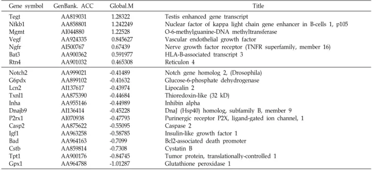
Table 1. Cell death-related genes
Gene symbol GenBank. ACC Global.M Title
TegtNfkb1 MgmtVegf NgfrBat3 Rtn4
AA819031 AA858801 AI044880 AA924335 AI500767 AA900362 AA901032
1.28322 1.242249 1.22528 0.845627 0.67439 0.591977 0.465308
Testis enhanced gene transcript
Nuclear factor of kappa light chain gene enhancer in B-cells 1, p105 O-6-methylguanine-DNA methyltransferase
Vascular endothelial growth factor
Nerve growth factor receptor (TNFR superfamily, member 16) HLA-B-associated transcript 3
Reticulon 4 Notch2
G6pdx Lcn2Txnl1 InhaDnajb9 P2rx1 Casp2 Igf1Bad CstbTpt1 Gpx1
AA999021 AA899102 AI137617 AA875390 AA955146 AI136414 AI070938 AA875622 AA963258 AA964163 AA859814 AA900176 AA964788
-0.41489 -0.41632 -0.43974 -0.44684 -0.44989 -0.45228 -0.47793 -0.55095 -0.58785 -0.7099 -0.7308 -0.84745 -1.01287
Notch gene homolog 2, (Drosophila) Glucose-6-phosphate dehydrogenase Lipocalin 2
Thioredoxin-like (32 kD) Inhibin alpha
DnaJ (Hsp40) homolog, subfamily B, member 9 Purinergic receptor P2X, ligand-gated ion channel, 1 Caspase 2
Insulin-like growth factor 1 Bcl2-associated death promoter Cystatin B
Tumor protein, translationally-controlled 1 Glutathione peroxidase 1
at 1 day after hypoxic shock was shown in Fig. 3. There were more downregulated genes (725 ea) than upregulated ones (472 ea). Lists of genes in each groups are shown in Table 1-7. In the following sections, meanings for only apop- tosis-, stress response-, and cell cycle-related genes are inves- tigated, because these genes are most important in cell survival.
Upregulated apoptosis-related genes
Genes that were up- or downregulated by larger than Global M value 0.2 (i.e., >15% increase or >15% decrease) by RR water extracts was shown in Table 1. Genes upregu- lated more than 2-fold are testis enhanced gene transcript (Tegt; Global M=1.28322), nuclear factor of kappa light chain gene enhancer in B-cells 1, p105 (Nfkb1; Global M=1.242249), and O-6-methylguanine-DNA methyltransferase (Mgmt;
Global M=1.22528).
Testis enhanced gene transcript (Tegt)
Tegt was upregulated by 2.4-fold. Tegt, also known as Bax inhibitor-1 (BI-1), is conserved in both animal and plant species [5,58]. Tegt can inhibit the endoplasmic reticulum (ER) stress proteins as well as the accumulation of ROS, thereby protecting the cells [4,33,61]. Tegt is a suppressor of apoptosis [61], and overexpression of Tegt in rat nigral CSM14.1 and human SH-SY5Y neuroblastoma cells mark- edly protected cell death induced by thapsigargine, a stress agent blocking the Ca2+-ATPase of the ER [8]. Moreover, Tegt was neuroprotective in oxygen-glucose as well as se-
rum deprivation [8]. Since Tegt functions as a suppressor of apoptosis, its upregulation by RR water extracts is ex- pected to be beneficial for cell survival in hypoxia.
Nuclear factor of kappa light chain gene enhancer in B-cells 1, p105 (Nfkb1)
Nfkb1 was upregulated by 2.4-fold. NF-κB is an evolutio- narily conserved signaling module that plays a critical role in the immune system among many biological processes [1].
In many cell types, the most abundant form of NF-κB is the p50/p65 heterodimer, which forms a ternary complex with the inhibitor of NF-κB, IκBα, and remains inactive in the cytoplasm. Upon stimulation, IκBα is rapidly degraded, allowing translocation of p50/p65 heterodimers into the nu- cleus [16,24,29,32] and activation of genes related to in- flammation and proliferation. p105 is the precursor of p50 component of NF-κB [24]. Increase in NFKB1 is expected to provide a beneficial environment for neuronal survival in hypoxia.
O-6-methylguanine-DNA methyltransferase (Mgmt) Mgmt was upregulated by 2.3-fold. O6-methylguanine (O6MeG) is the most critical DNA lesion. Another pre-muta- genic methylation lesion is O4-methylthymine (O4MeT).
O6MeG and O4MeT are repaired by MGMT (also referred to as ATase, AGT, AGAT; E.C. 2.1.1.63) [15,37]. If not re- paired, O6MeG can cause cell death, chromosomal aberra- tions, mutations and cancer. One MGMT molecule can repair only one alkyl adduct. Therefore, the cell's capacity for re- moving DNA O6-alkylguanine adducts depends on the total
Table. 2. Stress-related genes
Gene symbol GenBank. ACC Global.M Title
Defb3 Nfkb1 MgmtCygb AhsgAlox5 Sod3Crp ThbdPtafr Fen1Insig2 Gng7Xpo1 CluA2m Ptprc Tsc2Pros1 Pparg
AA819022 AA858801 AI044880 AA866399 AA955349 AI044102 AA963644 AA926359 AA818521 AA924637 AA819793 AA818627 AA925506 AA818465 AA818413 AA817954 AA924685 AA899998 AA925039 AI111890
1.458768 1.242249 1.22528 1.167176 1.125401 1.049389 0.860088 0.835698 0.826701 0.748363 0.705112 0.656895 0.633971 0.618395 0.602256 0.591385 0.513707 0.474074 0.428995 0.401628
Defensin beta 3
Nuclear factor of kappa light chain gene enhancer in B-cells 1, p105 O-6-methylguanine-DNA methyltransferase
Cytoglobin
Alpha-2-HS-glycoprotein Arachidonate 5-lipoxygenase Superoxide dismutase 3, extracellular C-reactive protein, petaxin related Thrombomodulin
Platelet-activating factor receptor Flap structure-specific endonuclease 1 Insulin induced gene 2
Guanine nucleotide binding protein, gamma 7 Exportin 1, CRM1 homolog (yeast)
Clusterin
Alpha-2-macroglobulin
Protein tyrosine phosphatase, receptor type, C Tuberous sclerosis 2
Protein S (alpha)
Peroxisome proliferator activated receptor, gamma Apex1
Ogg1Gm1960 Tlr4LOC81816 Slc2a1 Pros1 InhaDnajb9 FgaDefb1 Txnrd2 Ninj1 DdtEvl Erbb3 Cd81Camk2g Trip10 Prkab1 Itga1 Pla2g1b CrryC1qb Gpx1
AA900301 AA859654 AI045017 AI044119 AA859229 AA875020 AA875352 AA955146 AI136414 AA925421 AA998504 AA925357 AI044670 AA900788 AA997968 AA924236 AA964328 AA866334 AI059990 AA924247 AA964090 AA955059 AA925136 AA925356 AA964788
-0.40687 -0.41122 -0.41831 -0.42067 -0.42814 -0.44564 -0.44855 -0.44989 -0.45228 -0.46384 -0.46674 -0.46952 -0.47247 -0.47251 -0.504 -0.50868 -0.54156 -0.58588 -0.59428 -0.63369 -0.6439 -0.71966 -0.83107 -0.88625 -1.01287
Apurinic/apyrimidinic endonuclease 1 8-oxoguanine-DNA-glycosylase Gene model 1960, (NCBI) Toll-like receptor 4
Ubiquitin conjugating enzyme
Solute carrier family 2 (facilitated glucose transporter), member 1 Protein S (alpha)
Inhibin alpha
DnaJ (Hsp40) homolog, subfamily B, member 9 Fibrinogen, alpha polypeptide
Defensin beta 1 Thioredoxin reductase 2 Ninjurin 1
D-dopachrome tautomerase
RNB6V-erb-b2 erythroblastic leukemia viral oncogene homolog 3 (avian) CD 81 antigen
Calcium/calmodulin-dependent protein kinase II gamma Thyroid hormone receptor interactor 10
Protein kinase, AMP-activated, beta 1 non-catalytic subunit Integrin alpha 1
Phospholipase A2, group IB Complement receptor related protein
Complement component 1, q subcomponent, beta polypeptide Glutathione peroxidase 1
number of MGMT molecules per cell. Upregulation of Mgmt would be highly beneficial to neuronal survival in hypoxia.
Vascular endothelial growth factor (Vegf)
Vegf was upregulated by 1.8-fold. Vegf was also sig- nificantly upregulated (Global M=0.8456266). VEGF can acti- vate anti-apoptotic kinase and maintain survival signals in endothelial cells [19,36]. It has been shown that VEGF can prevent cell death in various conditions. For example, VEGF
prevented apoptosis of human umbilical vein endothelial cells (HUVEC) from high glucose exposure [63]. In high glu- cose exposure, pretreatment of VEGF lowered ROS gen- eration, calcium overload, Bax/Bcl-2 ratio, caspase-3 activa- tion in HUVEC. In vitro, VEGF-A stimulates axonal out- growth, improves neuronal survival, and prevents hippo- campal cells from apoptosis induced by serum withdrawal [26,51,52]. Recently, VEGF-A was proved to be a survival
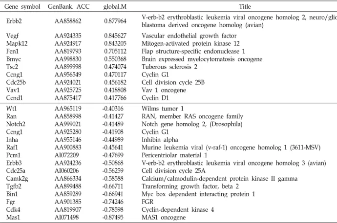
Table 3. Cell cycle-related genes
Gene symbol GenBank. ACC global.M Title
Erbb2 AA858862 0.877964 V-erb-b2 erythroblastic leukemia viral oncogene homolog 2, neuro/glio- blastoma derived oncogene homolog (avian)
VegfMapk12 Fen1Bmyc Tsc2Ccng1 Cdc25b Vav1Ccnd1
AA924335 AA924917 AA819793 AA998830 AA899998 AA956549 AA924021 AA925725 AA875417
0.845627 0.843205 0.705112 0.550368 0.474074 0.470117 0.456182 0.418808 0.417766
Vascular endothelial growth factor Mitogen-activated protein kinase 12 Flap structure-specific endonuclease 1 Brain expressed myelocytomatosis oncogene Tuberous sclerosis 2
Cyclin G1
Cell division cycle 25B Vav 1 oncogene Cyclin D1 Wt1Ran
Notch2 Ccng1 InhaRaf1 Pcm1Erbb3 Cdc25a Camk2g Tgfb2 Bin1Fgr Cdk4Mas1
AA965119 AA858998 AA999021 AA925280 AA955146 AA900883 AI072209 AA924236 AI060206 AA866334 AA899488 AA859289 AA901385 AA819907 AI071498
-0.40316 -0.41427 -0.41489 -0.41908 -0.44989 -0.45641 -0.47699 -0.50868 -0.56259 -0.58588 -0.66711 -0.66941 -0.74246 -0.78598 -0.87495
Wilms tumor 1
RAN, member RAS oncogene family Notch gene homolog 2, (Drosophila) Cyclin G1
Inhibin alpha
Murine leukemia viral (v-raf-1) oncogene homolog 1 (3611-MSV) Pericentriolar material 1
V-erb-b2 erythroblastic leukemia viral oncogene homolog 3 (avian) Cell division cycle 25A
Calcium/calmodulin-dependent protein kinase II gamma Transforming growth factor, beta 2
Myc box dependent interacting protein 1 FGRCyclin-dependent kinase 4
MAS1 oncogene
Table 4. Immune response genes
Gene symbol GenBank. ACC Global.M Title
Nfkb1 AhsgAlox5 CrpGbp2 Ptafr Znf179 A2mIgbp1 Cd5Ptprc Tsc2Vav1 Gbp2Pparg
AA858801 AA955349 AI044102 AA926359 AA819701 AA924637 AA997188 AA817954 AA997141 AA925584 AA924685 AA899998 AA925725 AA923928 AI111890
1.2422 1.1254 1.0494 0.8357 0.7649 0.7484 0.716 0.5914 0.5478 0.5264 0.5137 0.4741 0.4188 0.4061 0.4016
Nuclear factor of kappa light chain gene enhancer in B-cells 1, p105 Alpha-2-HS-glycoprotein
Arachidonate 5-lipoxygenase C-reactive protein, petaxin related
Guanylate binding protein 2, interferon-inducible Platelet-activating factor receptor
Zinc finger protein 179 Alpha-2-macroglobulin
Immunoglobulin (CD79A) binding protein 1 CD5 antigen
Protein tyrosine phosphatase, receptor type, C Tuberous sclerosis 2
Vav 1 oncogene
Guanylate binding protein 2, interferon-inducible Peroxisome proliferator activated receptor, gamma Fth1Gm1960
Tlr4Inha DdtHla-dmb Cd81Itga1 Mx2Bad CrryC1qb
AA818441 AI045017 AI044119 AA955146 AA900788 AI146187 AA964328 AA964090 AI030615 AA964163 AA925136 AA925356
-0.4014 -0.4183 -0.4207 -0.4499 -0.4725 -0.4949 -0.5416 -0.6439 -0.6496 -0.7099 -0.8311 -0.8862
Ferritin, heavy polypeptide 1 Gene model 1960, (NCBI) Toll-like receptor 4 Inhibin alpha
D-dopachrome tautomerase
Major histocompatibility complex, class II, DM beta CD 81 antigen
Integrin alpha 1
Myxovirus (influenza virus) resistance 2 Bcl2-associated death promoter
Complement receptor related protein
Complement component 1, q subcomponent, beta polypeptide
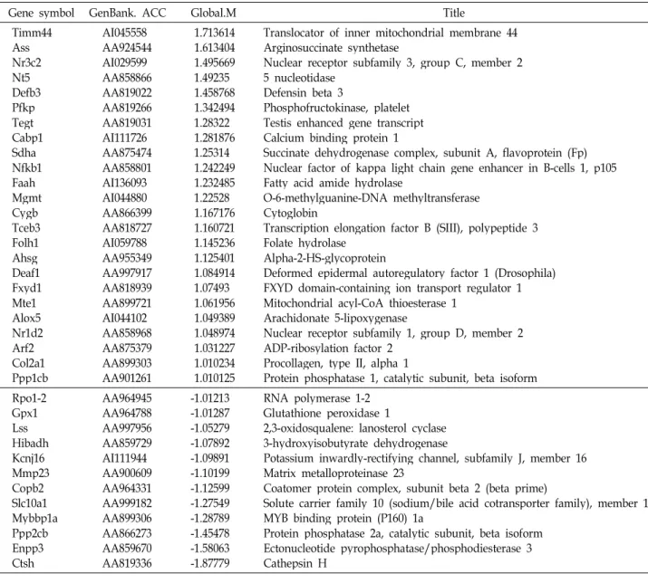
Table 5. Physiological process-related genes
Gene symbol GenBank. ACC Global.M Title
Timm44 AssNr3c2 Nt5Defb3 PfkpTegt Cabp1 SdhaNfkb1 FaahMgmt CygbTceb3 Folh1 AhsgDeaf1 Fxyd1 Mte1Alox5 Nr1d2 Arf2Col2a1 Ppp1cb
AI045558 AA924544 AI029599 AA858866 AA819022 AA819266 AA819031 AI111726 AA875474 AA858801 AI136093 AI044880 AA866399 AA818727 AI059788 AA955349 AA997917 AA818939 AA899721 AI044102 AA858968 AA875379 AA899303 AA901261
1.713614 1.613404 1.495669 1.49235 1.458768 1.342494 1.28322 1.281876 1.25314 1.242249 1.232485 1.22528 1.167176 1.160721 1.145236 1.125401 1.084914 1.07493 1.061956 1.049389 1.048974 1.031227 1.010234 1.010125
Translocator of inner mitochondrial membrane 44 Arginosuccinate synthetase
Nuclear receptor subfamily 3, group C, member 2 5 nucleotidase
Defensin beta 3
Phosphofructokinase, platelet Testis enhanced gene transcript Calcium binding protein 1
Succinate dehydrogenase complex, subunit A, flavoprotein (Fp) Nuclear factor of kappa light chain gene enhancer in B-cells 1, p105 Fatty acid amide hydrolase
O-6-methylguanine-DNA methyltransferase Cytoglobin
Transcription elongation factor B (SIII), polypeptide 3 Folate hydrolase
Alpha-2-HS-glycoprotein
Deformed epidermal autoregulatory factor 1 (Drosophila) FXYD domain-containing ion transport regulator 1 Mitochondrial acyl-CoA thioesterase 1
Arachidonate 5-lipoxygenase
Nuclear receptor subfamily 1, group D, member 2 ADP-ribosylation factor 2
Procollagen, type II, alpha 1
Protein phosphatase 1, catalytic subunit, beta isoform Rpo1-2
Gpx1Lss Hibadh Kcnj16 Mmp23 Copb2 Slc10a1 Mybbp1a Ppp2cb Enpp3 Ctsh
AA964945 AA964788 AA997956 AA859729 AI111944 AA900609 AA964331 AA999182 AA899306 AA866273 AA859670 AA819336
-1.01213 -1.01287 -1.05279 -1.07892 -1.09891 -1.10199 -1.12599 -1.27549 -1.28789 -1.45478 -1.58063 -1.87779
RNA polymerase 1-2 Glutathione peroxidase 1
2,3-oxidosqualene: lanosterol cyclase 3-hydroxyisobutyrate dehydrogenase
Potassium inwardly-rectifying channel, subfamily J, member 16 Matrix metalloproteinase 23
Coatomer protein complex, subunit beta 2 (beta prime)
Solute carrier family 10 (sodium/bile acid cotransporter family), member 1 MYB binding protein (P160) 1a
Protein phosphatase 2a, catalytic subunit, beta isoform Ectonucleotide pyrophosphatase/phosphodiesterase 3 Cathepsin H
factor for retinal neurons to ischemic injury [41]. Therefore, upregulation of Vegf would provide a beneficial environ- ment for neuronal survival in hypoxia.
Nerve growth factor receptor (Ngfr)
Ngfr was upregulated by 1.6-fold (Global M=0.6743903).
The receptors for neurotrophins are members of a family of highly similar transmembrane tyrosine kinases (TrkA, TrkB, and TrkC). Nerve growth factor (NGF) binds mainly TrkA. Binding of NGF to TrkA initiates activation of phos- phatidylinositol 3-kinase/Akt, MAPK, and phospholipase C- γ1 signaling pathways [28], leading to the prevention of apoptotic cell death and promotion of cellular differentia- tion. Therefore, upregulation of Ngfr would be highly bene- ficial to neuronal survival in hypoxia.
Downregulated apoptosis-related genes
Bcl2-associated death promoter (Bad). Bad was down- regulated by -64% (Global M=-0.7098992). The Bcl-2 family proteins play an essential role in cell death regulation. BAD is a pro-apoptotic member of the Bcl-2 family and a regu- latory target of survival signaling [65]. Among pro-apoptotic members, BAD belongs to BH3-only proteins that sense stress signals and initiate the death program [25]. Therefore, downregulation of Bad would prevent apoptosis and lead to cell survival.
Cystatin B (Cstb)
Cstb gene was downregulated by -66% (Global M=
-0.7308042). Cystatin-B-deficient homozygous mice show be- havioral defects such as poor balance while moving and a
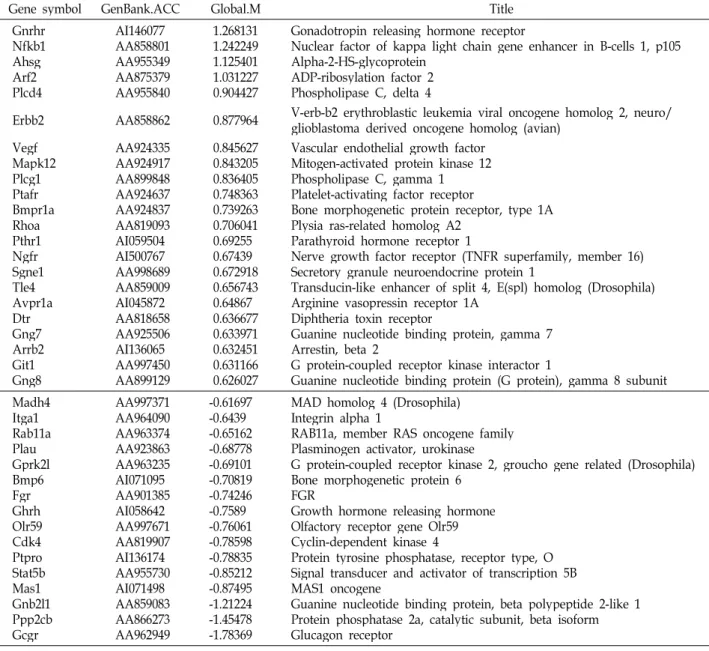
Table 6. signal transduction-related genes
Gene symbol GenBank.ACC Global.M Title
Gnrhr Nfkb1 AhsgArf2 Plcd4
AI146077 AA858801 AA955349 AA875379 AA955840
1.268131 1.242249 1.125401 1.031227 0.904427
Gonadotropin releasing hormone receptor
Nuclear factor of kappa light chain gene enhancer in B-cells 1, p105 Alpha-2-HS-glycoprotein
ADP-ribosylation factor 2 Phospholipase C, delta 4
Erbb2 AA858862 0.877964 V-erb-b2 erythroblastic leukemia viral oncogene homolog 2, neuro/
glioblastoma derived oncogene homolog (avian) VegfMapk12
Plcg1 Ptafr Bmpr1a RhoaPthr1 NgfrSgne1 Tle4Avpr1a DtrGng7 Arrb2 Git1Gng8
AA924335 AA924917 AA899848 AA924637 AA924837 AA819093 AI059504 AI500767 AA998689 AA859009 AI045872 AA818658 AA925506 AI136065 AA997450 AA899129
0.845627 0.843205 0.836405 0.748363 0.739263 0.706041 0.69255 0.67439 0.672918 0.656743 0.64867 0.636677 0.633971 0.632451 0.631166 0.626027
Vascular endothelial growth factor Mitogen-activated protein kinase 12 Phospholipase C, gamma 1 Platelet-activating factor receptor
Bone morphogenetic protein receptor, type 1A Plysia ras-related homolog A2
Parathyroid hormone receptor 1
Nerve growth factor receptor (TNFR superfamily, member 16) Secretory granule neuroendocrine protein 1
Transducin-like enhancer of split 4, E(spl) homolog (Drosophila) Arginine vasopressin receptor 1A
Diphtheria toxin receptor
Guanine nucleotide binding protein, gamma 7 Arrestin, beta 2
G protein-coupled receptor kinase interactor 1
Guanine nucleotide binding protein (G protein), gamma 8 subunit Madh4
Itga1 Rab11a PlauGprk2l Bmp6Fgr GhrhOlr59 Cdk4Ptpro Stat5b Mas1Gnb2l1 Ppp2cb Gcgr
AA997371 AA964090 AA963374 AA923863 AA963235 AI071095 AA901385 AI058642 AA997671 AA819907 AI136174 AA955730 AI071498 AA859083 AA866273 AA962949
-0.61697 -0.6439 -0.65162 -0.68778 -0.69101 -0.70819 -0.74246 -0.7589 -0.76061 -0.78598 -0.78835 -0.85212 -0.87495 -1.21224 -1.45478 -1.78369
MAD homolog 4 (Drosophila) Integrin alpha 1
RAB11a, member RAS oncogene family Plasminogen activator, urokinase
G protein-coupled receptor kinase 2, groucho gene related (Drosophila) Bone morphogenetic protein 6
FGRGrowth hormone releasing hormone Olfactory receptor gene Olr59 Cyclin-dependent kinase 4
Protein tyrosine phosphatase, receptor type, O Signal transducer and activator of transcription 5B MAS1 oncogene
Guanine nucleotide binding protein, beta polypeptide 2-like 1 Protein phosphatase 2a, catalytic subunit, beta isoform Glucagon receptor
lack of motor coordination (Pennacchio et al., 1998).
Previously, these mice exhibited neuronal loss in various brain regions including cerebellar granule cell layer, en- torhinal cortex, and hippocampus [43,50]. This phenotype is similar to the progressive myoclonus epilepsy of the Unverricht-Lundborg type (locus symbol EPM1) which is re- lated to the increased activity of cysteine proteins cathepsins [47]. Since Cystatin B is a cysteine protease inhibitor, cyto- plasmic cathepsins are free to activate the apoptotic path- ways in this pathogenesis [64]. However, the meaning of Cstb downregulation in hypoxia is not clear.
Tumor protein, translationally-controlled 1 (Tpt1) Tpt1 gene was downregulated by 80% (Global M=
-0.8474458). TPT1 is widely expressed and conserved throughout vertebrates. The tpt1 is the strongest differ- entially expressed gene between tumor and tumor-reversed states in human leukemia and breast-cancer cells [57]. The meaning of Cstb downregulation in hypoxia is not clear.
Glutathione peroxidase 1 (Gpx1)
Gpx1 gene was downregulated by 2-fold (Global M=
-1.012869). GPX1 is an anti-oxidant enzyme. Over-expression of Gpx1 or Cu/Zn-superoxide dismutase (SOD1) protects neuronal apoptosis after focal cerebral ischemia [11,30].
Knockout of SOD1 or Gpx1 increases neuronal cell damage after focal cerebral ischemia [7,31]. The reason why Gpx1, an anti-oxidant gene, is reduced is not clear.
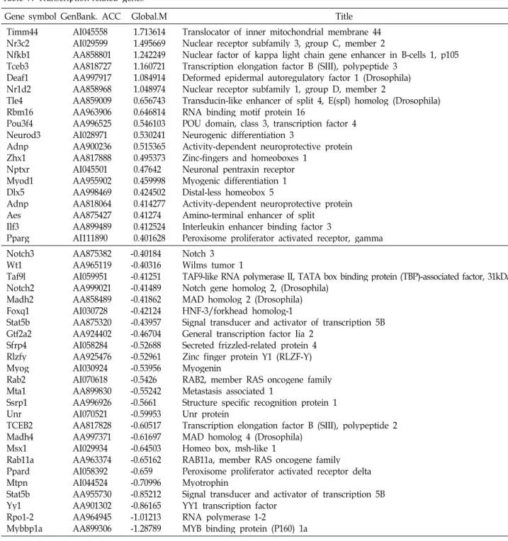
Table 7. Transcription-related genes
Gene symbol GenBank. ACC Global.M Title
Timm44 Nr3c2 Nfkb1 Tceb3 Deaf1 Nr1d2 Tle4Rbm16 Pou3f4 Neurod3 AdnpZhx1 Nptxr Myod1 Dlx5Adnp AesIlf3 Pparg
AI045558 AI029599 AA858801 AA818727 AA997917 AA858968 AA859009 AA963906 AA996525 AI028971 AA900236 AA817888 AI045501 AA955902 AA998469 AA818064 AA875427 AA899489 AI111890
1.713614 1.495669 1.242249 1.160721 1.084914 1.048974 0.656743 0.646814 0.546103 0.530241 0.515365 0.495373 0.47642 0.459998 0.424502 0.414277 0.41274 0.412524 0.401628
Translocator of inner mitochondrial membrane 44 Nuclear receptor subfamily 3, group C, member 2
Nuclear factor of kappa light chain gene enhancer in B-cells 1, p105 Transcription elongation factor B (SIII), polypeptide 3
Deformed epidermal autoregulatory factor 1 (Drosophila) Nuclear receptor subfamily 1, group D, member 2
Transducin-like enhancer of split 4, E(spl) homolog (Drosophila) RNA binding motif protein 16
POU domain, class 3, transcription factor 4 Neurogenic differentiation 3
Activity-dependent neuroprotective protein Zinc-fingers and homeoboxes 1
Neuronal pentraxin receptor Myogenic differentiation 1 Distal-less homeobox 5
Activity-dependent neuroprotective protein Amino-terminal enhancer of split
Interleukin enhancer binding factor 3
Peroxisome proliferator activated receptor, gamma Notch3
Wt1Taf9l Notch2 Madh2 Foxq1 Stat5b Gtf2a2 Sfrp4 Rlzfy MyogRab2 Mta1Ssrp1 UnrTCEB2 Madh4 Msx1Rab11a Ppard MtpnStat5b Yy1Rpo1-2 Mybbp1a
AA875382 AA965119 AI059951 AA999021 AA858489 AI030728 AA875320 AA924402 AI058284 AA925476 AI030924 AI070618 AA899830 AA996926 AI070521 AA817828 AA997371 AI029934 AA963374 AI058392 AI044524 AA955730 AA901302 AA964945 AA899306
-0.40184 -0.40316 -0.41251 -0.41489 -0.41862 -0.42124 -0.43957 -0.46704 -0.52688 -0.52961 -0.53956 -0.5426 -0.55242 -0.5661 -0.59953 -0.60517 -0.61697 -0.64503 -0.65162 -0.659 -0.70996 -0.85212 -0.86165 -1.01213 -1.28789
Notch 3 Wilms tumor 1
TAF9-like RNA polymerase II, TATA box binding protein (TBP)-associated factor, 31kDa Notch gene homolog 2, (Drosophila)
MAD homolog 2 (Drosophila) HNF-3/forkhead homolog-1
Signal transducer and activator of transcription 5B General transcription factor Iia 2
Secreted frizzled-related protein 4 Zinc finger protein Y1 (RLZF-Y) Myogenin
RAB2, member RAS oncogene family Metastasis associated 1
Structure specific recognition protein 1 Unr protein
Transcription elongation factor B (SIII), polypeptide 2 MAD homolog 4 (Drosophila)
Homeo box, msh-like 1
RAB11a, member RAS oncogene family Peroxisome proliferator activated receptor delta Myotrophin
Signal transducer and activator of transcription 5B YY1 transcription factor
RNA polymerase 1-2
MYB binding protein (P160) 1a
Stress response-related genes
Genes that were up- or downregulated larger than Global M value 0.2 (i.e., >15% increase or >15% decrease) by RR water extracts were shown in Table 2. Genes upregulated more than 2-fold are defensin beta 3 (Defb3; Global M=
1.458768), Nfkb1, Mgmt, cytoglobin (Cygb; Global M=
1.167176), alpha-2-HS-glycoprotein (Ahsg; Global M=
1.125401), and arachidonate 5-lipoxygenase (Alox5; Global M=1.049389).
Defensin beta 3 (Defb3)
The Defb3 gene was upregulated by 2.7-fold (Global M=1.458768). Mammalian cells express a number of peptide antibiotics as a innate host defense system [27]. Defensins and cathelicidin are the two major classes of antimicrobial peptides in humans [14,42]. Defensins are divided into α- and β-defensins [42]. α-Defensins are expressed in small in- testine (neutrophils and Paneth cells), whereas human β- defensins (hBDs) are mainly found in epithelial tissues [42].
hBD-1 is constitutively expressed in various epithelial tis- sues [12,66], while hBD-2 is mainly found in skin, respira- tory and gastrointestinal tracts [21,36]. hBD-3, the third type β-defensin, is inducible upon stimulation with bacteria and cytokines in both epithelial and non-epithelial tissues [21,13]. Therefore, upregulation of Defb3 would protect neurons in hypoxia.
Cytoglobin (Cygb)
The Cygb gene was upregulated by 2.2-fold (Global M=1.167176). In vertebrates, four types of globins have been reported: hemoglobin (Hb) in red blood cell [23], myoglobin (Mb) in muscle [59,60], neuroglobin (Ngb) and cytoglobin (Cygb). Cygb is ubiquitously expressed [3,20], and is upregu- lated in rat heart and liver following hypoxia [49] or in brain by hypoxia [10]. When Cygb mRNA was knocked down by siRNA, cell death was increased upon hydrogen per- oxide-treatment [35], suggesting that Cygb protects cells against oxidative stress. In addition, Ngb expression was up-regulated following hypoxic challenges [54] and in ische- mic-hypoxia brain injury [53]. Thus, Cygb that binds an oxy- gen (O2) molecule may be induced under oxidative stress to protect cells from death.
Alpha-2-Heremans-Schmid (HS)-glycoprotein (Ahsg) Ahsg was upregulated by 2.18-fold (Global M=1.125401).
AHSG is a serum glycoprotein. ahsg-KO mice demonstrated impaired tolerance to ischemia suggesting that AHSG exerts a protective effect against ischemia in the cardiomyocyte [39,45]. Thus, upregulation of Ahsg is expected to protect cells from death under hypoxic stress.
Arachidonate 5-lipoxygenase (Alox5)
Alox5 was upregulated by 2-fold (Global M=1.049389).
ALOX5 is a dual-function protein that converts arachidonate to 5-hydroperoxyeicosatetrenoic and subsequently to leuko- triene A4 [44]. Tonget al. [56] showed that 5-LOX inhibitor Rev-5901 blocked cell proliferation in pacreactic cancer and induced apoptosisin vivo and in vitro. This results indicate that downstream products of ALOX5 stimulate cell pro- liferation and promote cell survival. Therefore, upregulation of ALox5 would help cells to survive in hypoxia.
Cell cycle-related genes
The up- or downregulated cell cycle-related genes are shown in Table 3. The erythroblastic leukemia viral onco- gene homolog 2 (Erbb2), Vascular endothelial growth factor (Vegf) and Mitogen-activated protein kinase 12 (Mapk12)
were most highly increased.
Erbb2
The Erbb2 gene was upregulated by 1.84-fold (Global M=0.8779643). The ErbB-2 · ErbB-3 dimer signals through the mitogen-activated protein kinase (MAPK) pathway, which stimulates cell proliferation. This dimer also signals through the PI3K/Akt pathway. This pathway promotes cel- lular survival and antiapoptotic signals [6]. Recently, it has been reported that ErbB2 expression increased TNF-induced apoptosis [62]. These results indicate that Erbb2 is involved in cell proliferation and survival. Therefore, upregulation of Erbb2 is beneficial for cellular survival.
Mapk12
The Mapk12 gene (also called Erk6 or SAPK3) was upre- gulated by 1.8-fold (Global M=0.8432047). Mapk12 responds to environmental stress and pro-inflammatory cytokines and phosphorylate downstream targets. c-jun is one of the down- stream targets of Mapk12 [38]. Activation of c-jun stimulates cell proliferation. A stimulation to proliferate is expected to promote cell survival.
Summary
As indicated by MA-plot, most genes were up- or down- regulated within 2-fold by RR water extracts. Antiapoptosis genes such as Tegt (2.4-fold), Nfkb1 (2.4-fold) Veg (1.8-fold), Ngfr (1.6-fold) were upregulated, while pro-apoptosis genes such as Bad (-64%), Cstb (-66%) were downregulated. Genes for combating environmental stress (stress response genes) such as Defb3 (2.7-fold), Cygb (2.2-fold), Ahsg (2.18-fold), Alox5 (2-fold) were upregulated. Genes for cell proliferation (cell cycle-related genes) such as Erbb2 (1.84-fold), Mapk12 gene (1.8-fold) was upregulated. Therefore, RR water ex- tracts upregulate many pro-survival genes while down- regulating many pro-death genes. It is interpreted that these genes, in combination with other regulated genes, can pro- mote neuronal survival in a stress such as hypoxia.
Acknowledgment
This work was supported by the Dongguk University Research Fund.
References
1. Bonizzi, G. and M. Karin. 2004. The two NF-B activation pathways and their role in innate and adaptive immunity.
Trends Immunol. 25, 280-288.
2. Brewer, G. J., J. R. Torricelli, E. K. Evege and P. J. Price.
1993. Optimized survival of hippocampal neurons in B27-supplemented Neurobasal, a new serum-free medium combination. J. Neurosci. 35, 567-576.
3. Burmester, T., B. Ebner, B. Weich and T. Hankeln 2002.
Cytoglobin: a novel globin type ubiquitously expressed in vertebrate tissues. Mol. Biol. Evol. 19, 416-421.
4. Chae, H. J., H. R. Kim, C. Xu, B. Bailly-Maitre, M.
Krajewska, S. Krajewski, S. Banares and et. al. 2004. BI-1 regulates an apoptosis pathway linked to endoplasmic retic- ulum stress. Mol. Cell 15, 355-366.
5. Chae, H. J., N. Ke, H. R. Kim, S. Chen, A. Godzik, M.
Dickman and J. C. Reed. 2003. Evolutionarily conserved cy- toprotection provided by Bax Inhibitor-1 homologs from an- imals, plants, and yeast. Gene323, 101-113.
6. Citri, A., K. B. Skaria and Y. Yarden. 2003. The deaf and the dumb: the biology of ErbB-2 and ErbB-3.Exp. Cell Res.
284, 54-65.
7. Crack, P. J., J. M. Taylor, N. J. Flentjar, J. Haan, P. Hertzog, R. C. Iannello and I. Kola. 2001. Increased infarct size and exacerbated apoptosis in the Gpx-1 knockout mouse brain in response to ischemia/reperfusion injury.J. Neurochem.78, 1389-1399
8. Dohm, C. P., S. Siedenberg, J. Liman, A. Esposito, F. S.
Wouters, J. C. Reed, M. Bähr and P. Kermer. 2006, Bax in- hibitor-1 protects neurons from oxygen-glucose deprivation. J. Mol. Neurosci. 29, 1-8.
9. Dudoit, S., Y. H. Yang, M. J. Callow and T. P. Speed. 2002.
Statistical methods for identifying differentially expressed genes in replicated cDNA microarray experiments.Statistica Sinica 12, 111-140.
10. Fordel, E., E. Geuens, S. Dewilde, P. Rottiers, P. Carmeliet, J. Grooten and L. Moens, 2004. Cytoglobin expression is up- regulated in all tissues upon hypoxia: an in vitro and in vivo study by quantitative real-time PCR.Biochem. Biophys.
Res. Commun. 319, 342-348.
11. Fujimura, M., Y. Morita-Fujimura, N. Noshita, T. Sugawara, M. Kawase and P. H. Chan. 2000. The cytosolic antioxidant copper/zinc-superoxide dismutase prevents the early re- lease of mitochondrial cytochrome c in ischemic brain after transient focal cerebral ischemia in mice. J. Neurosci. 20, 2817-2824.
12. Fulton, C., G. M. Anderson, M. Zasloff, R. Bull and A.
Quinn. 1997. Expression of natural peptide antibiotics in hu- man skin. Lancet 350, 1750-1751
13. García, J. R, F. Jaumann, S. Schulz and A. Krause, J.
Rodríguez-Jiménez, U. Forssmann, K. Adermann, E. Klüver, C. Vogelmeier, D. Becker, R. Hedrich, W. G. Forssmann and R. Bals. 2001. Identification of a novel, multifunctional β- defensin (human β-defensin 3) with specific antimicrobial activity. Its interaction with plasma membranes of Xenopus oocytes and the induction of macrophage chemoattraction.
Cell Tissue Res. 306, 257-264.
14. Gennaro, R. and M. Zanetti. 2001. Structural features and biological activities of the cathelicidin-derived antimicrobial
peptides. Biopolymers55, 31-49.
15. Gerson, S. L. 2004. MGMT: its role in cancer aetiology and cancer therapeutics. Nat. Rev. Cancer 4, 296-307.
16. Ghosh, S. and D. Baltimore. 1990. Activation in vitro of NF-kappa B by phosphorylation of its inhibitor I kappa B.
Nature344, 678-682.
17. Goslin, K., H. Asmussen and G. Banker. 1998. Rat hippo- campal neurons in low-density culture, pp. 339-370, In Banker, G. and K. Goslin (eds.), 2nd eds. Culturing nerve cells. Cambridge, MA: MIT Press.
18. Green, D. R. and J. C. Reed. 1998. Mitochondria and apoptosis. Science281, 1309-1312.
19. Grosjean, J., S. Kiriakidis, K. Reilly, M. Feldmann and E.
Paleolog. 2006. Vascular endothelial growth factor signal- ling in endothelial cell survival: a role for NF-κB.Biochem.
Biophys. Res. Commun. 340, 984-994.
20. Hankeln, T., S. Wystub, T. Laufs, M. Schmidt, F. Gerlach, S. Saaler-Reinhardt, S. Reuss and T. Burmester. 2004. The cellular and subcellular localization of neuroglobin and cy- toglobin—A clue to their function?.IUBMB Life56, 671-679.
21. Harder, J., J. Bartels, E. Christophers and J. M. Schröder.
2001. Isolation and characterization of human β-defensin-3, a novel human inducible peptide antibiotic.J. Biol. Chem.
276, 5707-5713.
22. Harder, J., J. Bartels, E. Christophers and J. M. Schröder.
1997. A peptide antibiotic from human skin.Nature387, 861.
23. Hardison, R. C. 1996. A brief history of hemoglobins: plant, animal, protist, and bacteria.Proc. Natl. Acad. Sci. USA93, 5675-5679.
24. Hayden, M. S. and S. Ghosh. 2005. Signaling to NF-kappaB.
Genes Dev.18, 2195-2224.
25. Huang, D. C. and A. Strasser. 2000. BH3-Only proteins—
Essential initiators of apoptotic cell death.Cell103, 839-842.
26. Jin, K. L., X. O. Mao and D. A. Greenberg. 2000. Vascular endothelial growth factor: direct neuroprotective effect in in vitroischemia.Proc. Natl. Acad. Sci. USA97, 10242-10247.
27. Kaiser, V. and G. Diamond. 2001. Expression of mammalian defensin genes. J. Leukoc. Biol. 68, 779-784.
28. Kaplan, D. R. and R. M. Stephens. 1994. Neurotrophin sig- nal transduction by the Trk receptor. J. Neurobiol. 25, 1404-1417.
29. Karin, M. and Y. N. Ben. 2000. Phosphorylation meets ubiq- uitination: the control of NF-kappaB activity.Annu. Rev.
Immunol. 18, 621-663.
30. Kinouchi, H., C. J. Epstein, T. Mizui, E. Carlson, S. F. Chen and P. H. Chan. 1991. Attenuation of focal cerebral ischemic injury in transgenic mice overexpressing CuZn superoxide dismutase.Proc. Natl. Acad. Sci. USA 88, 11158-11162.
31. Kondo, T., A. G. Reaume, T. T. Huang, E. Carlson, K.
Murakami, S. F. Chen, E. K. Hoffman, R. W. Scott, C. J.
Epstein and P. H. Chan. 1997. Reduction of CuZn-super- oxide dismutase activity exacerbates neuronal cell injury and edema formation after transient focal cerebral ischemia.
J. Neurosci. 17, 4180-4189.
32. Krappmann, D. and C. Scheidereit. 2005. A pervasive role of ubiquitin conjugation in activation and termination of