서 론
는 바텐병 Neuronal ceroid lipofuscinoses (NCLs) (Batten
이라 불리며 명 당 명의 발병률을 가진 신경 disease) , 12,500 1
퇴행 축적 질환(neurodegenerative storage disease)이다[9].
영아형 바텐병(early onset of Batten disease, EBD)은 Ceroid 유전자의 산물인 Lipofuscinosis, Neuronal 1 (CLN1) palmi- toyl-protein thioesterase 1 (PPT1)의기능상실로 인해 신경 퇴 행 질환인 영아형 바텐병으로 유도되며[12], 다양한 형태의 중에서도 가장 치명적이고 파괴적인 질병으로 인구 NCLs
명 당 명의 발병률과 명 중 명이 보인자 빈도를 보
10,000 1 70 1
이며 핀란드 사람에게서 많이 보고되고, [1,9,10,17,20,22],출생 후 약6~24 개월 이내에 발병한다[4].영아형 바텐병은 리소좀
에 라는
(lysosome) ceroid (thioesterified polypeptides) waste 가 비정상적으로 축적 되어지는 증상을 보인다 product
현재까지 다양한 연구 결과를 통해 영아형 바텐 [1,7,11,21,22].
병 환자에서PPT1효소의 불활성과 수송의 중단을 이끄는 다 수의missense mutation 및nonsense mutation들이 보고 되어
졌다[6,7,16,17,20].
은 수용성 리소좀 가수분해효소
PPT1 (soluble lysosomal
로 생체내의 거대분자 들을 분
hydrolases) (macromolecules) 해하는 것으로 예상되지만 현재까지 PPT1의 기질은 명확하 게 확인되지 않았고[3-6,15,20], 또한 PPT1 knockout mice (EBD KO 와 이의 대조군인) wild type mice (W T)를 이용한 실험에서, PPT1은S-acylated 단백질의 분해대사에 관여하는 soluble lysosomal enzymes으로 S-acylated (palmitoylated) 된 단백질 분해를 촉진하기 위해서 단백질에서 thioester 결 합의 절단을 촉진하여 palmitate잔기를 제거하는 역할을 수 행한다[8,19,20]. 이러한 PPT1의 기능상실로 인해 소포체 (endoplasmic reticulum, ER)내에 unfolding protein re-
이 발생하고 소포체 스트레스 를 유
sponse (UPR) (ER stress)
발하며 발생된 소포체 스트레스 는 소포체 내, Ca2+의 항상 성을 붕괴하여 Ca2+ 방출을 유도하고 최종적으로 세포사멸 을 유도한다는 사실이 보고되었으나 아직 그
(apoptosis) [9],
명확한 발병기전 및 치료 방법이 발견되어지지 않고 있다.
은 결합 단백질로서 또
Neurogranin calmodulin RC3, p17 는BICK라 불리며cerebral cortex, hippocampus, amygdale 및 basal ganglia 등 뇌에서 풍부하게 발현된다[2].
은 개의 아미노산으로 이루어져 있으며
Neurogranin 78 , pro-
과산화수소에 의한 산화스트레스가 영아형 바텐병에서 neurogranin 의 인산화에 미치는 영향
윤동호 김한복 박주훈․ ․ 1․김성조*
호서대학교 생명공학과, 1한방화장품과학과
Received March 18, 2009 /Accepted April 16, 2009
E ffect of N eurogranin Phosphorylation on Oxidative Stress by H ydrogen Peroxide in Early Onset of Batten Disease. D ong-H o Yoon, H an Bok Kim , Joo-H oon Park1 and Sung-Jo Kim *. Department of biotechnology, Hoseo university, 165, Baebang-Myun, Asan, Chungnam, Republic of Korea, 336-795,
1Department of cosmetic science, Hoseo university, 165, Baebang-Myun, Asan, Chungnam, Republic of Korea, 336-795 - Early onset of Batten disease (EBD), one of the most lethal neurodegenerative storage dis- orders of childhood, is caused by inactivating mutations in the Ceroid Lipofuscinosis, Neuronal (CLN1) gene. Neurogranin, a calmodulin-binding protein, is expressed in the brain and participates in the protein kinase C (PKC) signaling pathway. While oxidative stress is the suggested cause of neu- rodegeneration in EBD, its molecular mechanism(s) remains obscure. In this research, we examined the levels of neurogranin in the brain mRNA of wild-type (W T) mice and EBD knockout (KO) mice, as well as the proteins. W e also performed neuronal cultures to measure the expression levels of neu- rgranin and phosphorylated-neurogranin with or without oxidative stress inducers and anti-oxidants.
Results showed that neurogranin in both EBD KO mice brain mRNA and protein extracts decreased in an age dependent manner. However, high amounts of phosphorylated-neurogranin were detected in the 6-month brain. This pattern was also confirmed by cultured neurospheres samples. Moreover, neurospheres treated with H2O2, an oxidative stress inducer, showed increased phosphorylated-neuro- granin patterns. Interestingly, this pattern returned to normal status when treated with N-acetyl-L-cys- tein, an anti-oxidant, after H2O2 treatment was performed. Our results suggest that the phosphor- ylation of neurogranin is affected by oxidative stress status in EBD, and appropriate anti-oxidant treat- ment will relieve hyper-phosphorylation of neurogranin.
Key w ords : N eurogranin, oxidative stress, anti-oxidant, early onset of Batten disease (EBD )
*Corresponding author
*Tel +82-41-540-5571, Fax +82-41-548-6231: :
*E-mail : [email protected]
신호전달 경로에 관여하며
tein kinase C (PKC) , dendritic
및 시냅스 가소성 형성과정에 참여
spine (Synaptic Plasticity)
하며 시냅스 말단에서 Ca2+부족 시 calmodulin과 결합하는 것으로 알려져 있다[14].또한 인산화된neurogranin은 neuro- granin과 결합하는calmodulin과의 친화력을 감소시키며, is-
국소빈혈 혈중산소감소 등의 조절자로서
chemic ( ), hypoxic ( )
역할을 수행할 뿐 아니라, calmodulin에 의존적인nitric oxide synthase (NOS), calmodulin protein kinase II (CaMkII) 및
등의 활성에 영향을 미친다 adenylate cyclase (AC) [13].
최근 EBD KO에서 소포체 스트레스 발생이 활성산소 의 생산을 증가 시켜 (reactive oxygen species, ROS) Ca2+ 의 항상성에 혼란을 야기하며 미토콘드리아 막(mitochondria 을 불안정 하게 하여 이 활성화 되어 최 membrane) caspase-9
종적으로 세포사멸을 유도 또는 가속화 한다는 결과가 보고되 어 있다[9]. 이를 바탕으로 본 논문에서는Ca2+ 부족에 의해 결합하는 calmodulin 결합 단백질인 neurogranin이 W T과 EBD KO 쥐의 뇌에서 발현되는 수준과 인산화된 neurogranin 의 수준 그리고 산화스트레스와 항산화제가 인산화된, neuro- granin에 어떠한 효과를 나타내는지 알아보고자 한다.
재료 및 방법
신경세포 배양 및 약품 처리
EBD KO와 정상 쥐 각각에서 임신 후 15일 경과한 태아 쥐의 뇌를 적출하고 NeuroCult NSC proliferation supple- ments (Stem Cell Technologies, Vancouver, BC, Canada)와 human epidermal growth factor (Invitrogen, Carlsbad, CA,
가 함유된
USA) NeuroCult NSC Basal Medium (Stem Cell 에서 신경세포 Technologies, Vancouver, BC, Canada)
를 배양하였다 이후
(Neurosphere) . 100 µM H2O2를 처리하고 시간 경과 후 대조군에
24 4 mM N-acetyl-L-cysteine (Sigma, 을 처리하고 시간 경과 후 실험에 Saint Louis, MO, USA) 24
사용하였다.
Reat-time PCR 분석
EBD KO 쥐와 정상 쥐의 뇌를 적출하여 TriZol (Invitrogen, 을 사용하여 전체 를 분리하였고 Carlsbad, CA, USA) RNA
과 를
RNeasy Mini Kit (QIAGEN, Hilden, Germany) DNase 처리하여 RNA를 정제하였다 이후. SuperScript III first- strand synthesis system (Invitrogen, Carlsbad, CA, USA)역 전사효소를 처리하고 정량한 후 각 시료별로, 10 ng의 전체 RNA와 RNASYBR Green PCR Master Mix (Applied
를 혼합 한 뒤
Biosystems, Foster City, CA, USA) , neurogranin primer를 통해ABI Prism 7000 Sequence Detection System (Applied Biosystems, Foster City, CA, USA)을 통해real-time
을 수행하였다 얻어진 결과는
PCR . ABI Prism Software ver-
sion 1.01 (Applied Biosystems, Foster City, CA, USA)을 통해 분석하였고 최종결과는 β-actin과 비교하여 정량분석을 실시 하였다 각각 회의 반복실험을 통해 얻어진 실험결과는 평균. 3
표준편차로 표기하였고 통계적 유의성 검정은
± , Student’s
를 통해 실시하였으며 값이 미만일 경우 통계적으
t-test , p 0.05
로 유의하다고 판단하였다.
Western blot 분석
각 연령대 별로 쥐의 뇌를 적출하여 단백질 추출용 용해액 인 PhosphoSafe extraction reagent (EMD Biosciences, San Diego, CA, USA)와protease-inhibitor cocktails (Sigma, Saint 를 섞고 조직파쇄기를 통해 단백질을 추출하 Louis, MO, USA)
였다 배양된 신경세포는. PhosphoSafe extraction reagent 와 섞은 뒤 얼음에서 (EMD Biosciences, San Diego, CA, USA)
시간 반응시켜 단백질 추출물을 얻었다 각 시료별로
1 . 20 µg
의 단백질을 4-15% sodium dodecyl sulfate-polyacryamaide gel electrophoresis (SDS-PAGE) (Bio-Rad, Hercules, CA,
를 실시하였고 이후 전기 이동을 통해
USA) nitrocellulose
에 단백질을 이동시 membrane (Bio-Rad, Hercules, CA, USA)
켜western blot에 사용하였다 단백질을 보유한. membranes 은5% non-fat dry milk (Bio-Rad, Hercules, CA, USA)를 통해
차 처리를 한 후 과
1 anti-neurogranin anti-phospho-neuro-
차 항체 및
granin 1 (Millipore, Billerica, MA, USA) anti-β 항체를 처리하였다 차 -actin (Sigma, Saint Louis, MO, USA) . 2 항체는 goat anti-rabbit IgG (Santa Cruz Biotechnology, Santa
를 사용하였고
Cruz, CA, USA) Supersignal west pico lumi- 을 통해 단 nol/enhancer solution (Pierce, Rockford, IL, USA)
백질 밴드를 확인하였다.
결 과
본 논문에서는 월령1 , 3월령, 6월령으로 분리되는 W T 및 EBD KO 쥐의 뇌에서 neurogranin의 상대적인 발현 수준을 알아보기 위해mRNA를 이용한real-time PCR 및 추출된 단 백질을 이용한 western blot 을 수행하였고, neurogranin의 인산화정도 배양된 신경세포인, neurospheres를 이용한neu- rogranin 및 인산화 된 neurogranin의 수준을 확인하였다 나. 아가 산화스트레스와 항산화제가 neurogranin의 인산화에 미 치는 영향을 확인하기 위하여 neurogranin에 산화스트레스 유발 물질인 H2O2를 처리하였고, 24시간 경과 후 일부 대조군 에는 항산화제인 N-acetyl-L-cysteine (NAC)을 처리하여 그 결과를 확인하였다.
을 통한 및 쥐 뇌의
Real-time PCR WT EBD KO 상대적 발현수준
neurogranin
월령 월령 월령
1 , 3 , 6 W T, EBD KO 쥐에서 적출한 뇌의
KO 1M KO 3M KO 6M
Fig 1. Expression levels of neurogranin m RNA in the brain of wild-type (WT) mice and EBD KO (KO) mice by real-time PCR. 1 M : 1 m onth, 3 M : 3 m onths, 6 M : 6 months.
를 이용하여
mRNA neurogranin 발현 수준을 real-time PCR 을 수행하여 확인하였다. W T 쥐에서는 월령1 , 3월령, 6월령 등 노화가 진행되는 상황에서도 neurogranin mRNA의 생성 수준에는 차이가 없었으나, EBD KO쥐에서는 월령1 , 3월령,
월령과 같이 노화가 진행됨에 따라
6 neurogranin mRNA의
생성 수준은 감소되었다(Fig. 1).이 결과를 통해, 1월령EBD KO 쥐에서는 neurogranin mRNA가WT쥐와 동일하게 정상 적으로 생성되지만 노화가 진행됨에 따라 neurogranin 의 생성 수준이 현격히 감소한다는 것을 발견하여
mRNA ,
결핍이 정상적인
PPT1 neurogranin의 발현에 영향을 주는 것 을 확인 하였다.
및 쥐의 뇌를 이용한 분석
WT EBD KO western blot
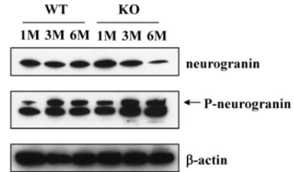
월령 월령 월령의 과
1 , 3 , 6 WT EBD KO 쥐에서 적출한 뇌의 단백질을 이용하여western blot 분석을 수행 하였다 그 결과. 의 결과와 유사하게 월령 월령 월령으로 구 real-time PCR 1 , 3 , 6
분된WT 쥐의 뇌에서는neurogranin의 발현량에 차이가 없었 으나 월령 월령 월령으로 구분된, 1 , 3 , 6 EBD KO 쥐의 뇌에서는 노화가 진행될수록 neurogranin의 발현이 점차 감소하는 것 을 확인 하였다.
또한 인산화된, neurogranin은 월령 월령 월령으로 구1 , 3 , 6 성된W T 및EBD KO 쥐에서 노화가 진행될수록 그 수준이 어느정도 증가하는 형태를 확인하였으며 흥미롭게도 월령, 6 의EBD KO 쥐의 뇌에서는 감소된 neurogranin의 양에 비교 하여 볼 때 인산화된 neurogranin의 양이 증가해 있음이 확인 되었다(Fig. 2).
배양 신경세포인neurospheres western blot의 분석 WT과EBD KO 쥐의 태아에서 추출한neurospheres를 배 양하여 neurogranin의 양을 확인한 결과, W T 및 EBD KO
내의 수준은 앞서 실험 결과와 동
neurospheres neurogranin
일하게EBD KO 쥐에서 감소하는 형태를 확인하였으며 인산, 화된neurogranin의 수준은WT 쥐 보다EBD KO 쥐에서 현격 하게 수준이 증가한 형태를 확인하였다(Fig. 3).이렇듯neural
인 에서도 감소하는 의 발
stem cell neurospheres neurogranin
Fig 2. W estern blot analysis for non-phosphorylated and phos- phorylated neurogranin in the brain of wild-type (W T) and EBD KO (KO) mice. β-actin was used as a loading control. 1 M : 1 month, 3 M : 3 months, 6 M : 6 months.
P-neurogranin; phospho-neurogranin.
Fig 3. W estern blot analysis for non-phosphorylated and phos- phorylated neurogranin from the cultured brain neuro- spheres of wild-type (W T) and EBD KO (KO) m ice. β- actin was used as a loading control. P-neurogranin; phos- pho-neurogranin.
현수준에 비해 인산화된 neurogranin의 양이 증가하는 것이 확인 되었다 이전의 다양한 연구를 통해. H2O2및 nitric oxide 와 같은 활성산소들에 의해 산화스트레스가 활성화 되고
(NO) ,
노화와 관련이 있는 신경 퇴행 질환에 영향을 미친다는 결과가 보고된 바 있다[18]. 또한 영아형 바텐병에서도 신경세포 내 산화스트레스 정도가 상승되어있음이 이전 연구를 통해 밝혀 진 바[9],산화스트레스의 정도 변화가neurogranin의 인산화 정도를 변화시키는지의 여부를 확인 할 필요성이 대두되었다.
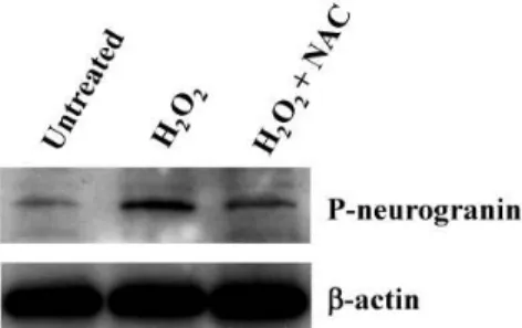
Neurospheres에 H2O2 및 N-acetyl-L-cysteine (NAC) 의 처리
이전의 연구결과를 통해 밝혀진 바와 같이 영아형 바텐병 에서 소포체 스트레스에 의해 활성산소가 발생하고 Ca2+ 항 상성에 붕괴가 발생하여 최종적으로 세포사멸이 발생한다면
또한 결합 단백질로서
[9], neurogranin calmodulin Ca2+ 항상 성 붕괴에 의한 영향을 받을 것으로 사료되어 활성산소가, 의 인산화에 미치는 영향을 조사하기 위해 다량
neurogranin ,
의 활성산소를 지닌EBD KO 쥐의neurospheres [9]에 산화 스트레스 유발물질인 H2O2를 처리하고, 24시간 경과 후 항산 화제인NAC 을 처리하여western blot 분석을 수행하였다.
Fig 4. W estern blot analysis for the treated to H2O2 and N-ace- tyl-L-cysteine (NAC) from cultured neurosphrers in the EBD KO mice brain. Control: M ock treated. β-actin was used as a loading control.
를 실시한 의 인산화된 수준과
Mock treat control neurogranin 비교하여 산화스트레스 유발물질인 H2O2를 처리하자 그 수 준이 현격히 상승하는 것을 확인하였으며, H2O2처리 후 항산 화제인NAC을 투여한 샘플에서는neurogranin의 인산화 수 준이 H2O2를 처리하였을 당시보다 감소된 결과를 확인하였 다(Fig. 4).따라서 이 결과를 통해neurogranin의 인산화 정도 는 세포가 가진 산화스트레스 정도에 의해 변화함을 확인 할 수 있었다.
고 찰
영아형 바텐병은PPT1 결핍 및 기능상실에 의해 발생하는 신경 퇴행 질환이다 현재까지 다양한 연구를 통해 영아형 바. 텐병의 발병원인을 밝혀내기 위한 시도가 이루어지고 있으나 아직 명확한 발병원인 및 치료방법이 알려지고 있지 않다 본. 연구에서는 Ca2+ 부족 조건에서calmodulin과 결합하는 것으 로 알려진neurogranin이WT 및EBD KO 쥐 뇌에서 발현되는 수준을 확인하였고 더불어 인산화된, neurogranin의 수준 산, 화스트레스와 항산화제가 인산화된neurogranin에 어떠한 효 과를 나타내는지 검토하였다. real-time PCR을 통해WT, EBD
쥐 뇌에서
KO neurogranin mRNA의 상대적 생성수준을 확인 할 결과W T쥐에서는 노화의 진행과 무관하게neurogranin
생성수준은 동일하였지만
mRNA ,EBD KO 쥐에서는 노화가
진행되면서 점차적으로neurogranin mRNA 생성수준이 감소 하는 것이 관찰되었는데 이를 통해, PPT1결핍이 뇌에서 풍부 하게 발현되는 neurogranin의 정상적인 발현을 저해시키는 것으로 사료된다 이와 같은. mRNA 의 감소가 단백질의 생성 감소를 의미하는지 확인하기 위하여WT 와EBD KO 쥐 뇌의 단백질을 사용하여western blot분석을 수행한 결과real-time 의 결과와 유사하게 쥐의 경우 노화가 진행됨에 있어
PCR WT
의 발현 양에는 큰 차이가 없었으나 이 제
neurogranin , PPT1
기능을 수행하지 못하는EBD KO 쥐에서는 노화가 진행될수 록neurogranin의 발현 양이 감소하는 것을 확인하였다 인산. 화된neurogranin의 경우WT 및EBD KO 쥐 모두에서 노화가
진행될수록 그 수준이 어느 정도 증가함이 관찰되었으나 월, 6 령의 EBD KO 쥐에서는 감소된neurogranin의 발현 양과 비 교할 경우 인산화된neurogranin이 증가해 있음을 알 수 있었 다 이는. PPT1결핍이neurogranin의 인산화를 촉진 혹은 탈 인산화를 저해하여, neurogranin과 결합하는 calmodulin과의 반응성에 영향을 미칠 수 있으며 이를 통해 NOS, CaMK-II,
활성에도 영향을 줄 것으로 사료된다
AC [13].
W T과EBD KO 쥐의 태아에서 배양한neurospheres를 사 용하여western blot 분석을 수행한 결과neurospheres내의
수준 및 인산화된 의 수준은 앞서
neurogranin neurogranin
실험결과와 동일한 양상으로 관찰되었다 이를 통해 배양된. 신경세포에서도 in vivo 상태와 동일한 현상이 발생하고 있음 을 확인하였고 이를 통해 배양 중인, neurospheres에 직접 산 화스트레스 유발 물질 및 항산화제를 투여할 수 있음을 확인 하였다 이를 바탕으로. EBD KO쥐의neurospheres에 산화스 트레스 유발물질인 H2O2를 처리하였고24시간 경과 후 대조 군에는 항산화제인NAC 를 처리하고 인산화된neurogranin 의 양적 변화를western blot분석을 통해 확인한 결과H2O2를 처리하자mock control과 비교하여 인산화된neurogranin의 수준이 증가한 것을 확인하였으며, H2O2를 처리 후 다시 NAC 을 투여한 샘플에서는 H2O2만을 처리한 샘플보다 neuro- 의 인산화 정도가 감소되는 결과를 확인하였다 이는
granin .
인산화된 neurogranin의 수준이 세포가 가진 산화스트레스 정도에 의해 변화함을 의미하며 항산화제에 의한 산화스트레, 스의 감소를 통해neurogranin의 기능을 정상적으로 회복시 킬 수 있을 것으로 사료되는 바이다.
현재 전 세계적으로 영아형 바텐병에 대한 연구가 활발히 진행 중 이지만 아직까지는 정확한 발병 원인 및 치료법이 밝혀지지 않아 보다 많은 연구가 수행되어야 할 것이라 사료 되며, PPT1결핍 및 기능장애에 의해 야기되는neurogranin의 정상적인 발현 장애와 인산화된neurogranin의 과다 존재에 의한 세포 내 이상 기작은 추가 연구를 통해 보다 명확하게 밝혀져야 할 것으로 생각되는 바이다.
요 약
영아형 바텐병은PPT1결핍 및 기능장애로 인해 발병하며, 명 당 명의 발병률을 가진 신경 퇴행 질환이다 전 세계
12,500 1 .
적으로 수많은 연구가 진행 중 이지만 아직 명확하게 밝혀진, 발병원인 및 치료방법에 대해서는 알려지고 있지 않다 본 연. 구에서는 뇌에서 풍부하게 발현되는neurogranin의 발현수준 이 W T과 EBD KO 쥐에서 어떤 변화를 보이는지 확인하기 위해mRNA, 단백질 배양된, neurospheres를 이용하여 실험 을 수행하였다. real-time PCR을 통한neurogranin의 발현수 준 비교 결과WT에서는 노화와 무관하게neurogranin mRNA 수준에 차이가 없었으나, EBD KO 쥐에서는 노화가 진행됨에
따라neurogranin mRNA 발현수준이 감소하였으며 뇌에서, 추출된 단백질을 이용한western blot 분석에서도 real-time
과 동일한 결과를 확인할 수 있었다 또한
PCR . WT, EBD KO
쥐의 태아로 부터neural stem cell 인neurospheres를 배양하 여western blot 분석을 수행한 결과PPT1결핍에 의해neuro- 의 정상적인 인산화에 문제가 발생함을 확인하였다 이
granin .
러한 결과들을 바탕으로neurospheres에 산화스트레스 유발 물질인 H2O2를 처리하였고, 24시간 경과 후 항산화제인NAC 을 처리하자 H2O2를 처리한 시료에서는mock control인 인산 화된neurogranin에 비해 그 수준이 증가하였으며, H2O2처리 후NAC을 투여한 시료의 인산화 수준은mock control보다는 높았지만 H2O2만을 처리한 시료 수준보다neurogranin의 인 산화 정도가 감소하는 결과를 확인하였다 이러한 결과들을. 통해PPT1 결핍으로 인해 신경세포 내에 과다하게 인산화된
이 존재하며 인산화 정도는 세포가
neurogranin , neurogranin
지닌 산화스트레스 정도에 의해 변화함을 알 수 있었다 또한. 항산화제를 사용하여 세포의 산화스트레스 수준을 감소시킬 경우neurogranin의 기능을 정상적으로 회복시킬 수 있는 가 능성을 확인하였다.
감사의 글
본 연구는 호서대학교2008년도 교내학술연구비 지원으로 수행되었으며(20080128), 이에 감사드립니다.
References
1. Ahtiainen, L., J. Kolikova, A. L. Mutka, K. Luiro, M. Gentile, E. Ikonen, L. Khiroug, A . Jalanko, and O. Kopra. 2007.
Palmitoyl protein thioesterase 1 (Ppt1)-deficient mouse neu- rons show alterations in cholesterol metabolism and cal- cium homeostasis prior to synaptic dysfunction. Neurobiol.
Dis. 28, 52-64.
2. Dominguez-Gonzalez, I., S. N. Vazquez-Cuesta, A. Algaba, and F. J. Diez-Guerra. 2007. Neurogranin binds to phospha- tidic acid and associates to cellular membranes. Biochem. J.
404, 31-43.
3. Gupta, P., A. A. Soyombo, A. Atashband, K. E. W isniewski, J. M . Shelton, J. A . Richardson, R. E. Hamm er, and S. L.
Hofmann. 2001. Disruption of PPT1 or PPT2 causes neuro- nal ceroid lipofuscinosis in knockout mice. Proc. Natl. Acad.
Sci. 98, 13566-13571.
4. Hobert, J. A . and G. Dawson. 2006. Neuronal ceroid lip- ofuscinoses therapeutic strategies: past, present and future.
Biochim. Biophys. Acta. 1762, 945-953.
5. Isosomppi, J., J. Vesa, A . Jalanko, and L. Peltonen. 2002.
Lysosomal localization of the neuronal ceroid lipofuscinosis CLN5 protein. Hum. Mol. Genet. 11, 885-891.
6. Jalanko, A., J. Vesa, T. Manninen, C. von Schantz, H. Minye, A. L. Fabritius, T. Salonen, J. Rapola, M . Gentile, O. Kopra,
and L. Peltonen. 2005. M ice with Ppt1Deltaex4 mutation replicate the INCL phenotype and show an inflamm ation- associated loss of interneurons. Neurobiol. Dis. 18, 226-241.
7. Kalviainen, R., K. Eriksson, M . Losekoot, I. Sorri, I.
Harvima, P. Santavuori, I. Jarvela, T. A utti, R. Vanninen, T. Salmenpera, and O. P. van Diggelen. 2007. Juvenile-onset neuronal ceroid lipofuscinosis with infantile CLN1 muta- tion and palmitoyl-protein thioesterase deficiency. Eur. J.
Neurol. 14, 369-372.
8. Kim, S. J., Z. Zhang, E. Hitomi, Y. C. Lee, and A. B.
M ukherjee. 2006. Endoplasmic reticulum stress-induced caspase-4 activation m ediates apoptosis and neuro- degeneration in INCL. Hum. Mol. Genet. 15, 1826-1834.
9. Kim, S. J., Z. Zhang, Y. C. Lee, and A. B. M ukherjee. 2006.
Palmitoyl-protein thioesterase-1 deficiency leads to the acti- vation of caspase-9 and contributes to rapid neuro- degeneration in INCL. Hum. Mol. Genet. 15, 1580-1586.
10. Filippo, M . Santorelli., B. Enrico, P. Vittoria, D. C. M atteo, C. Stefano, G. Paolo, and Z. M assimo. 1998. A Novel Insertion M utation (A169i) in the CLN1 Gene Is A ssociated with Infantile Neuronal Ceroid Lipofuscinosis in an Italian Patient. Biochem. Biophys. Res. Commun. 245, 519-522.
11. Kyttala, A., U . Lahtinen, T. Braulke, and S. L. Hofmann.
2006. Functional biology of the neuronal ceroid lip- ofuscinoses (NCL) proteins. Biochim. Biophys. Acta. 1762, 920-933.
12. Lehtovirta, M ., A. Kyttala, E. L. Eskelinen, M . Hess, O.
Heinonen, and A. Jalanko. 2001. Palmitoyl protein thioester- ase (PPT) localizes into synaptosomes and synaptic vesicles in neurons: implications for infantile neuronal ceroid lip- ofuscinosis (INCL). Hum. Mol. Genet. 10, 69-75.
13. Li, J., C. Yang, S. Han, P. Zu, J. W u, Q. Xu, and L. Fang.
2006. Increased phosphorylation of neurogranin in the brain of hypoxic preconditioned mice. Neurosci. Lett. 391, 150-153.
14. Martinez de Arrieta, C., B. Morte, A. Coloma, and J. Bernal.
1999. The human RC3 gene homolog, NRGN contains a thy- roid hormone-responsive element located in the first intron.
Endocrinology 140, 335-343.
15. Qiao, X., J. Y. Lu, and S. L. Hofmann. 2007. Gene expression profiling in a mouse model of infantile neuronal ceroid lip- ofuscinosis reveals upregulation of immediate early genes and mediators of the inflammatory response. BMC.
Neurosci. 8, 95.
16. Salonen, T., O. Heinonen-Kopra, J. Vesa, and A . Jalanko.
2001. Neuronal trafficking of palmitoyl protein thioesterase provides an excellent model to study the effects of different m utations which cause infantile neuronal ceroid lipofuscinocis. Mol. Cell Neurosci. 18, 131-140.
17. Salonen, T., I. Jarvela, L. Peltonen, and A. Jalanko. 2000.
Detection of eight novel palm itoyl protein thioesterase (PPT) m utations underlying infantile neuronal ceroid lip- ofuscinosis (INCL;CLN1). Hum. Mutat. 15, 273-279.
18. W atson, J. B., H. Khorasani, A. Persson, K. P. Huang, F.
L. Huang, and T. J. O'Dell. 2002. A ge-related deficits in long-term potentiation are insensitive to hydrogen per- oxide: coincidence with enhanced autophosphorylation of
Ca2+/calmodulin-dependent protein kinase II. J. Neurosci.
Res. 70, 298-308.
19. Zhang, J. P., W . Y. Liang, Z. H. Luo, Z. C. Yang, H. C.
Chan, and Y. S. Huang. 2007. Involvement of p38 MAP kin- ase in burn-induced degradation of m embrane phospholi- pids and upregulation of cPLA2 in cardiac myocytes. Shock 28, 86-93.
20. Zhang, Z., J. D. Butler, S. W . Levin, K. E. W isniewski, S.
S. Brooks, and A. B. M ukherjee. 2001. Lysosomal ceroid de- pletion by drugs: therapeutic im plications for a hereditary neurodegenerative disease of childhood. Nat. Med. 7,
478-484.
21. Zhang, Z., Y. C. Lee, S. J. Kim , M . S. Choi, P. C. Tsai, A.
Saha, H. W ei, Y. Xu, Y. J. Xiao, P. Zhang, A. Heffer, and A. B. M ukherjee. 2007. Production of lysophosphatidylcho- line by cPLA2 in the brain of mice lacking PPT1 is a signal for phagocyte infiltration. Hum. Mol. Genet. 16, 837-847.
22. Zhang, Z., Y. C. Lee, S. J. Kim, M . S. Choi, P. C. Tsai, Y.
Xu, Y. J. Xiao, P. Zhang, A . Heffer, and A . B. M ukherjee.
2006. Palmitoyl-protein thioesterase-1 deficiency mediates the activation of the unfolded protein response and neuro- nal apoptosis in INCL. Hum. Mol. Genet. 15, 337-346.