439
Copyright © 2015 The Korean Society of Fisheries and Aquatic Science pISSN:0374-8111, eISSN:2287-8815
서 론
면역반응에작용하는세포로써염증및숙주의방어에대식세 포
(macrophages)
의역할은잘알려져있다.
또한병원체의감 염및조직손상에의해유발된염증반응을해소하고손상된조 직을회복하는과정에도대식세포는중요한역할을한다.
이러 한다양한생리학적기능을수행할수있는근거는대식세포가주변의국소적인환경에따라다른형태로변형
(macrophage
polarization)
될수있는특성에따른것이다(Ferrante and Lei- bovich, 2012).
최근이러한대식세포표현형
(phenotypes)
의변화에대해많 은연구들이이루어지고있고,
활성화된두가지의형태의주요 표현형,
즉M1
및M2
의형태를가지고있음이확인되었다(Fer- rante and Leibovich, 2012).
고전적활성화(classically activat-
복강대식세포의 염증성 표현형에 대한 곰피(Ecklonia stolonifera) 유래 Phlorotannins의 효과
최민우·최준형·김형락·김재일*
부경대학교 식품영양학과
Effect of Phlorotannins Isolated from the Ethyl Acetate Fraction of Ecklonia stolonifera on Peritoneal Macrophage Polarization
Min-Woo Choi, Jun-Hyeong Choi, Hyeung-Rak Kim and Jae-Il Kim*
Department of Food Science and Nutrition, Pukyong National University, Busan 48513, Korea
Inflammation is a protective response to infection or injury. However, prolonged inflammation can contribute to the pathogenesis of many diseases, such as cancer, diabetes, arthritis, atherosclerosis, and Alzheimer’s disease. Recent studies have shown that activated macrophages, inflammatory effector cells, can react to tissue insults in a polar- ized manner, in which their phenotypes are polarized into two major subtypes, categorized as M1 or M2. Classical M1 activation involves the production of pro-inflammatory cytokines, such as interleukin (IL)-6 and tumor necrosis factor (TNF)-α, and free radicals, while M2 or alternative activation is an anti-inflammatory phenotype involved in homeostatic processes, such as wound healing, debris scavenging, and the dampening of inflammation via the production of very low levels of pro-inflammatory cytokines and high levels of anti-inflammatory mediators, includ- ing IL-10. As part of our ongoing effort to isolate anti-inflammatory compounds from seaweeds, we investigated the effects of phlorotannins isolated from the brown alga Ecklonia stolonifera on macrophage polarization. Mouse peritoneal macrophages were treated with various concentrations of the extracts, and real-time RT-PCR analyses were performed to examine the expression of polarization markers: IL-1β, IL-6, and TNF-α for M1 and arginase-1, peroxisome proliferator-activated receptor (PPAR)-γ, found inflammatory zone-1 (Fizz-1), chitinase 3-like 3 (Ym1), and Krüppel-like factor 4 (Klf-4) for M2. The pretreatment of cells with eckol, dieckol, and phlorofucofuroeckol-A (PFF-A), isolated from the ethyl acetate fraction of E . stolonifera ethanolic extract, potentiated the anti-inflammatory M2 phenotype of the macrophages. These results indicate that phlorotannins derived from E . stolonifera can be used to enrich macrophages with markers of the M2 anti-inflammatory state.
Key words: Ecklonia stolonifera , Phlorotannins, Macrophage polarization, Alternative activation, Anti-inflammatory phenotype
This is an Open Access article distributed under the terms of the Creative Commons Attribution Non-Commercial Licens (http://creativecommons.org/licenses/by-nc/3.0/) which permits unrestricted non-commercial use, distribution, and reproduction in any medium, provided the original work is properly cited.
http://dx.doi.org/10.5657/KFAS.2015.0439 Korean J Fish Aquat Sci 48(4) 439-446, August 2015 Received 18 August 2015; Accepted 23 August 2015
*Corresponding author: Tel: +82. 51. 629. 5849 Fax: +82. 51. 629. 5842
E-mail address: [email protected]
ed)
형태인M1
표현형은세균의lipopolysaccharides (LPS)
및peptidoglycan
과같은외인성pathogen-associated molecular patterns (PAMPs)
및괴사된세포에서유리되는단백질및핵 산과같은damage-associated molecular patterns (DAMPs),
그 리고T helper 1 (Th1) cell
에서분비되는cytokine
인interferon (IFN)-γ
에의한자극에의해서유도되고,
이에의해염증촉진성cytokines
및매개체[nitric oxide (NO)
및superoxide radical]
의생성량이 많아지는염증촉진성
(pro-inflammatory)
특성을 가진다(Mosser, 2003; Bystrom et al., 2008; Colin et al., 2014).
다른형태인
M2
표현형은염증을억제(anti-inflammatory)
하 는특성을가지고있고,
이는IL-4
및IL-13
과같은Th2 cyto- kines
에의해서유도된다(Chang et al., 2015).
이들cytokines
이외에glucocorticoids
및resolvins
과같은항염증성및염증 해소성자극또한M2 phenotype
을유도한다(Titos et al., 2011;
Zhang et al., 2009). M2
대식세포는IL-10
과같은항염증성cytokine
을분비하고염증반응부산물의소거에관련된CD36
의발현이증가하게되고
,
그에따라상처의회복및조직의리 모델링과같은항상성을유지하는데작용함으로써염증을해 소하는데주된역할을한다.
따라서염증반응동안대식세포의 활성화상태는염증의지속여부를결정짓는데중요한역할을 하고,
표현형의적절한변화(macrophage polarization)
가이루 어지지않을경우만성염증상태로발전하게된다(Ferrante and Leibovich, 2012; Bystrom et al., 2008).
병리학적으로만성적 인염증을동반하는죽상동맥경화(atherosclerosis),
천식,
관절 염과같은질환에서염증촉진성M1
표현형이주로관찰되는 것이보고되었다.
최근이러한염증성질환의예방및치료를위 한새로운접근법으로써약물및식이성분등을이용하여대식 세포를M2
표현형으로의유도하거나M2
표현형을더욱증강 시키는방법들이연구되고있다(Camell and Smith, 2013; Li et al., 2013; Chen et al., 2014; Chang et al., 2015).
해조류는육상식물에서는발견되지않는물질들을함유하고 있어다양한생리활성을나타내는것으로알려져있다
(Kim et al., 2009; Lee et al., 2012; Wijesinghe and Jeon YJ, 2012).
그 중갈조류는엽록소와갈색을띄는xanthophyll
계caroteinods
의 일종인fucoxanthin
과fucoidan
등의 다당류, fucosterol, phlorotannins
과같은다양한생리활성물질들을가지고있는 것으로보고되었다(Kang et al., 2003; Hosokawa et al., 2004;
Kim et al., 2012;Brown et al., 2014).
본연구실에서는이전 에수행한실험에서다양한갈조류의주정추출물및유기용매 획분들을분획하였고,
또한분리한화합물을이용하여다양한 활성을분석하여보고하였다.
특히,
곰피(Ecklonia stolonifera)
주정추출물의ethyl acetate (EtOAc)
획분및그획분에서분 리한phlorotannins [eckol, dieckol, phlorofucofuroeckol-A
(PFF-A)]
의경우뛰어난항산화및항염증효과가있는것으로확인되었다
(Kim et al., 2009; Lee et al., 2012).
본연구에서 는복강대식세포(mouse peritoneal macrophages, PEM)
를이용하여
LPS
및IL-4
처리하고M1
및M2 marker genes
의발 현을분석하여M1
및M2
표현형이유도됨을확인하고자하 였고,
이들시스템에곰피-
유래phlorotannins
을처리하여대식 세포표현형의변화를분석하였다.
본연구를통해향후항염증 성물질의개발에대한새로운접근법을제시하고,
또한해조류 를이용한대식세포표현형의변화연구에대한기초자료를제 공하고자한다.
재료 및 방법
실험재료
부산기장에서채취한곰피
(E. stolonifera)
로부터95%
주정 추출물의 제조및그로부터ethyl acetate (EtOAc)
획분의분 리,
그리고EtOAc
획분으로부터phlorotannins (eckol, dieckol, PFF-A)
의분리및동정은전보의방법(Kim et al., 2009; Lee et al., 2012)
에따라수행하였다.
세포배양 및 처리
ICR
마우스를이용하여Alleva et al. (1998)
의방법에따라복 강대식세포(peritoneal macrophages, PEM)
을분리하였다.
즉10
주령마우스를이용하여대식세포분리3-4
일전에복강에4% thioglycollate (w/v) 3 mL
을주사하였다.
이후마우스를마 취하여경추탈골한뒤복강의내피를노출시키고,
주사기를이 용하여3% bovine calf serum (BCS, Gibco, Rockville, MD, USA)
을주입하여복강의대식세포를회수하였다.
이과정을2-3
회 반복한 이후 원심분리하고phosphate-buffered saline (PBS)
로 세척하였다.
분리한복강대식세포는10% fetal bo- vine serum (FBS)
와penicillin (100 units/mL), streptomycin (100 μg/mL)
을첨가한Dulbecco’s modified Eagle’s medium (DMEM, WellGene, Daegu, Korea)
을 사용하여5% CO
2, 37℃
에서1
일배양한뒤이후시료를처리하였다.
각시료들은100% dimethyl sulfoxide (DMSO)
에녹여사용하였고, IL-4
와IL-13 (R&D systems, Minneapolis, MN, USA), LPS (Sigma- Aldrich, St. Louis, MO, USA)
는PBS
에녹여사용하였다. 세포독성 시험
복강대식세포를
96-well plate
에2×10
5cells/well
로분주하 고37℃
에서24
시간동안배양하였다.
이후에각시료들이농도별로희석된
DMEM
배지로교체하여다시24
시간배양하였다
.
이후CellTiter96
®Aqueous 3-(4,5-dimethylthiazol-2-yl)-
5-(3-carboxymethoxyphenyl)-2-(4-sulfophenyl)-2H-tetrazoli-
um (MTS)
시험키트(Promega, Madison, WI, USA)
를사용하 여제조사의방법에따라세포생존율을분석하였다. MTS
용액 은FBS-free DMEM
에5% (v/v)
의농도로섞어100 μL
씩처 리하였다. 1
시간후에microplate reader (Glomax Multi Detec-
tion System, Promega, Madison, WI, USA)
를이용하여490
nm
파장에서흡광도를측정하였다. mRNA 발현양의 분석
복강대식세포
(1×10
6cells/well)
에시료들을4
시간동안처 리한이후에Quiazol
시약(Quiagen, Valencia, CA, USA)
을이 용하여total RNA
를분리하였고, 1 μg
의total RNA
에GoS- cript™ Reverse transcriptase (Promega, Madison, WI, USA)
를 사용하여 제조사의 방법대로
cDNA
합성을 진행하였다.
합성된
cDNA
와TOPreal™ qPCR 2× PreMIX (Enzynom- ics, Daejeon, Korea),
표적유전자특이적인primers
를사용하 여real-time reverse transcription-polymerase chain reaction (real-time RT-PCR, Rotor-Gene Q, Quiagen, Valencia, CA, USA)
방법을통해유전자발현양을분석.비교하였다.
유전자발현의비교는상대적인비교를위해
ΔΔCt
방법을이용하였으며
, real-time RT-PCR
에 이용된각primers
의 염기서열은Table 1
에나타내었다.
통계 처리
본연구의모든실험은세번이상반복하였으며
,
얻어진결과들을평균값과표준편차
(mean±SD)
를계산하여나타내었다.
실험군간의유의성검증은
P<0.05
수준에서Student’s t-test
로 검증하였다.
결과 및 고찰
복강대식세포에 대한 세포독성 시험
곰피
(E. stolonifera)
주정 추출물에서 분획한ethyl acetate (EtOAc)
획분(ESA)
과분리한eckol, dieckol, PFF-A
의세포독 성을평가하기위해,
각각의시료들을마우스의복강대식세포에농도별로처리하고세포생존율을분석하였다
. Fig. 1
에나타 내었듯이, eckol
과dieckol
의경우100 µM
의농도까지, PFF-A
의경우50 µM, ESA
의경우200 µM
의농도까지독성이나타 나지않았다.
이전대식세포세포주인RAW264.7 cell
에대한PFF-A
및dieckol
의세포독성을분석하였을때에도유사한경 향이관찰되었다(Kim et al., 2009).
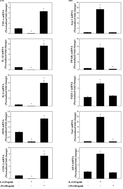
복강대식세포의 염증촉진성(M1) 및 항염증성(M2) 표 현형의 유도
상기에서분리한복강대식세포
(PEM)
에LPS
또는IL-4
를4
시간동안처리하고,
이들세포들이M 1
및M2
형태로유도되 는지알아보기위해서RNA
를추출하여real-time RT-PCR
방 법을통해서각각의marker
유전자들의발현profile
을분석하 였다. M1
에대한markers (Mosser., 2003; Bystrom et al., 2008;
Colin et al., 2014)
로는IL-1β, IL-6, TNF-α
와같은cytokines
이외에NO
및prostaglandin E
2(PGE
2)
와같은염증촉진성매 개체를생성하는inducible nitric oxide synthase (iNOS)
및cy- clooxygenase-2 (COX-2)
를분석하였다. M2
에대한marker
유 전자는arginase (Arg)-1
과found in inflammatory zone (Fizz)- 1, chitinase-like 3 (Ym1), peroxisome proliferator-activated receptor (PPAR)-γ, Krüppel-like factor (Klf)-4
와같은염증상 태개선및조직복구에관여하는 유전자들(Liao et al., 2011;
Ferrante and Leibovich, 2012; Liu et al., 2014)
을분석하였다. Fig. 2
에 나타내었듯이, LPS
처리에의해M1
의 특징적인marker
유전자들의 발현이 현저하게 증가하였고(P<0.05),
IL-4
처리에 의해 그 발현양이 감소하는 것으로 나타났다(P<0.05) (Fig. 2A).
반면, M2 marker
유전자들의경우IL-4
처 리에의해모두유의적으로발현이증가하는것으로나타났고(P<0.05), LPS
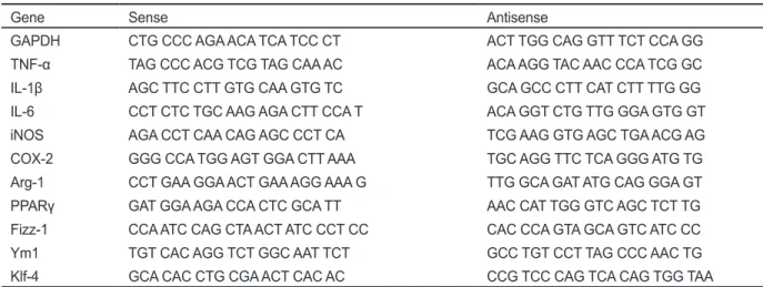
처리에의해서는큰변화가없거나일부감소하Table 1. Primers used for real-time RT-PCR analysis
Gene Sense Antisense
GAPDH CTG CCC AGA ACA TCA TCC CT ACT TGG CAG GTT TCT CCA GG
TNF-α TAG CCC ACG TCG TAG CAA AC ACA AGG TAC AAC CCA TCG GC
IL-1β AGC TTC CTT GTG CAA GTG TC GCA GCC CTT CAT CTT TTG GG
IL-6 CCT CTC TGC AAG AGA CTT CCA T ACA GGT CTG TTG GGA GTG GT
iNOS AGA CCT CAA CAG AGC CCT CA TCG AAG GTG AGC TGA ACG AG
COX-2 GGG CCA TGG AGT GGA CTT AAA TGC AGG TTC TCA GGG ATG TG
Arg-1 CCT GAA GGA ACT GAA AGG AAA G TTG GCA GAT ATG CAG GGA GT
PPARγ GAT GGA AGA CCA CTC GCA TT AAC CAT TGG GTC AGC TCT TG
Fizz-1 CCA ATC CAG CTA ACT ATC CCT CC CAC CCA GTA GCA GTC ATC CC
Ym1 TGT CAC AGG TCT GGC AAT TCT GCC TGT CCT TAG CCC AAC TG
Klf-4 GCA CAC CTG CGA ACT CAC AC CCG TCC CAG TCA CAG TGG TAA
GAPDH, glyceraldehyde 3-phosphate dehydrogenase; TNF-α, tumor necrosis factors-α; IL-1β, interleukin-1β; IL-6, interleukin-6; iNOS, inducible nitric oxide synthase; COX-2, cyclooxygenase-2; Arg-1, arginase-1; PPARγ, peroxisome proliferator-activated receptor-γ; Fizz-1, found in inflammatory zone-1; Ym1, chitinase-3-like protein 3; Klf-4, Kruppel-like factor-4
는것으로확인되었다
(Fig. 2B).
이러한결과는복강대식세포 는각각의자극에의해염증촉진성및항염증성표현형으로유 도된다는것을확인할수있었고,
이러한조건은향후대식세포 표현형의polarization
분석을위한기초시스템으로이용될수 있을것이다.
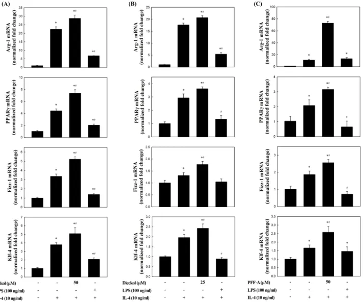
복강대식세포의 항염증성 표현형에 대한 곰피-유래 시 료의 효과
복강대식세포의
M2 polarization
에대한곰피-
유래eckol, di- eckol, PFF-A
의직접적인유도효과를분석한결과,
일부의시료에서
M2 marker
유전자의발현이증가하는것으로나타났지만유의적인효과는관찰되지않았다
(data not shown).
그러 나,
복강대식세포를이들화합물로전처리한이후IL-4
로자극하였을경우
M2 marker
유전자의발현이현저하게증가하는것으로나타났다
(Fig. 3). Fig. 2
와마찬가지로, IL-4
만을처리하였을때
M2 marker
유전자의발현이유의적으로증가하는것이확인되었고
(P<0.05),
이러한증가는LPS
전처리에의해 서는변화가없거나감소또는증가함으로써일정한경향은없 는것으로확인되었다.
그러나,
곰피-
유래화합물을전처리한 경우에는IL-4
단독으로자극한경우와비교하여M2 marker
유전자의발현양은현저하게더높은수준으로증가하는것으 로나타났다(Fig. 3, P<0.05).
특히이러한상승효과는eckol, dieckol, PFF-A
세가지화합물모두공통적으로관찰되었고(Fig. 3A, B, and C, P<0.05),
유전자발현양의증가폭을기준으 로세화합물의효과를비교하였을때, dieckol<eckol<PFF-A
의순으로나타났다.
M2 marker
유전자의 하나인Arg-1
은 가장 잘알려진M2 marker
이다(Munder et al., 1999).
이효소는Arg
을L-ornithine
으로변환시키고이후상처회복및matrix
침착에사용될수있 는polyamines
과proline
의전구체를제공해주는역할을한다(Munder, 2009). Arg
은iNOS
의기질이기도하기때문에Arg-1
의발현이증가할경우
iNOS
에의한NO
의생성을경쟁적으로억제하는역할을하게된다
(Corraliza et al., 1995; Morris, 2007). Fizz-1
의경우세포외부(extracelluar) matrix
의침착을 촉진시킴으로써손상된조직의회복을돕는것으로알려져있 다(Raes et al., 2002). Klf-4
는최근연구에서M2
대식세포에 서현저하게증가하고반면M1
형태에서는감소하는것으로 확인됨으로써대식세포의M1/M2 polarization
에중요한조절 인자로작용한다는것이보고되었다(Liao et al., 2011). Ym1
은extracellular matrix
성분의분해를저해함으로써조직손상을 막는것으로보고되었다(Chang et al., 2001, Hung et al., 2002).
이상과같이
M2 marker
유전자들의공통적인특성은염증상태의해소및조직손상의회복에관련된항염증활성을나타낸다
.
따라서이들의발현이곰피-
유래phlorotannins
에의해더욱더 높은수준으로증가한다는것은M2
표현형이강화되고그에의 해강력한염증억제효과를나타낼수있음을의미하는것이다.
Fig. 1. Cytotoxicity of the ethyl acetate fraction of Ecklonia stolonifera (ESA) or phlorotannins isolated from ESA in mouse peritoneal mac- rophages (PEM). Cells were treated with various concentrations of the samples including ESA, eckol, dieckol and phlorofucofuroeckol-A (PFF-A) for 24 hrs. Viabilities of the cells were measured by MTS assay and their relative viabilities are shown.
최근대식세포의
M2
표현형유도를위해식이지방산의이용 가능성을검토한연구가보고되었다. Oleic acid (C18:1 n-9)
처리에의해대식세포에서
Klf-4
및Arg-1
의전사수준이유의 적으로증가하는것이관찰되었고(Camell and Smith., 2013),
(B) (A)
Fig. 2. Changes in mRNA expression profile induced by lipopolysacchrarides (LPS) or IL-4 treatment in mouse peritoneal macrophages (PEM). M1 (A) or M2 (B) marker gene expressions in the cells treated with LPS or IL-4 were analyzed by real-time RT-PCR. All data are presented as mean±SD of three independent experiments. *P<0.05 indicates significant difference compared to non-treated control.
n-3
고도불포화지방산인docosahexaenoic acid (DHA, C22:6 n-3)
처리에의해서RAW264.7
세포의M1
표현형이억제되는 반면, M2
표현형이증가하는것으로확인되었다(Chang et al., 2015). DHA
의경우PPARγ ligand
로작용함으로써PPARγ
의 활성화를일으키고M2 polarization
을유도하는것으로확인되 었다.
또한항산화및항염증등의다양한생리활성을가진것 으로알려져있는curcumin
의경우RAW264.7
세포에처리하 였을때Klf-4
및Fizz-1
의발현을증가시키는것으로나타났고,
이러한M2 phenotype
유도는PPARγ
의활성화를통해서일어 나는것으로보고되었다(Chen et al., 2014). PPARγ
는M2
형태로의변환을유도하는데중추적인역할을하는전사인자이고
(Bouhlel et al., 2007),
본연구에서관찰된PPARγ
의발현증가 는곰피-
유래화합들이M2 polarization
유도에효과적임을의 미하는것이다(Fig. 3).
이상의결과에서만성염증을병리학적특성으로하는염증성 질환의치료및예방을위한새로운접근법으로써대식세포의 표현형을항염증성으로유도하는데곰피
-
유래phlorotannins
이 효과적임을확인할수있었다.
향후보다광범위한marker
유전 자들의분석및관련신호전달기전의구명이필요한것으로판 단된다.
(A) (B) (C)
Fig. 3. Effect of eckol, dieckol or phlorofucofuroeckol-A (PFF-A) isolated from ethyl acetate fraction of Ecklonia stolonifera on M2 polar- ization in IL-4-treated mouse peritoneal macrophages (PEM). Cells pretreated with eckol (A), dieckol (B), PFF-A (C) or lipopolysaccha- rides (LPS) for 1 h were stimulated with IL-4 for 4 hr, and then changes in their M2 marker gene expressions were analyzed by real-time RT- PCR. All data are presented as mean±SD of three independent experiments. *P<0.05 or #P<0.05 indicates significant differences compared to non-treated control or IL-treated group, respectively.
사 사
이논문은부경대학교자율창의학술연구비
(2014
년)
에 의하 여연구되었음.
References
Alleva DG, Kaser SB and Beller DI. 1998. Intrinsic defects in macrophage IL-12 production associated with immune dys- function in the MRL/++ and New Zealand Black/White F1 Lupus-prone mice and the Leishmania major-susceptible BALB/c strain. J Immunol 161, 6878-6884.
Bouhlel MA, Derudas B, Rigamonti E, Dièvart R, Brozek J, Haulon S, Zawadzki C, Jude B, Torpier G, Marx N, Staels B and Chinetti-Gbaguidi G. 2007. PPARgamma activation primes human monocytes into alternative M2 macrophages with anti-inflammatory properties. Cell Metab 6, 137-143.
Brown ES, Allsopp PJ, Magee PJ, Gill CI, Nitecki S, Strain CR and McSorley EM. 2014. Seaweed and human health. Nutr Rev 72, 205-216.
Bystrom J, Evans I, Newson J, Stables M, Toor I, van Rooi- jen N, Crawford M, Colville-Nash P, Farrow S and Gil- roy DW. 2008. Resolution-phase macrophages possess a unique inflammatory phenotype that is controlled by cAMP. Blood 112, 4117-127. http://dx.doi.org/10.1182/
blood-2007-12-129767.
Camell C and Smith CW. 2013. Dietary oleic acid increases M2 macrophages in the mesenteric adipose tissue. PLoS One 8, e75147. http://dx.doi.org/10.1371/journal.pone.0075147.
Chang HY, Lee HN, Kim W and Surh YJ. 2015. Docosahexae- noic acid induces M2 macrophage polarization through per- oxisome proliferator-activated receptor γ activation. Life Sci 120, 39-47. http://dx.doi.org/10.1016/j.lfs.2014.10.014.
Chang NC, Hung SI, Hwa KY, Kato I, Chen JE, Liu CH and Chang AC. 2001. A macrophage protein, Ym1, transient- ly expressed during inflammation is a novelmammalian lec- tin. J Biol Chem 276, 17497-17506.
Chen F, Guo N, Cao G, Zhou J and Yuan Z. 2014. Molecu- lar analysis of curcumin-induced polarization of murine RAW264.7 macrophages. J Cardiovasc Pharmacol 63, 544- 552. http://dx.doi.org/10.1097/FJC.0000000000000079.
Colin S, Chinetti-Gbaquidi G and Staels B. 2014. Macrophage phenotypes in atherosclerosis. Immunol Rev 262, 153-166.
http://dx.doi.org/10.1111/imr.12218.
Corraliza IM, Soler G, Eichmann K and Modolell M. 1995.
Arginase induction by suppressors of nitric oxide synthe- sis (IL-4, IL-10 and PGE2) in murine bone-marrow-de- rived macrophages. Biochem Biophys Res Commun 206, 667-673.
Ferrante CJ and Leibovich SJ. 2012. Regulation of macro- phage polarization and wound healing. Adv Wound Care (New Rochelle)1, 10-16.
Hosokawa M, Kudo M, Maeda H, Kohno H, Tanaka T and Miyashita K. 2004. Fucoxanthin induces apoptosis and enhances the antiproliferative effect of the PPARγ ligand, troglitazone, on colon cancer cells. Biochim Biophys Acta 1675, 113-119.
Hung SI, Chang AC, Kato I and Chang NC. 2002. Transient ex- pression of Ym1, a heparin-binding lectin, during develop- mental hematopoiesis and inflammation. J Leukoc Biol 72, 72-82.
Kang HS, Chung HY, Jung JH, Son BW and Choi JS. 2003. A new phlorotannin from the brown alga Ecklonia stolonifera.
Chem Pharm Bull 51, 1012-1014.
Kim AR, Shin TS, Lee MS, Park JY, Park KE, Yoon NY, Kim JS, Choi JS, Jang BC, Byun DS, Park NK and Kim HR.
2009. Isolation and identification of phlorotannins from Eck-
lonia stolonifera with antioxidant and anti-inflammatory
properties. J Agric Food Chem 57, 3483-3489. http://dx.doi.org/10.1021/jf900820x.
Kim KJ, Yoon KY, and Lee BY. 2012. Low molecular weight fucoidan from the sporophyll of Undaria pinnatifida sup- presses inflammation by promoting the inhibition of mitogen-activated protein kinases and oxidative stress in RAW 264.7 cells. Fitoterapia 83, 1628-1635. http://dx.doi.
org/10.1016/j.fitote.2012.09.014.
Laskin DL, Sunil VR, Gardner CR and Laskin JD. 2011. Macro- phages and tissue injury: agents of defense or destruction?
Annu Rev Pharmacol Toxicol 51, 267-288. http://dx.doi.
org/10.1146/annurev.pharmtox.010909.105812.
Lee MS, Kwon MS, Choi JW, Shin T, No HK, Choi JS, Byun DS, Kim JI and Kim HR. 2012. Anti-inflammatory activities of an ethanol extract of Ecklonia stolonifera in lipopolysac- charide-stimulated RAW 264.7 murine macrophage cells.
J Agric Food Chem 60, 9120-9129. http://dx.doi:10.1021/
jf3022018.
Li QZ, Sun J, Han JJ and Qian ZJ. 2013. Anti-inflammation of simvastatin by polarization of murine macrophages from M1 phenotype to M2 phenotype. Zhonghua Yi Xue Za Zhi 93, 2071-2074.
Liao X, Sharma N, Kapadia F, Zhou G, Lu Y, Hong H, Paru- churi K, Mahabeleshwar GH, Dalmas E, Venteclef N, Flask CA, Kim J, Doreian BW, Lu KQ, Kaestner KH, Clement K and Jain MK. 2011. Kruppel-like factor 4 regulates mac- rophage polarization. J Clin Invest 121, 2736-2749. http://
dx.doi.org/10.1172/JCI45444.
Liu YC, Zou XB, Chai YF and Yao YM. 2014. Macrophage polarization in inflammatory diseases. Int J Biol Sci 10, 520- 529. http://dx.doi.org/10.7150/ijbs.8879.
Morris SM Jr. 2007. Arginine metabolism: boundaries of our knowledge. J Nutr 137, 1602S-1609S.
Mosser DM. 2003. The many faces of macrophage activation. J Leukoc Biol 73, 209-212.
Munder M, Eichmann K, Morán JM, Centeno F, Soler G
and Modolell M. 1999. Th1/Th2-regulated expression of ar- ginase isoforms in murine macrophages and dendritic cells. J Immunol 163, 3771-3777.
Munder M. 2009. Arginase: an emerging key player in the mam- malian immune system. Br J Pharmacol 158, 638-651. http://
dx.doi.org/10.1111/j.1476-5381.2009.00291.x.
Raes G, Noël W, Beschin A, Brys L, de Baetselier P and Hassan- zadeh GH. 2002. FIZZ1 and Ym as tools to discriminate be- tween differentially activated macrophages. Dev Immunol 9, 151-159.
Titos E, Rius B, González-Périz A, López-Vicario C, Morán- Salvador E, Martínez-Clemente M, Arroyo V and Clària J. 2011. Resolvin D1 and its precursor docosahexae- noic acid promote resolution of adipose tissueinflamma- tion by eliciting macrophage polarization toward an M2- like phenotype. J Immunol 187, 5408-5418. http://dx.doi.
org/10.4049/jimmunol.1100225.
Wijesinghe WA and Jeon YJ. 2012. Exploiting biological activi- ties of brown seaweed Ecklonia cava for potential industrial applications: a review. Int J Food Sci Nutr 63, 225-235.
http://dx.doi.org/10.3109/09637486.2011.619965.
Zhang S, Shen Z, Hu G, Liu R and Zhang X. 2009. Effects of endogenous glucocorticoids on allergic inflammation and T(H)1/T(H)2 balance in airway allergic disease. Ann Allergy Asthma Immunol 103, 525-534. http://dx.doi.org/10.1016/
S1081-1206(10)60270-0.