Received on June 13, 2014. Revised on July 18, 2014. Accepted on July 24, 2014.
CC This is an open access article distributed under the terms of the Creative Commons Attribution Non-Commercial License (http://creativecommons.org/licenses/by-nc/3.0) which permits unrestricted non-commercial use, distribu- tion, and reproduction in any medium, provided the original work is properly cited.
*Corresponding Authors. Byungsuk Kwon, School of Biological Sciences, University of Ulsan, 93 Daehak-ro, Nam-gu, Ulsan, Korea. Tel: 82-52-259-2860; Fax: 82-52-259-2740; E-mail: [email protected], Hong R. Cho, Department of Surgery, Ulsan University Hospital, School of Medicine, University of Ulsan, 877 Bangeojinsunwhando-ro, Dong-gu, Ulsan, Korea. Tel: 82-52-250-7100; Fax: 82-52-250-8071; E-mail: [email protected]
Abbreviations: IL-1RAP, IL-1 receptor accessory protein; HK, heat-killed; KO, knock-out; MOI, multiplicity of infection;
ROS, reactive oxigen species; ST2, suppression of tumorigenicity 2; WT, wild-type
IL-33 Priming Enhances Peritoneal Macrophage Activity in Response to Candida albicans
Vuvi G. Tran1, Hong R. Cho2,3* and Byungsuk Kwon1,2*
1School of Biological Sciences, University of Ulsan, Ulsan 680-749, 2Biomedical Research Center, 3Department of Surgery, Ulsan University Hospital, School of Medicine, University of Ulsan, Ulsan 682-714, Korea
IL-33 is a member of the IL-1 cytokine family and plays a role in the host defense against bacteria, viruses, and fungi. In this study, we investigated the function of IL-33 and its re- ceptor in in vitro macrophage responses to Candida albicans. Our results demonstrate that pre-sensitization of isolated peritoneal macrophages with IL-33 enhanced their pro-inflammatory cytokine production and phagocytic activ- ity in response to C. albicans. These macrophage activities were entirely dependent on the ST2-MyD88 signaling pathway. In addition, pre-sensitization with IL-33 also in- creased ROS production and the subsequent killing ability of macrophages following C. albicans challenge. These results indicate that IL-33 may increase anti-fungal activity against Candida through macrophage-mediated resistance mechani- sms.
[Immune Network 2014;14(4):201-206]
Keywords: Peritoneal macrophage, IL-33, Candida albicans, Pro-inflammatory cytokines, Phagocytosis, Fungicidal activity
INTRODUCTION
IL-33 is a multifaceted, multifunctional cytokine. It was ini- tially found to be highly expressed in the nuclei of endothe- lial and epithelial cells (1). Binding of IL-33 to a hetero-
dimeric receptor complex consisting of ST2 and IL-1RAP re- sults in the recruitment of an adapter protein, MyD88, and the onset of signal transduction (2). IL-33 signaling induces the production of various mediators involved in inflammation and tissue repair by nonhematopoietic and hematopoietic cells, including endothelial cells, epithelial cells, macro- phages, basophils, eosinophils, and mast cells (3).
Candida albicans is a commensal organism of the gastro- intestinal tract and vagina (4); however, it is also the most common pathogen associated with mucosal and systemic infections. Invasive candidiasis is the fourth leading cause of nosocomial bloodstream infection in the USA (5), and it is estimated to occur worldwide in over 400,000 people every year with mortalities ranging from 46% to 75% despite admin- istration of antifungal therapy in modern intensive care unit facilities (6). The resistance mechanism of mice to C. albicans infection relies mainly on the phagocytic and killing ability of innate immune cells, such as neutrophils and macrophages (7,8). Our previous studies have shown that IL-33 enhances neutrophil recruitment to site of infection and their phagocytic activities in peritoneal C. albicans infection (9,10). Little is known about the role of IL-33 in macrophages during Candida infection. In this study, we demonstrate that pre-sen- sitizing peritoneal macrophages with IL-33 enhances various activities that facilitate fungal clearance.
Figure 1. Pre-treatment of peritoneal macrophages with IL-33 increases the production of pro-inflammatory cytokines in response to C. albicans infection. Peritoneal macrophages were pre-treated with 100 ng/ml of IL-33 or PBS for 2 h prior to infection with heat-killed (HK) C. albicans (MOI=10). Levels of IL-6 (A) and TNFα (B) were measured 2 h, 6 h, 12 h, and 24 h after challenge with C. albicans. Data are presented as the mean±SEM of 2-3 trials with similar results. *p<0.05; **p<0.01.
MATERIALS AND METHODS Mice
C57BL/6 mice were purchased from Orient Bio-Charles River.
MyD88 KO mice with a C57BL/6 genetic background were maintained in a specific pathogen-free facility and used when they were 7 to 8 weeks old. All experiments were conducted according to the regulations of the Animal Committee of the University of Ulsan.
Production of recombinant IL-33 protein
Recombinant IL-33 protein was produced as previously de- scribed (9-11).
Isolation of peritoneal macrophages
Mice were intraperitoneally injected with 3 ml of sterile 3%
thioglycollate broth (Difco). After 3 days, mice were sacrificed and peritoneal exudate cells (PECs) were harvested from the peritoneal cavities. The cell pellet was washed and re- suspended in DMEM medium supplemented with 10% fetal bovine serum (FBS). PECs were seeded into 24-well tissue culture plate (1×10⁶ cells/ml) and incubated at 37oC for 2 h, at which point the adherent cells were harvested.
Measurement of cytokines
Cytokines present in culture supernatants were measured by ELISA (eBioscience or R&D systems), according to manu- facturers’ protocols.
Real-time RT-PCR
Total RNA was extracted from isolated peritoneal macro- phages using TRIzol (Invitrogen), according to the manu-
facturer’s instructions. cDNA was synthesized from 2μg of total RNA using SuperScript reverse transcriptase (Invitrogen).
Real-time PCR was performed using SYBR Green PCR Master Mix (Qiagen) in the ABI 7500 Fast Real-Time PCR System (Applied Biosystems). The primers used to measure levels of ST2 mRNA were 5’-TGA CGG CCA CCA GAT CAT TCA CAG-3’ (forward) and 5’-GCC AAA GCA AGC TGA ACA GGC AAT AC-3’ (reverse).
Phagocytosis assay
In vitro phagocytosis assays were performed as previously de- scribed (7). Briefly, peritoneal macrophages were incubated with IL-33 (100 ng/ml) at 37oC for 2 h. Heat-killed C. albicans was labeled with FITC, opsonized, and challenged against IL-33-primed macrophages at 37oC for 20 min (MOI=10).
Phagocytosis was stopped by transferring cells to ice and washing thoroughly with cold FACS buffer. Extracellular fluo- rescence was quenched by adding 200μl of PBS containing 0.04% trypan blue and 1% formaldehyde. Fungus-containing cells were then counted by flow cytometry and the degree of phagocytosis expressed as the percentage of FITC-positive macrophages.
Killing assay
Macrophages were mixed with opsonized live C. albicans (MOI=1) and incubated at 37oC for 20 min with continuous rotation. Cells were washed thoroughly in cold PBS, re- suspended in warm DMEM medium, and further incubated at 37oC. At the indicated times, 200 ml samples were har- vested and cells lysed in PBS containing 0.1% Triton X-100.
CFUs were quantified by plating lysates on agar. Percent kill- ing was calculated as [1−(CFUs after incubation/phagocytosed
Figure 2. The effect of IL-33 pre- sensitization on pro-inflammatory cytokine production by peritoneal macrophages is dependent on the ST2-MyD88 signaling axis. (A) Peritoneal macrophages were pre- treated with 100 ng/ml of IL-33 or PBS for 2 h prior to infection with heat-killed (HK) C. albicans (MOI=
10). ST2 mRNA expression was analyzed by performing qRT-PCR 24 h post-challenge. (B) Peritoneal macrophages were pre-treated with 5μg/ml of anti-mouse ST2 mAb or control IgG for 1 h, at which point 100 ng/ml IL-33 was added to the medium. After a 2 h incubation period, macrophages were then challenged with HK-Candida (MOI
=10). Levels of IL-6 and TNFα were determined by ELISA with culture supernatants harvested 24 h post-challenge. (C) The same ex- periments were repeated with per- itoneal macrophages isolated from WT and MyD88 KO mice. Data are presented as the mean±SEM of 2-3 independent experiments. *p<0.05;
**p<0.01.
CFUs at the start of incubation)]×100.
Determination of reactive oxygen species (ROS) generation
Macrophages were pre-sensitized with IL-33 (100 ng/ml) or PBS for 2 h and were then further incubated in the presence of 2μM 2',7'-dichlorodihydrofluorescein diacetate (Molecular Probes) for 20 min in the dark. Cells were washed twice with PBS, challenged with heat-killed C. albicans (MOI=10) for 10 or 30 min, and analyzed by FACS.
Flow cytometry
Prepared cells were blocked with 2.4G2 mAb in staining buf- fer (PBS containing 0.2% BSA and 0.1% sodium azide) at 4oC for 20 min, incubated with rat anti-mouse T1/ST2 (clone DJ8)-FITC mAb or rat IgG1κ isotype control at 4oC for 20 min, and then washed twice with staining buffer. Flow cyto-
metric analysis was performed using a FACS Canto II unit (BD Biosciences). Data were analyzed with FACS Diva (BD Biosci- ences) and FlowJo software (TreeStar).
Statistical analysis
All data were analyzed in GraphPad Prism5. Survivals and un- paired data were analyzed with log rank- and t-tests, re- spectively. Results are expressed as the mean±SEM. Statisti- cal significance was accepted for p-values<0.05.
RESULTS
IL-33 enhances the production of pro-inflammatory cy- tokines by macrophages via the ST2/MyD88 signaling axis
To investigate whether IL-33 can affect cytokine production by macrophages in response to C. albicans, peritoneal macro-
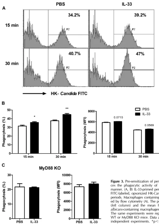
Figure 3. Pre-sensitization of peritoneal macrophages with IL-33 enhan- ces the phagocytic activity of macrophages in a MyD88-dependent manner. (A, B) IL-33-primed peritoneal macrophages were mixed with FITC-labeled, opsonized HK-Candida (MOI=10) for the indicated time periods. Macrophages containing FITC-labeled C. albicans were analyz- ed by flow cytometry (A). The percentage of FITC-positive macrophages (left column) and the mean fluorescence intensity for FITC of C.
albicans-containing macrophages (right column) are quantified in B. (C) The same experiments were repeated with peritoneal macrophages of WT or MyD88 KO mice. Data are presented as the mean±SEM of 2-3 independent experiments. *p<0.05; **p<0.01.
phages were stimulated with heat-killed Candida (MOI=10).
We found that C. albicans markedly increased the secretion of IL-6 and TNFα by macrophages at 6 h after challenge and thereafter (Fig. 1). Pre-sensitization of macrophages with IL-33 enhanced the production of these pro-inflammatory cy- tokines following challenge with C. albicans (Fig. 1).
Since IL-33 signaling is mediated by ST2 (11), we examined whether the expression of ST2 in peritoneal macrophages is regulated by C. albicans. RT-PCR analysis demonstrated that ST2 expression was significantly increased in macrophages 24 h after stimulation with C. albicans (Fig. 2A). We next sought to determine whether the pre-sensitization effect of IL-33 was
Figure 4. IL-33 pre-sensitization enhances ROS production and fungi- cidal activity in peritoneal macrophages. (A) ROS production was measured as described in Materials and Methods. (B) Peritoneal macrophages were pre-incubated with IL-33 (100 ng/ml) for 3 h before challenge with live, opsonized Candida (MOI=1). Fungal killing activities were determined 15 and 30 min after challenge with C. albicans. Data are presented as the mean±SEM of 2-3 independent experiments. *p<0.05; **p<0.01.
dependent on ST2. Notably, pre-treatment with a ST2 neutral- izing antibody abolished the effect of pre-sensitization with IL-33 on IL-6 and TNFα production following C. albicans in- fection (Fig. 2B). As ST2 signaling is dependent upon MyD88, we further analyzed the effect of pre-sensitization with IL-33 on pro-inflammatory cytokine production in MyD88 KO peri- toneal macrophages. As expected, IL-33 pre-sensitization had no effect on the secreted levels of IL-6 and TNFα following C. albicans challenge in MyD88 KO macrophages (Fig. 2C).
These data clearly establish that the effect of pre-sensitization with IL-33 on IL-6 and TNFα production by peritoneal mac- rophages after C. albicans challenge is mediated through the ST2-MyD88 signaling axis.
IL-33 pre-sensitization enhances the phagocytic activity of macrophages
We have previously shown that IL-33 pre-sensitization can in- crease the phagocytic activity of neutrophils against C. albi- cans (9,10), though little is known about the effect of IL-33 on macrophage phagocytosis. As seen in Fig. 3A, IL-33 pri- ming results in a significant increase in the overall percentage of macrophages containing FITC-conjugated C. albicans at 15 and 30 min post-challenge, yet this was accompanied by only a marginal increase in the FITC intensity observed in C. albi- cans-engulfed macrophages (Fig. 3B). Notably, IL-33 priming had no effect on the phagocytic activity of MyD88 KO peri- toneal macrophages against C. alibicans (Fig. 3C). These re- sults indicate that IL-33 priming enhanced macrophage phag- ocytosis in a MyD88-dependent manner by expanding the
population of phagocytic macrophages targeting C. albicans, rather than elevating the phagocytic ability of the individual macrophages.
In addition to phagocytosis, the ability of macrophages to kill engulfed pathogens is also critical for the innate immune response to fungal infection (12,13). Notably, the capacity to generate and utilize intracellular reactive oxygen species (ROS) is crucial for killing C. albicans in phagocytes (14,15).
To determine the role of IL-33 priming on ROS production, we analyzed the fluctuations of ROS levels in IL-33-primed peritoneal macrophages in response to fungal challenge. As shown in Fig. 4A, IL-33-primed macrophages displayed high- er intracellular ROS levels following challenge when com- pared to mock-primed controls. Consistent with this result, phagocytosed C. albicans were more rapidly cleared inside IL-33 primed macrophages (Fig. 4B).
DISCUSSION
We have previously shown that administration of IL-33 prior to peritoneal challenge with a lethal dose of C. albicans pre- vents sepsis-induced mortality (9,10). In that model, pre-sen- sitization of the host with IL-33 increases neutrophil responses at multiple stages. Notably, IL-33-primed peritoneal macro- phages play a critical role in neutrophil recruitment by pro- ducing the CXCR1/2 necessary for neutrophil chemotaxis.
The data presented in this study further indicate that peri- toneal macrophages may be critical in neutrophil activation, as IL-33-primed macrophages are sufficient to provide the pro-inflammatory cytokines prerequisite for full neutrophil activation. Our results also suggest that IL-33-primed macro- phages may contribute to fungal clearance by directly phag- ocytosing C. albicans, though in vivo experiments are neces- sary to confirm our in vitro observations.
Although neutrophils play a key role in the anti-fungal de- fense against C. albicans, macrophages appear to be of equal importance in fungal clearance (16,17). A recent study dem- onstrates that treatment with IL-13 or PPARγ ligand enhances dectin-1 receptor expression, resulting in induction of phag- ocytic activity in peritoneal macrophages (18). Interestingly, IL-33 can strengthen M2 macrophage activation to enhance fungal clearance in response to Pneumocystis murina (19).
Similarly, our unpublished data revealed that IL-33 pre-treat- ment can result in a significant increased M2 macrophage po- larization in the kidney following C. albicans systemic in- fection at 3 d post-challenge and thereafter; however, the ma-
jority of macrophages present in the kidney at 1 d post-in- fection display a M1 phenotype. Taken together, this suggests that IL-33 promotes the activation of M1 macrophages during early C. albicans infection, and then slowly converts them to- ward a M2 macrophage phenotype. Nevertheless, both types of macrophages are known to mediate activities necessary for fungal clearance. This characteristic of IL-33 is unique among cytokines and has a merit in its ability to enhance both resist- ance and tolerance to C. albicans infection (our unpublished data). In summation, our in vitro data indicate that IL-33 may function as an important mediator of anti-fungal host defenses early after C. albicans infection by promoting fungal clear- ance.
ACKNOWLEDGEMENTS
This work was supported by grants from the National Research Foundation of Korea (NRF) funded by the Ministry of Education, Science and Technology (2009-0094050 and NRF-2012R1A12008653).
CONFLICTS OF INTEREST
The authors have no financial conflict of interest.
REFERENCES
1. Moussion, C., N. Ortega, and J.-P. Girard. 2008. The IL-1lLike cytokine IL-33 is constitutively expressed in the nucleus of endothelial cells and epithelial cells: a novel ‘alarmin’? PLoS One 3: e3331.
2. Liew, F. Y., N. I. Pitman, and I. B. McInnes. 2010.
Disease-associated functions of IL-33: the new kid in the IL-1 family. Nat. Rev. Immunol. 10: 103-110.
3. Le, H., W. Kim, J. Kim, H. R. Cho, and B. Kwon. 2013.
Interleukin-33: a mediator of inflammation targeting hema- topoietic stem and progenitor cells and their progenies.
Front. Immunol. 4:104.
4. Romani, L. 2011. Immunity to fungal infections. Nat. Rev.
Immunol. 11: 275-288.
5. Lionakis, M. S., and M. G. Netea. 2013. Candida and host determinants of susceptibility to invasive candidiasis. PLoS Pathog. 9: e1003079.
6. Brown, G. D., D. W. Denning, N. A. Gow, S. M. Levitz, M.G. Netea, and T. C. White. 2012. Hidden killers: human fungal infections. Sci. Transl. Med. 4: 165rv13.
7. Lionakis, M. S., B. G. Fischer, J. K. Lim, M. Swamydas, W.
Wan, C. C. Richard Lee, J. I. Cohen, P. Scheinberg, J. L.
Gao, and P. M. Murphy. 2012. Chemokine receptor Ccr1 drives neutrophil-mediated kidney immunopathology and mortality in invasive candidiasis. PLoS Pathog. 8: e1002865.
8. Majer, O., C. Bourgeois, F. Zwolanek, C. Lassnig, D.
Kerjaschki, M. Mack, M. Müller, and K. Kuchler. 2012. Type I interferons promote fatal immunopathology by regulating inflammatory monocytes and neutrophils during Candida infections. PLoS Pathog. 8: e1002811.
9. Le, H. T., V. G. Tran, W. Kim, J. Kim, H. R. Cho, and B.
Kwon. 2012. IL-33 priming regulates multiple steps of the neutrophil-mediated anti-Candida albicans response by mod- ulating TLR and dectin-1 signals. J. Immunol. 189: 287-295.
10. Kim, J., W. Kim, H. T. Le, U. J. Moon, V. G. Tran, H. J.
Kim, S. Jung, Q.-T. Nguyen, B.-S. Kim, J.-B. Jun, H. R. Cho, and B. Kwon. 2014. IL-33-induced hematopoietic stem and progenitor cell mobilization depends upon CCR2. J. Immunol.
doi: 10.4049/jimmunol.1400176.
11. Schmitz, J., A. Owyang, E. Oldham, Y. Song, E. Murphy, T.
K. McClanahan, G. Zurawski, M. Moshrefi, J. Qin, X. Li, D.
M. Gorman, J. F. Bazan, and R. A. Kastelein. 2005. IL-33, an interleukin-1-like cytokine that signals via the IL-1 re- ceptor-related protein ST2 and induces T helper type 2-asso- ciated cytokines. Immunity 23: 479-490.
12. Brown, G. D. 2011. Innate antifungal immunity: the key role of phagocytes. Annu. Rev. Immunol. 29: 1-21.
13. Cheng, S.-C., L. A. Joosten, B. J. Kullberg, and M. G. Netea.
2012. Interplay between Candidaalbicans and the mamma- lian innate host defense. Infect. Immun. 80: 1304-1313.
14. Missall, T. A., J. K. Lodge, and J. E. McEwen. 2004. Mechani- sms of resistance to oxidative and nitrosative stress: im- plications for fungal survival in mammalian hosts. Eukaryot.
Cell 3: 835-846.
15. Wellington, M., K. Dolan, and D. J. Krysan. 2009. Live Candidaalbicans auppresses production of reactive oxygen species in phagocytes. Infec. Immun. 77: 405-413.
16. Lewis, L. E., J. M. Bain, C. Lowes, C. Gillespie, F. M. Rudkin, N. A. Gow, and L. P. Erwig. 2012. Stage specific assessment of Candidaalbicans phagocytosis by macrophages identifies cell wall composition and morphogenesis as key determi- nants. PLoS Pathog. 8: e1002578.
17. Marcil, A., D. Harcus, D. Y. Thomas, and M. Whiteway.
2002. Candidaalbicans Killing by RAW 264.7 mouse macro- phage cells: effects of Candida genotype, infection ratios, and gamma interferon treatment. Infec. Immun. 70: 6319-6329.
18. Galès, A., A. Conduché, J. Bernad, L. Lefevre, D. Olagnier, M. Béraud, G. Martin-Blondel, M. D. Linas, J. Auwerx, A.
Coste, and B. Pipy. 2010. PPARγ controls Dectin-1 ex- pression required for host antifungal defense against Candida albicans.PLoS Pathog. 2010. 6: e1000714.
19. Nelson, M.P., B. S. Christmann, J. L. Werner, A. E. Metz, J. L. Trevor, C. A. Lowell, and C. Steele. 2011. IL-33 and M2a Alveolar Macrophages Promote Lung Defense against the Atypical Fungal Pathogen Pneumocystis murina. J. Immunol.
186: 2372-2381.