섬오가피 추출물의 항암관련 사이토카인 분비활성
유수연·박원봉*,#
대구한의대학교 한약재 약리학과, *서울여자대학교 자연과학대학 화학과 (Received February 16, 2010; Revised May 12, 2010; Accepted May 12, 2010)
Effects of Acanthopanax koreanum Extracts on Anticancer Related Cytokine Secretions
Su-Yun Lyu and Won-Bong Park*,#
Dept. of Herbal Medicinal Pharmacology, Daegu Haany University, Kyeongbuk 712-715, Korea
*Dept. of Chemistry, College of Natural Sciences, Seoul Women’s University, Seoul 139-774, Korea
Abstract — Stems and roots of Acanthopanax koreanum Nakai were extracted with water and treated on immune cells in order to determine their immunomodulatory activites. Various Th-1 type cytokines were measured using ELISA including interleukin (IL)-2, IL-12, interferon-gamma (IFN-γ), and tumor necrosis factor-alpha (TNF-α) secreted by dendritic cells, T- cells, intestinal epithelial cells, natural killer cells, and macrophages. As a result, there was a significant increase in IL-12 and IFN-γ, secretion, but there was no change in the secretion of TNF-α. Additionally T-cells slightly increased the secretion of IL-2, but there was a significant increase of IL-2 in intestinal epithelial cells. Therefore, our results suggest that A. kore- anum Nakai may act as an immunomodulator by stimulating the cell-mediated immunity which can help the immune system defend against infections or cancer cells.
Keywords □ Acanthopanax koreanum, Cytokines, IL-2, IL-12, IFN-γ
인삼과 같은 과(오가과, Araliaceae)에 속해있는 오가피는 잎사 귀의 모양이 인삼과 매우 흡사할 뿐 아니라 효능 면에서도 인삼 못지않기 때문에 나무인삼, 또는 나무산삼이라고 부르기도 한다.1) 이 식물은 전통적으로 강정, 강장 및 진통목적으로 사용되고 있 으며, 민간에서 신경통, 류마티스, 고혈압, 중풍, 당뇨, 습진, 거 담 등에 사용되어 왔다.1,2)오가피는 우리나라 전역과 중국 동북 부 지방, 러시아의 시베리아, 일본의 북해도 등에 분포하며, 국 내에는, 섬오가피(Acanthopanax koreanum Nakai), 흰털오가피 (A. divaricatus var. albeofructus), 가시오가피(A. senticosus, Maxim) 등 10여종이 자생 또는 재배되고 있다.1)가시오가피는 1960년대 러시아 과학자 Breckman에 의해서 범 적응적 효과가 보고된 이래 세계적으로 주목 받기 시작하였으나 우리나라에는 분포도가 그리 높지 않은 것으로 알려져 있다. 제주지역에 주로 자생하는 섬오가피의 성분으로는 acanthoside D, syringoside, ariensin, chiisanoside 등이 보고되어 있다.3-5)
암의 면역학적 치료는 인체 내의 면역세포들이 암세포 표면에
발현되는 정상세포와 다른 구조를 인식함으로써 항암 면역반응 을 일으킨다는 데 기초를 두고 있다. 생체 내 정상적으로 존재하 는 면역감시기구 중 종양세포를 인지하고 소멸시키는 것은 T-세 포나 자연살해세포(natural killer cell, NK cell) 등에 의한 세포 성 면역기능의 활성화가 중요한 역할을 한다.6) T-세포의 활성화 과정은 이를 조절하는 항원제시세포(antigen-presenting cell, APC)의 역할이 선행되어야만 하는데, 수지상 세포(dendritic cell) 는 고도로 전문화된 항원제시세포로서 resting/naive T-세포의 강 력한 activator이며, 여러 가지 cytokine을 분비한다.7-10)
T-세포는 크게 CD8+, cytotoxic T(Tc)-세포와 CD4+ helper T (Th)-세포로 구분된다.6,11)항암 면역반응은 주로 Tc-세포가 암세 포상의 MHC class I 항원과 결합된 종양 항원을 인식함으로써 이루어지며 이러한 Tc-세포의 완전한 활성화를 위해 Th-세포로 부터 생성된 cytokine(interleukin-2)의 도움이 필요하다. Th-세 포는 분비되는 cytokine에 의해 다시 Th-1 type(IL-2, IL-12, IFN-γ, TNF-α 등)과 Th-2 type(IL-4, IL-6, IL-8, TGF-β 등)으 로 나뉜다.12) Th-1/Th-2 세포는 공통의 naive T-세포로부터 분 화하며, naive T-세포가 항원자극 후에 IL-12 존재 하에서 분화 하면 Th-1 세포로, IL-4 존재 하에서 분화하면 Th-2 세포로, 분 화한다.12)
#본 논문에 관한 문의는 저자에게로 (전화) 02-970-5655 (팩스) 02-975-3159 (E-mail) [email protected]
종설
Proinflammatory cytokine을 생산하는 Th-1 세포는 세포매개 성 면역반응(cell-mediated immune response)을 촉진한다.13) IL- 2는 CD4+ T-세포에 의해 생산되는 T-세포 성장인자로서, NK- 세포 및 B-세포의 성장을 촉진하며, Th-2에서 Th-1으로 면역체 계를 유도하고 NK-세포를 활성화하여 종양의 전이를 억제시킨 다. 또한 활성화된 대식세포나 수지상 세포에 의하여 생산되는 IL-12는 NK-세포를 활성화하며, T-세포나 NK-세포에 의하여 IFN-γ를 생성하게 하는 강력한 유도체이다. IFN-γ는 대식세포를 활성화하며, IL-12의 양을 증가시키고, Th-1 type 반응을 촉진한 다. 또한 IFN-γ는 B-세포에 작용하여 IgG급의 항체 생산을 촉진 하며, cytotoxic T-세포의 생산을 도와준다. IL-12는 IFN-γ가 생 성하는 Th-1 세포의 분화를 증가시켜 특이성 면역의 개발을 촉 진한다.13)따라서 IL-12 및 IFN-γ는 virus 등 세포 내 감염과 종 양세포에 대한 숙주의 방어에 중요한 영향을 미친다.14) TNF-α 는 대식세포에 작용하여 염증반응을 촉진하고, T-세포와 B-세포 의 활성화에 co-stimulator로 작용하며, 종양세포에 작용하여 세 포자살(apoptosis)을 유도하기도 한다.15,16) 한편, Th-2 세포는 anti-inflammatory cytokine인 Th-2 type cytokine(IL-4, IL-6, IL-8, IL-10 등)을 분비하여 T-세포의 활성을 억제하면서 Th-1 type cytokine의 생산을 억제시킨다.16,17)따라서 Th-1 세포의 활 성화 및 Th-2 세포의 억제는 항암면역 기능에 중요한 영향을 미 칠 수 있다.
오가피의 면역조절에 관련된 연구로는 가시오가피(Acanthopanax senticosus)의 당단백 분획이 NK-세포 및 대식세포를 활성화시키 며, IL-1, IL-12, IFN-γ의 분비를 촉진시킨다는 보고가 있다.18,19) 또한 흰털오가피 열매 추출물이 Th-1 cytokine의 분비를 촉진시 키며, Th-2 cytokine의 분비를 억제시켜 virus 등 세포 내 감염 과 종양세포에 대한 숙주의 방어에 도움이 될 가능성이 있다고 보고된바 있다.20) 또한, 섬오가피로부터 분리한 다당체 (polysaccharide)가 B-세포를 활성화시키나 T-세포에는 영향이 없 으며,21)섬오가피로부터 분리한 acanthoic acid가 IL-1과 TNF-α 의 분비를 억제시키며,22,23)장내피세포(human colon epithelial cells, IECs)에서 mitogen-activated protein kinases(MAPKs)와 nuclear factor-kappa B(NF-κB) 경로를 통한 IL-8의 생산을 억제 한다고 보고된 바 있다.24)
본 연구에서는 국내자생 섬오가피의 줄기 및 뿌리 추출물의 Th-1 type cytokine인 IL-2, IL-12, IFN-γ, TNF-α의 분비에 미 치는 영향을 확인하고, 섬오가피의 암 예방 및 치료물질로서의 가능성을 알아보고자 하였다.
실험방법
시료
섬오가피 뿌리의 물 추출물(AKR) 및 줄기의 물 추출물(AKS)
은 대전생명공학연구소의 이정준 박사팀에서 준비한 시료를 사 용하였다. 추출물은 증류수에, 50 mg/ml의 농도로 용해시킨 후, -20oC에 보관하고, 실험 시, 적정 농도로 세포 배양 배지로 희석 하여 사용하였다.
세포주 및 배양
Mouse dendritic 세포인 lymphoid 계열의 DC1 세포와 myeloid 계열의 DC2 세포를 서울대학교 의과대학 면역학교실의 강재승 교수로부터 분양 받았다. 그리고 rat 장 상내피세포인 IECs(intestinal epithelial cells)-6 세포, mouse 대식세포인 RAW 264.7 세포, 사람 T-세포인 Jurkat-T 세포는 한국세포주은 행(KCLB, Seoul, Korea)에서 분양 받았다. 또한 Human IL-2 cDNA를 transfection 시킨 사람 자연살해세포인 NK92MI 세포 는 American Type Culture Collection(ATCC, Rockville, MD, USA)에서 분양 받았다.
DC1, DC2 및 Jurkat-T 세포는 RPMI 1640(GibcoTM Invitrogen Coporation, Carlsbad, CA, USA)에 10%(v/v) fetal bovine serum(FBS, GibcoTM)과 1%(v/v) penicillin-streptomycin을 가 해 37oC(5% CO2/air)에서 배양하였다. RAW 264.7 및 IEC-6 세 포는 modified Eagle's medium(DMEM, GibcoTM)에 10%(v/v) FBS와 1%(v/v) penicillin-streptomycin을 가해 37oC에서 배양하 였다. NK92MI는 ribonucleosides와 deoxyribonucleosides를 포 함하지 않은 Alpha minimum essential medium에 2 mM L- glutamine, 1.5 g/l, sodium bicarbonate (GibcoTM), 0.2 mM inositol(Sigma, St. Louis, MO, USA), 0.1 mM 2-mercapto- ethanol(Sigma), 0.02 mM folic acid(Sigma), 12.5% horse serum (GibcoTM) 12.5% FBS, 1% penicillin-streptomycin을 가해 배양 하였다.
MTT assay
배지로 희석시킨 시료를 세포에 농도 별로 처리하여, MTT(3- [4,5-dimethylthiazol-2-yl]-2,5-diphenyltetrazolium bromide, 5 mg/ml) assay로 측정하여 세포에 대한 시료의 독성을 확인하였 다. 즉, 1×10⁴cells/well로 넣은 세포와 시료를 96 well plate에 분주한 다음 48시간 동안 배양하고, MTT 용액을 50 µl씩 첨가 하여, 4시간 동안 37oC, 5% CO2 배양기에서 배양하였다. 배양 액을 제거한 후, DMSO(dimethylsulfoxide, Duksan)용액 200 µl 씩 첨가하고 10분간 흔들어준 후, 595 nm에서 ELISA(enzyme- linked immunosorbent assay) reader(Molecular Devices Co.) 로 흡광도를 측정하였다.
ELISA(Enzyme-Linked Immunosorbent Assay)
Jurkat-T 세포는 50 ng/ml PMA와 1 µg/ml PHA로 그리고 그 이외의 세포는 1 µg/ml의 LPS로 자극 시킨 후. 배지로 희석시킨
섬오가피 추출물을 농도 별로 세포에 처리하여 24시간 배양 후, 상등액 100 µl를 취하여 cytokine 측정용 시료로 사용하였다. 96 well plate에 purified anti-mouse interleukin(IL)-12, anti- human IL-2, anti-mouse TNF-α, anti-human IFN-γ, anti-rat IL-2 capture antibody(eBiosciences, San Diego, CA, USA), biotin-conjugated anti-mouse IL-12, anti-human IL-2, anti- mouse TNF-α, anti-human interferon-γ, anti-rat IL-2 monoclonal antibody(eBiosciences, USA)를 사용하여 세포배양 상등액 중의 cytokine을 측정하였다. Anti-cytokine capture antibody를 binding buffer(0.1 M sodium phosphate buffer, pH 9.0)에 2µg/ml 되도록 희석해서 96 well plate(NUNC Co., Rochester, NY, USA)에 50 ml/well씩 넣은 후, 4oC에서 하룻밤 방치하여 coating하였다. Tween 20(Sigma, Poole, UK)을 phosphate buffered saline(PBS)에 0.5%(v/v)가 되도록 가한 PBS/Tween 용 액으로 plate를 3회 세척 후, blocking buffer인 1%(w/v) bovine serum albumin(BSA)(Sigma, Poole, UK)을 가하여 상온에서 2 시간 동안 방치하였다. PBS/Tween 용액으로 3회 세척 후, 세포 배양액 50 ml를 가하고 4oC에서 하룻밤 방치하였다. PBS/Tween 으로 4회 세척 후, biotinylated antibody를 blocking buffer/PBS/
Tween에 희석하여 100 µl/well로 처리하여 37oC에서 1시간 동안 방치하였다. 다시 세척하고 1 : 1000으로 희석한 streptavidin- peroxidase(eBiosciences, USA)을 100 µg/well로 blocking buffer/
PBS/Tween에 희석하여 넣고 1시간 방치 후, PBS/Tween으로 세 척하였다. 0.03% hydrogen peroxide를 함유한 0.05 M phosphate- citrate 완충용액(pH 5.0)에 3,3',5,5'-tetramethylbenzidine(TMB, Sigma, Poole, UK)를 0.1 mg/ml가 되도록 제조한 기질용액을 100µg/well씩 넣었다. Stop reagent로 2.5 M sulfuric acid을 50µl/well로 넣어 15분간 방치하여 효소반응을 중지시킨 후 96- well plate reader(Molecular Devices, Sunnylvale, CA, USA)를 이용하여 450 nm에서 흡광도를 측정하였다.
통계처리
모든 실험결과는 평균±표준오차로 나타내었으며 자료분석은 ANOVA test를 이용하여 유의성을 검정하였다.
결과 및 고찰
섬오가피 추출물의 세포독성
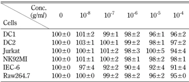
Cytokine 측정을 위한 전 단계로 시료의 세포에 대한 영향을 평가하기 위하여 사용된 6종류의 세포에 대한 시료의 독성여부 를 MTT assay에 의하여 확인하였다. 10-4g/ml에서 시작하여 10 배수로 희석한 뿌리(AKR) 및 줄기(AKS) 물 추출물을 각 세포 에 처리하여 세포의 생존율을 측정하였다. 그 결과, 사용된 모든 시료의 농도에서 사용된 세포들의 생존율이 90% 이상인 것으로
보아 세포의 생존에 큰 영향이 없는 것을 알 수 있었다(Table I, II). 따라서 이 결과를 토대로, 세포의 생존에 큰 영향이 없는 10-6g/ml 이하의 추출물을 세포에 처리하여 cytokine 분비실험 을 수행하였다.
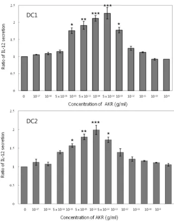
섬오가피 추출물을 처리한 수지상 세포의 IL-12분비 경향 T-세포의 활성화 과정은 항원제시세포의 역할이 선행되어야만 하는데, 수지상세포는 전문화된 항원제시 세포이다.7-10)또한 이 세포는, Tc-세포, Th-1 type-세포 및 NK-세포를 활성화 시키는 것으로 확인된 바 있다.25-27)본 연구에서는 수지상 세포의 IL-12 분비에 미치는 섬오가피 추출물의 영향을 알아보기 위하여 LPS 를 처리하여 자극시킨 생쥐의 림프계(lymphoid) 수지상세포인 DC1과 골수계(myeloid) 수지상세포인 DC2에 추출물을 처리하 여 배양시킨 후 배양액 중의 IL-12의 농도를 측정하였다. 그 결 과, 매우 낮은 농도(5×10-17g/ml)의 추출물을 처리한 DC1 및 DC2 세포에서 IL-12의 분비가 증가하기 시작하는 것으로 나타 났다. 10-13g/ml 농도의 AKS를 처리한 경우 DC1 및 DC2 세포 모두에서 IL-12 분비가 가장 높았으며, 그 이상의 농도로 처리하 면 IL-12의 분비가 다시 감소하는 bell-shape type의 peak 형태 를 나타냈다(Fig. 1). 또한 AKR을 처리한 경우에도 유사한 분비 경향을 보였으며, 5×10-14g/ml의 추출물을 처리한 DC1세포의 경우 IL-12의 분비가 2.3배 현저하게 증가하였으며, DC1 및 DC2 세포 모두 줄기 추출물을 처리한 경우보다 더욱 낮은 농도에서 Table I − Cell viability (%) of stem extract of Acanthopanax koreanum (AKS) against cells. Cell viability was measured by MTT assay
Conc.
(g/ml) Cells
0 10-8 10-7 10-6 10-5 10-4
DC1 100±0 101±2 099±1 98±2 096±1 96±2
DC2 100±0 103±1 100±1 99±2 098±1 97±2
Jurkat 100±0 100±1 101±2 98±3 100±5 94±4 NK92MI 100±0 101±1 100±2 98±1 098±2 98±1 IEC-6 100±0 097±4 092±2 90±4 092±4 91±4 Raw264.7 100±0 100±0 099±2 98±2 096±2 95±0
Table II − Cell viability (%) of root extract of Acanthopanax koreanum (AKR) against cells. Cell viability was measured by MTT assay
Conc.
(g/ml) Cells
0 10-8 10-7 10-6 10-5 10-4
DC1 100±0 100±1 099±2 098±1 97±2 097±1 DC2 100±0 101±2 099±2 099±1 99±1 098±2 Jurkat 100±0 101±2 100±3 100±3 99±4 096±2 NK92MI 100±0 102±3 100±2 099±1 97±2 096±1 IEC-6 100±0 099±1 106±2 105±1 97±4 101±3 Raw264.7 100±0 101±2 100±3 099±1 97±2 097±3
최대치를 보였으며, 분비 증가 정도가 더욱 큰 것을 알 수 있었 다(Fig. 2).
Steinman 등은 수지상 세포는 분화되기 전의 전구세포의 계통 에 따라 형태학적으로 뚜렷이 구분되며, 이들을 각각 lymphoid DC와 myeloid DC로 구분할 수 있다고 보고하였다.28-30)그런데 수지상 세포의 기능은 동물과 사람에서 서로 상반되는 면역학적 인 조절기능을 수행한다는 주장도 있다.31)즉, 생쥐에서는 림프구 계통에서 분화된 lymphoid DC는 주로 Th1-세포를 유도하고 골 수구 계통에서 분화된 myeloid DC는 주로 Th2-세포를 유도하는 데 비하여,31)사람의 면역체계에서는 lymphoid DC나 형질세포 계통에서 분화된 plasmacytoid DC는 Th2 반응을 초래하고 myeloid DC는 주로 Th1 면역반응을 유도하는 것으로 밝혀져 있 다.32)그러나 수지상 세포가 활성화된 후에 매우 짧은 기간 동안 만 IL-12를 생산할 수 있으므로 nave T-세포를 Th1-세포로 유도 할 수 있는 능력은 이때만 존재하며, 그 후에는 수지상 세포가 단 지 Th2 반응만을 유도하거나 central memory T-세포를 생성하
도록 할 수 있다고 주장도 있다.33)또한 이러한 면역반응의 유형 을 결정할 수 있는 다른 요인들로서 항원의 투여량, 그리고 T-세 포와 항원제시세포가 접촉하는 기간 등을 들 수 있다고 하였다.33) 본 연구 결과에서는 Maldonado-Lopez 등31)의 주장과는 달리 섬오가피 추출물을 전구세포가 다른 생쥐의 림프계 수지상세포 (DC1)와 골수계 수지상세포(DC2)에 처리한 결과, 줄기 및 뿌리 추출물 모두 Th1 계열의 세포를 유도하는 IL-12의 분비를 촉진 시키는 것으로 나타났다. 또한 뿌리 추출물이 줄기 추출물보다 IL-12의 분비에 미치는 영향이 더욱 컸으며, 10-14~10-13g/ml 농 도의 추출물을 처리하였을 때 분비증가 정도가 가장 큰 것으로 나타났다. 따라서 Sallusto 등33)의 주장대로 면역반응의 유형은 수지상세포로 유도되는 전구세포의 기원이나 분화를 유도하는 자극의 특성 때문이 아니라 항원의 종류 및 양이나 수지상세포 가 활성화되고 성숙되는 정도에 따라서 결정되는 것으로 추측된다.
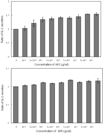
섬오가피 추출물을 처리한 T-세포 및 장상피세포의 IL-2 분비 경향 IL-2는 T-세포에 의해 생산되는 T-세포 성장인자로서, NK-세 포 및 B-세포의 성장을 촉진하며, Th-2에서 Th-1으로 면역체계 Fig. 1 − Secretion of IL-12 from DC1 and DC2 cells treated with
stem extract of Acanthopanax koreanum (AKS). The extracts were diluted with complete cell culture medium (CTCM) and then tested in triplicate wells. The cells stimulated with LPS were incubated for 24 hr at 37oC and the concentration of IL-12 in culture supernatants was measured by ELISA.
Fig. 2 − Secretion of IL-12 from DC1 and DC2 cells treated with root extract (AKR) of Acanthopanax koreanum. The extracts were diluted with CTCM and then tested in triplicate wells. The cells stimulated with LPS were incubated for 24 hr at 37oC and the concentration of IL-12 in culture supernatants was measured by ELISA.
를 유도하고 NK-세포를 활성화하여 종양의 전이를 억제시킨다.
또한, 항암 면역반응은 cytotoxic T-세포가 종양 항원을 인식함 으로써 이루어지며 이러한 Tc-세포의 완전한 활성화를 위해 IL- 2의 도움이 필요하다.6,11)본 연구에서는 사람 T-세포인 Jurkat-T 세포와 쥐의 장상피세포인 IEC-6 세포에 섬오가피 추출물을 처 리하여 배양한 배양액 중의 IL-2의 농도를 측정하였다. 그 결과, AKS은 Jurkat-T-세포의 IL-2 분비를 약간 증가시키는 것으로 나 타났다. 추출물의 농도를 5×10⁻⁷g/ml까지 증가시킬 경우 IL-2 분비가 1.5배 증가하였으며, 그 이상의 농도에서는 IL-2 분비가 더 이상 증가하지 않았다. 그러나 AKR은 Jurkat-T 세포의 IL-2 분비에 거의 영향이 없는 것을 알 수 있었다(Fig. 3). 본 연구에 서 사용한 물추출물은 다량의 다당체를 함유할 것으로 미루어 볼 때, 섬오가피로부터 분리한 다당체(polysaccharide)가 B-세포를 활성화시키나 T-세포에는 영향이 없다는 보고21)와는 다소 차이 가 있는 것을 알 수 있었다.
IL-1β, TNF-α와 같은 cytokine 또는 항원과의 직접적인 반응 에 의해서 활성화되는 장상피세포는 IL-8을 생산하여 염증반응
을 촉진시키는데, 섬오가피 추출물은 장상피세포에서 MAPKs와 NF-κB 경로를 통하여 IL-8의 생산을 억제한다고 보고된 바 있 다.24)본 연구에서는 IEC-6 세포에 섬오가피 추출물을 처리하여 IL-2의 분비여부를 확인한 결과, 농도의존적으로 IL-2의 분비가 증가하는 것을 알 수 있었다. 10-11g/ml 농도의 AKS 및 AKR 추 출물을 처리한 결과, IL-2의 분비가 증가하기 시작하였으며, 특 히 10-6g/ml의 AKR을 처리한 경우 IL-2의 분비가 2.03배 현저 히 증가하는 것을 알 수 있었다(Fig. 4). 현재, 대부분의 오가피 를 경구로 복용하고 있는 점을 고려하면, 복용 후 흡수단계에서 1차적으로 만나는 장상피세포에서의 IL-2의 분비촉진은 매우 의 미 있는 결과라고 볼 수 있다.
섬오가피 추출물을 처리한 NK-세포의 IFN-γ 분비 경향 Th1-세포 혹은 NK-세포에 의해서 생산되는 IFN-γ는 대식세포 를 활성화하며, IL-12의 양을 증가시키고, Th-1 type 반응을 촉 진한다. 또한 IFN-γ는 B-세포에 작용하여 IgG급의 항체 생산을
Fig. 4 − Secretion of IL-2 from IEC-6 cells treated with stem (AKS) and root (AKR) extract of Acanthopanax koreanum. The extracts were diluted with CTCM and then tested in triplicate wells. The cells stimulated with LPS were incubated for 24 hr at 37oC and the concentration of IL-2 in culture supernatants was measured by ELISA.
Fig. 3 − Secretion of IL-2 from Jurkat-T cells treated with stem (AKS) and root (AKR) extract of Acanthopanax koreanum.
The extracts were diluted with CTCM and then tested in triplicate wells. The cells stimulated with 50 ng/ml PMA and 1µg/ml PHA were incubated for 24 hr at 37oC and the concentration of IL-2 in culture supernatants was measured by ELISA.
촉진하며, cytotoxic T-세포의 생산을 도와 준다.14) 가시오가피의 당단백 분획이 NK-세포를 활성화하여 IFN-γ의 분비를 촉진시킨 다는 보고18,19)가 있으나 섬오가피에 관련된 자료는 보고된 바 없 다. 본 연구에서는 LPS로 자극시킨 NK(NK92MI)-세포에 섬오 가피 추출물을 처리하여 배양액 중의 IFN-γ의 농도를 측정하였 다. 그 결과, AKS를 처리한 경우 IFN-γ의 분비가 농도의존적으 로 크게 증가하였으며, 10-7g/ml 농도의 추출물을 처리하였을 때 IFN-γ의 분비가 2.4배 현저히 증가하는 것으로 나타났다. AKR 을 처리한 경우에도 IFN-γ의 분비가 약간 증가하였으나, AKS보 다 증가 정도가 다소 낮았다. 또한 10-12g/ml 농도 이상의 농도 에서는 IFN-γ의 분비가 더 이상 증가하는 것을 알 수 있었다 (Fig. 5).
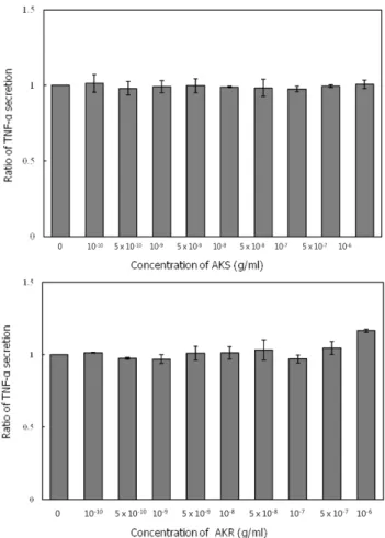
섬오가피 추출물을 처리한 대식세포의 TNF-α 분비 경향 Th-1 type의 cytokine 중의 하나인 TNF-α는 대식세포에 작용 하여 염증반응을 촉진하고, T-세포와 B-세포의 활성화에 co- stimulator로 작용하며, 종양세포에 작용하여 세포자살(apoptosis)
을 유도하기도 한다.15,16)섬오가피로부터 분리한 acanthoic acid 가 IL-1과 TNF-α의 분비를 억제시킨다는 보고22,23)가 있으나 그 이상은 보고된 바 없다. 본 연구에서는 대식세포의 TNF-α 분비 에 미치는 섬오가피 추출물의 영향을 알아보기 위하여 LPS로 자 극시킨 RAW263.7 세포에 추출물을 처리 후 배양액의 TNF-α의 양을 측정하였다. 그 결과, 줄기 및 뿌리 추출물 모두 대식세포의 TNF-α의 분비에 거의 영향을 미치지 않는 것을 알 수 있었고, 섬 오가피의 정제된 성분인 acanthoic acid를 처리한 Kang 등22)의 결 과와는 차이가 있었다.
결 론
암의 면역학적 치료는 인체 내의 면역세포들이 암세포 표면에 발현되는 정상세포와 다른 구조를 인식함으로써 항암 면역반응 을 일으킨다는 데 기초를 두고 있다. 여러 가지 중요한 면역반응 들을 조절하는 생리활성 cytokine은 암의 면역학적 치료에 상당 Fig. 5 − Secretion of IFN-γ from NK92MI cells treated with stem
(AKS) and root (AKR) extract of Acanthopanax koreanum.
The extracts were diluted with CTCM and then tested in triplicate wells. The cells stimulated with LPS were incubated for 24 hr at 37oC and the concentration of IFN-γ in culture supernatants was measured by ELISA.
Fig. 6 − Secretion of TNF-α from RAW263.7 cells treated treated with stem (AKS) and root (AKR) extract of Acanthopanax koreanum. The extracts were diluted with CTCM and then tested in triplicate wells. The cells stimulated with LPS were incubated for 24 hr at 37oC and the concentration of TNF-α in culture supernatants was measured by ELISA.
한 효과가 있는 것으로 알려져 있다. 세포매개성 면역반응을 촉 진하는 Th-1 세포는 proinflammatory cytokine인 Th-1 type (IL-2, IL-12, IFN-γ, TNF-α 등)을 생산하며, 체액성 면역을 촉 진하는 Th-2 세포는 anti-inflammatory cytokine인 Th-2 type cytokine(IL-4, IL-6, IL-8, IL-10)을 생산한다. 따라서 Th-1 세포 의 활성화 및 Th-2 세포의 억제는 항암면역기능에 중요한 기능 을 미칠 수 있다.13) 본 연구에서는 섬오가피의 줄기 및 뿌리 추 출물의 Th-1 type cytokine인 IL-2, IL-12, IFN-γ, TNF-α의 분 비에 미치는 영향을 확인하고, 섬오가피의 암 예방 및 치료물질 로서의 가능성을 알아보고자 하였다.
섬오가피 추출물을 전구세포가 다른 수지상세포에 처리한 결 과, 줄기 및 뿌리 추출물 모두 동일하게 Th1 계열의 세포를 유 도하는 IL-12의 분비를 촉진시키는 것으로 나타났다. 따라서 Sallusto 등33)의 주장대로 면역반응의 유형은 수지상세포로 유도 되는 전구세포의 기원이나 분화를 유도하는 자극의 특성 때문이 아니라 항원의 종류 및 양이나 수지상세포가 활성화되고 성숙되 는 정도에 따라서 결정되는 것으로 추측된다. 또한, 섬오가피 추 출물은 T-세포의 IL-2 분비에는 거의 영향이 없으나, IEC-6 세 포의 IL-2의 분비를 농도의존적으로 현저히 증가시키는 것으로 나타났다. 따라서, 대부분의 오가피를 경구로 복용하고 있는 점 을 고려하면, 복용 후 흡수단계에서 1차적으로 만나는 장상피세 포에서의 IL-2의 분비촉진은 매우 의미 있는 결과라고 볼 수 있 다. 또한, 섬오가피 줄기추출물은 NK-세포로부터 IFN-γ의 분비 를 크게 증가시켰으며, 뿌리 추출물은 IFN-γ의 분비증가 정도가 낮았다. 그러나 섬오가피 추출물은 대식세포의 TNF-α의 분비에 거의 영향을 주지 않았다.
결론적으로 섬오가피 추출물은 Th-1 type cytokine 중 IL-2, IL-12, IFN-γ의 분비를 증가시키나 TNF-α의 분비에는 영향을 주지 않았다. 또한 T-세포에 의한 IL-2 분비에는 큰 영향이 없었 으나 장상피세포 IEC-6 세포에 의한 IL-2의 분비에는 크게 영향 을 주는 것을 알 수 있었다. 따라서 섬오가피 추출물은 Th-1 type cytokine의 분비를 증가시켜 세포매개성 면역반응을 촉진시켜 세 포 내 감염과 종양세포에 대한 숙주의 방어에 도움을 줄 수 있 을 것으로 기대된다.
감사의 말씀
이 논문은 2010년도 서울여자대학교 교내학술특별연구비 지 원에 의하여 수행되었으며, 이에 감사를 드립니다.
참고문헌
1) Yook, C. S., Lee, D. H. and Seo, Y. K. : A new form of Acanthopanax species (I). Kor. J. Pharmacogn. 7, 179 (1978).
2) Lee, Y. S., Lee, E. B. and Kim, Y. H. : Some pharmacological activities of acanthoic acid isolated from Acanthopanax koreanum root bark. J. Appl. Pharmacol. 9, 176 (2001).
3) Kim, Y. H., Chung, B. S. and Kim, H. J. : Studies on the chemical constituents of Acanthopanax koreanum. Kor. J.
Pharmacogn. 16, 151 (1985).
4) Kim, Y. H., Chung, B. S. and Kim, H. J. : Studies on the constituents of Acanthopanax koreanum. Arch. Pharm. Res. 11, 159 (1988).
5) Kang, J. S., Linh, P. T., Cai, X. F., Lee, J. J. and Kim, Y. H. : Determination of chiisanoside in Acanthopanax species by high performance liquid chromatography. Nat. Product Sci. 9, 45 (2003).
6) Oppenheim, J. J. : Cytokines: past, present and future. Int. J.
Hematol. 74, 3 (2001).
7) Steinman, R. M. and Cohn, Z. A. : Identificaion of a novel cell type in peripheral lymphoid organs of mice. I. Morphology, quantitation, tissue distribution. J. Exp. Med. 137, 1142 (1973).
8) Shurin, M., R. : Dendritic cells presenting tumor antigen.
Cancer Immunol. Immunother. 43, 158 (1996).
9) Banchereau, J. and Steinman, R. M. : Dendritic cells and the control of immunity. Nature 392, 245 (1998).
10) Palucka, K. and Banchereau, J. : Dendritic cells: a link between innate and adaptive immunity. J. Clin. Immunol. 19, 12 (1999).
11) Beutler, B., Biron, C., Carroll, M., Cyster, J., Gerard, C., Gordon, S., Lanier, L., Lehrer, R., McGhee, J., Medzhitov, R., Springer, T. and Yokoyama, W. : Innate immunity. In: Janeway Jr., C. A., Travers, P., Walport, M. and Shlomchik, M. J. (eds.), Immunobiology, 6th ed. Garland Science Publishing, New York, NY, p. 76 (2005).
12) Lucey, D. R., Clerici, M. and Shearer, G. M. : Type 1 and type 2 cytokine dysregulation in human infectious, neoplastic, and inflammatory diseases. Clin. Microbiol. Rev. 9, 532 (1996).
13) Mossman, T. R. and Sad, S. : The expanding universe of T-cell subset: Th1, Th2 and more. Immunology Today 17, 138 (1996).
14) Podhajcer, O. L., Lopez, M. V. and Mazzolini, G. : Cytokine gene transfer for cancer therapy. Cytokine & Growth Factor Reviews 18, 183 (2007).
15) Yoshioka, Y., Tsutsumi, Y., Ikemizu, S., Yamamoto, Y., Shibata, H., Nishibata, T., Mukai, Y., Okamoto, T., Taniai, M.., Kawamura, M., Abe, Y., Nakagawa, S., Nagata, S., Yamagata, Y.
and Mayumi, T. : Optimal site-specific PEGylation of mutant TNF-α improves its antitumor potency. Biochem. and Biophysic. Res. Comm. 315, 808 (2004).
16) Trinchieri, G. : Interleukin-12: a cytokine produced by antigen- presenting cells with immunoregulatory functions in the generation of T-helper cell type 1 and cytokine lymphocytes.
Blood 84, 4008 (1995).
17) Nishimura, T., Nakui, M., Sato, M., Iwakabe, K., Kitamura, H.,
Sekimoto, M., Ohta, A., Koda, T. and Nishimura, S. : The critical role of Th1-dominant immunity in tumor immunology.
Cancer Chemother. Pharmacol. 46, S52 (2000).
18) Yoon, T. J., Yoo, Y. C., Lee, S. W., Shin, K. S., Choi, W. H., Hwang, S. H., Ha, E. S., Jo, S. K., Kim, S. H. and Park, W. M. : Anti-metastatic activity of Acanthopanax senticosus extract and its possible immunological mechanism of action. J. of Ethnopharmacol. 93, 247 (2004).
19) Ha, E. S., Hwang, S. H., Shin, K. S., Yu, K. W., Lee, K. H., Choi, J. S., Park, W. M. and Yoon, T. J. : Anti-metastatic activity of glycoprotein fractionated from Acanthopanax senticosus, involvement of NK-cell and macrophage activation. Arch.
Pharm. Res. 27, 217 (2004).
20) Lyu, S., Noh, B. and Park, W. B. : Modulation of Th1/Th2 cytokine secretion in human peripheral blood mononuclear cells by water extract of Acanthopanax divaricatus var.
albeofructus fruits. Yahak Hoeji 52, 27 (2008).
21) Han, S. B., Park, S. K., Ahn, H. J., Yoon, Y. D., Kim, Y. H., Lee, J. J., Lee, K. H., Moon, J. S., Kim, H. C. and M., K. H. : Characterization of B cell membrane receptors of polysaccharide isolated from the root of Acanthopanax koreanum. Int.
Immunopharmacol. 3, 683 (2003).
22) Kang, H. S., Kim, Y. H., Lee, C. S., Lee, J. J., Choi, I. and Pyun, K. H. : Suppression of interleukin-1 and tumor necrosis factor- alpha production by acanthoic acid, (-)-pimara-9(11),15-dien- 19-oic acid and its antifibrotic effects in vivo. Cellular Immunol.
170, 212 (1996).
23) Kang, H. S., Song, H. K., Lee, J. J., Pyun, K. H. and Choi, I. : Effects of acanthoic acid on TNF-alpha gene expression and haptoglobin synthesis. Mediators Inflamm. 7, 257 (1998).
24) Kim, J. A., Kim, D. K., Tae, J., Kang, O. H., Choi, Y. A., Choi, S. C., Kim, T. H., Nah, Y. H., Choi, S. J., Kim, Y. H., Bae, K. H.
and Lee, Y. M. : Acanthoic acid inhibits IL-8 production via MAPKs and NF-κB in a TNF-α-stimulated human intestinal epithelial cell line. Clinica. Chimica. Acta 342, 193 (2004).
25) Inaba, K., Young, J. W. and Steinman, R. M. : Direct activation of CD8+ cytotoxic T lymphocytes by dendritic cells. J. Exp.
Med. 166, 182 (1987).
26) Inaba, K., Metlay, J. P., Crowley, M. T. and Steinman, R. M. : Dendritic cells pulsed with protein antigens in vitro can prime antigen-specific, MHC-restricted T cells in situ. J. Exp. Med.
172, 631 (1990).
27) Fernandez, N. C., Flament, C., Crpineau, F., Angevin, E., Vivier, E. and Zitvogel, L. : Dendritic cells (DC) promote natural killer (NK) cell functions: dynamics of the human DC/
NK cell cross talk. Eur. Cytokine Netw. 13, 17 (2002).
28) Steinman, R. M. and Banchereau, J. : Dendritic cells and the control of immunity. Nature 392, 245 (1998).
29) Steinman, R. M. : The dendritic cell system and its role in immunogenicity. Annu. Rev. Immunol. 9, 271 (1991).
30) Reid, C. D. : Dendritic cells and immunotherapy for malignant disease. Br. J. Haematol. 112, 874 (2001).
31) Maldonado-Lopez, R., De Smedt, T., Mechel, P., Godfroid, J., Pajak, B., Heirman, C., Thielemans, K., Leo, O., Urbain, J. and Moser, M. : CD8 alpha+ and CD8 alpha subclasses of dendritic cells direct the development of distinct T helper cells in vivo.
J. Exp. Med. 189, 587 (1999).
32) Viera, P. L., de Jong, E. C., Wierenga, E. A., Kapsenberg, M. L.
and Kalinski, P. : Development of the Th1-inducing capacity in myeloid dendritic cells requires environmental instruction. J.
Immunol. 164, 4507 (2000).
33) Sallusto, F. and Lanzavecchia, A. : The instructive role of dendritic cells on T-cell responses. Arthritis Res. 4, Suppl 3, S127 (2002).