전사체 프로파일을 이용한 고려 홍삼의 항당뇨 기전 연구
원해단·신은정·정성현# 경희대학교 약학대학 약물학 임상약학교실
(Received May 1, 2008; Revised August 14, 2008)
Anti-diabetic Mechannism Study of Korean Red Ginseng by Transcriptomics
Hai Dan Yuan, En Jung Shin and Sung Hyun Chung#
Pharmacology and Clinical Pharmacy Lab., College of Pharmacy, Kyung Hee University, Seoul 130-701, Korea
Abstract
— This study was designed to investigate the anti-diabetic effect and mechanism of Korean red ginseng extract through transcriptomics in C57BL/KsJ db/db mice. The db/db mice were randomly divided into six groups: diabetic control group (DC), red ginseng extract low dose group (RGL, 100 mg/kg), red ginseng extract high dose group (RGH, 200 mg/kg), metformin group (MET, 300 mg/kg), glipizide group (GPZ, 15 mg/kg) and pioglitazone group (PIO, 30 mg/kg), and treated with drugs once per day for 10 weeks. At the end of treatment, we measured blood glucose, insulin, hemoglobin A1c (HbA1c), triglyceride (TG), adiponectin, leptin, non-esterified fatty acid (NEFA). RGL-treated group lowered the blood glu- cose and HbA1c levels by 19.6% and 11.4% compared to those in diabetic control group. In addition, plasma adiponectin and leptin levels in RGL-treated groups were increased by 20% and 12%, respectively, compared to those in diabetic control.
Morphological analyses of liver, pancreas and epidydimal adipose tissue were done by hematoxylin-eosin staining, and pan- creatic islet insulin and glucagon levels were detected by double-immunofluorescence staining. RGL-treated group revealed higher insulin contents and lower glucagon contents compared to diabetic control. To elucidate an action mechanism of Korean red ginseng, DNA microarray analyses were performed in liver and fat tissues, and western blot and RT-PCR were conducted in liver for validation. According to hierarchical clustering and principal component analysis of gene expression Korean red ginseng treated groups were close to metformin treated group. In summary, Korean red ginseng lowered the blood glucose level through protecting destruction of islet cells and shifting glucose metabolism from hepatic glucose pro- duction to glucose utilization and improving insulin sensitivity through enhancing plasma adiponectin and leptin levels.
Keywords □
Korean red ginseng, C57BL/KsJ db/db mice, diabetes, DNA microarray
당뇨병
(diabetes mellitus)
은대사성질환으로서고혈당을특징으로하며췌장에서인슐린분비가부족하거나인슐린에대한감 수성이 떨어져탄수화물대사에이상이 생기는질환이다
.
1,2)최근음식의서구화와더불어비만환자가급증하면서제
2
형당뇨병이급증하는추세는사회적인문제로대두되고있는데현 재한국의경우도
2030
년에는전체당뇨병환자수가약722
만 명으로늘어날것이라고추정한바있다.
3,4)현재임상에서많이사용하고있는경구용당뇨제제들로는 α
-glucosidase
억제제, sulfonylurea
계약물, biguanide
계약물, peroxisome proliferator- activated receptor-
γ(PPAR-
γ)
효능제등이있다.
5,6)하지만이런약물들을장기적으로사용할때저혈당
,
체중증가및간독성등부작용들이있다고보고되어있다
.
7-10)한국을비롯한여러아시아의나라들에서는민간요법으로여러 가지약초들을당뇨병및여러질병의치료에많이사용하여왔 다
.
다양한연구에따르면,
고려인삼은허약한체질,
숙취,
폐경,
월경불순
,
초기당뇨병,
빈혈,
간기능악화,
각종독성물질에기 인한중독,
피로,
추위,
스트레스,
산후조리,
체력쇠퇴등에효능이있음이알려져있다
.
11-13)고려홍삼은인삼뿌리(Panax ginseng
C.A. Meyer)
을쪄서건조시킨것으로서고혈압,
동맥경화,
항암,
항당뇨작용이있다고보고가되어있다
.
14-18)하지만고려홍삼의항당뇨작용은명확히밝혀지지않고있다
.
따라서본연구에서는고려홍삼물추출물과기존의당뇨치료제들인
metformin, glipizide, pioglitazone
을db
마우스에투여한후간에서의유전자발현양상을비교분석하여고려홍삼의항당뇨기전을살펴보았다
.
#본논문에관한문의는저자에게로
(
전화) 02-961-0373 (
팩스) 02-957-0384
(E-mail) [email protected]
실험재료 및 방법
실험재료
고려홍삼물추출물은
KT
&G(Seoul, Korea)
에서공급받아사용하였다
.
고려홍삼물추출물10 g
을2
l물에희석하여동결건 조하였다.
실험동물및약물투여
C57BL/KsJ db/db 5
주령의마우스를한국오리엔트사로부터구입하여
2
주동안실험실환경에적응시킨후실험에사용하였다
.
실험군은Diabetic control(DC);
고려홍삼엑스100 mg/kg
투 여군(RGL);
고려홍삼엑스200 mg/kg
투여군(RGH); metformin 300 mg/kg
투여군(MET); glipizide 15 mg/kg
투여군(GPZ);
pioglitazone 30 mg/kg
투여군(PIO)
으로나누며각약물은경구 로10
주간투여하였다.
경구당부하시험
실험동물을
12
시간절식한다음1.5 g/kg
의glucose
를경구투여한후
0
분, 30
분, 60
분, 90
분그리고120
분대에안와정맥으로부터혈액을채취하였다
.
19)5,000 rpm
에서5
분간원심분리하여 얻어진혈장을이용하여혈중포도당농도를glucose oxidase method
로측정하였다.
혈액지표분석
혈액지표분석을위한혈액채취는
12
시간절식후실시하였 다.
안와정맥을통해얻어진전혈을5,000 rpm
에서5
분간원심 분리한후혈청을분석에사용하였다.
혈중포도당농도는glucose oxidase method(Trinder method)
을사용하여측정하였으며흡 광도 측정은UV Spectrophotometer(U-3210, HITACHITM, Japan)
을사용하였다.
혈중인슐린농도는마우스insulin ELISA kit(Shibayagi, Japan)
을 구입하여ELISA reader(Labsystems, Finland)
로 측정하였다.
당화혈색소는Hemoglobin A1c kit (BioSystem S.A., Spain)
을이용하여측정하였고혈중중성지방(triglyceride, TG)
은영동제약에서kit
를구입하여측정하였다.
혈중
adiponectin
은 마우스adiponectin ELISA kit(Adipogen, Korea)
을구입하여측정하였으며혈중leptin
은마우스leptin ELISA kit(LINCO research, USA)
을구입하여측정하였다.
혈 중유리지방산(non-esterified fatty acid, NEFA)
은kit(
에이켄화 학(
주), Japan)
을구입하여측정하였다.
조직의형태학적관찰
쥐에서 적출한 간
,
부고환지방,
췌장조직을10% neutral buffered formalin
을사용하여고정하였다.
이후탈수및포매과 정을거쳐파라핀블럭을제작하고두께5
µm
의관상절편으로제작한후
xylene
으로파라핀을제거시키고100%, 95%, 90%, 80%, 70%
알코올로친수화시켰다.
염색방법으로는hematoxylin
과
eosin
염색으로탈수과정을거쳐Canada balsam
으로봉입 하고광학현미경(Olympus, Japan)
으로관찰하였다.
면역형광염색방법으로췌장
islet
에서insulin
과glucagon
의관찰
두께
5
µm
관상절편을xylene
으로 파라핀을제거시키고, 100%, 95%, 90%, 80%, 70%
알코올로친수화시켰다. PBS
로 씻은후0.1% tripsin
으로단백질구조를회복시키고primary antibody
인goat anti-insulin(Santa Cruz, USA)
을1 : 75
로희석 하여 붙이고 이어서secondary antibody
인donkey anti-goat IgG-FITC(Santa Cruz, USA)
을1 : 200
으로희석하여붙였으며같은 조직에 두 번째
primary antibody
인rabbit anti-mouse (Santa Cruz, USA)
를 붙이고 두 번째secondary antibody
인goat anti-rabbit IgG-TRITC(Santa Cruz, USA)
을붙인후형광현미경
(Olympus, Japan)
으로관찰하였다. RNA
분리조직에서
total RNA
는guanidine thiocyanate-water saturated phenol/chroloform
분리방법을이용하였다.
20)물층에있는total RNA
는이소프로판올을이용하여침전시켜분리한RNA
는260nm
와
280nm
의파장에서흡광도를측정하여정량하였다. Mouse
로 부터추출한total RNA
의상태를확인하는방법으로pH 8.0
의RNase-free water
에서OD260/280
비율이1.8
이상, 28S rRNA
와18S rRNA
비율이1.6
이상일경우가장최적의RNA
로판단하였다. Microarray analysis
Agilent 60-mer oligo Microarray protocol
에따라cy3
또는cy5
가결합된aminoallyl-UTP
로준비된RNA
를증폭시킨뒤oligo micaroarray(Mouse 44 K)
와60
oC
에서16
시간 동안hybridization
시켰다. Hybridization
된chip
은non-confocal laser scanner
인GenePix4000B(Axon Instruments, CA, USA)
로scanning
하였다. Scanning
하여얻은image
는16 bit TIFF file
로 저장한후GenePix v6.0 software
를사용하여image
분석을하 였다. Locally weighted scatter-plot smoother(LOWESS) nor- malization
방법을사용하여normalization
하였다. Microarray
데이터의 분석을 위해서
hierarchical clustering, principal component analysis
방법을사용하였으며유전자의군집화를통해유사한발현양상을보이는그룹을확인하였다
.
분석프로그 램은multiexperiment viewer(MeV) software
를이용하였다.
간조직에서
AMPK
발현측정간 조직에서
5'-AMP activated protein kinase(AMPK)
발현을 확인하고자
Western blot
방법으로phospho-5'-AMP activated protein kinase(P-AMPK), AMPK, phospho-acetyl CoA carboxylase(P-ACC), acetyl CoA carboxylase(ACC)
를 측 정하였다.
단백질분석을위해간조직을lysis buffer
를이용하여균질화하였다
.
단백질정량은Bio-Rad assay reagent(Bio- Rad, USA)
를이용하여측정하였으며,
정량한단백질20
µg
을8%
SDS-PAGE
로분리하였다.
이후gel
을membrane(Milipore, Cat.
No: IPVH00010)
에transfer
하고5% skim milk
로상온에서1
시 간blocking
하였으며1 : 3000
비율로희석시킨primary antibody (P-AMPK, AMPK, P-ACC, ACC)
와4
oC
에서overnight
하였다. Tris-buffered saline tween-20(TBST)
로4
번washing
한 후1 : 5000
의비율로희석시킨secondary antibody
와상온에서1
시간반응시켰다
.
이후TBST
로4
번washing
하고ECL solution (Amersham, Sweden)
을이용하여X-ray
필름에developing
하 였다.
간조직에서
RT-PCR
DNA microarray
실험을통하여특이적인발현을보이는유전자를선별하여
RT-PCR
을진행하였다.
총RNA 10
µg
을Moloney murine leukemia virus transcriptase
와Oligo(dT) 15 primer
를 이용하여역전사하였다. Primer
의종류및서열은다음과같다. SREBP1a(Sterol regulatory element-binding proteins)
의 주형사서열은
GCG CTA CCG GTC TTC TAT CA,
비주형사서 열은TGC TGC CAA AAG ACA AGG G; FAS(Fatty acid synthesis)
의주형사서열은GAT CCT GGA ACG AGA ACA C,
비주형사 서열은AGA CTG TGG AAC ACG GTG GT;
SCD1(Stearoyl CoA desaturase)
의주형사서열은CGA GGG TTG GTT GTT GAT CTG T,
비주형사 서열은ATA GCA
CTG TTG GCC CTG GA; GPAT(Glycerol-3-phosphate acyltransferase)
의주형사서열은GGT AGT GGA TAC TCT GTC GTC CA,
비주형사 서열은CAT CAG CAA CAT CAT TCG GT; CD36
의주형사서열은TCC TCT GAC ATT TGC AGG TCT ATC,
비주형사서열은GTG AAT CCA GTT ATG GGT TCC AC; PPAR-
α(Peroxisome proliferator activated receptor-
α)
의주형사서열은CCC TGA ACA TCG AGT GTC GA,
비주형사서열은CTT GCC CAG AGA TTT GAG GTC CT; CPN
의주형사서열은ATG GTC AAC CCC ACC GTG,
비주형사서열은
TTA GAG TTG TCC ACA GTC GGA GA;
PEPCK(phosphoenolpyruvate carboxykinase)
의주형사서열은ATG CCT CCT CAG CTG CAT A,
비주형사 서열은TTA CAT CTG GCT GAT TCT CTG TT
이다. PCR
반응조건은SREBP-1a, FAS, PPAR-
α, CPN, PEPCK
의경우95
oC
에서30
초동안변성
, 57
oC
에서30
초동안붙임, 72
oC
에서30
초동안연 장을하여총30 cycle
하였고SCD1, GPAT
의경우는95
oC
에서30
초동안변성, 57
oC
에서30
초동안붙임, 72
oC
에서30
초동안연 장을하여총26 cycle
하였으며CD36
의경우는95
oC
에서30
초동안변성
, 51
oC
에서30
초동안붙임, 72
oC
에서30
초동안연장을하여총
30 cycle
하였다.
이후반응생성물을0.5
µg/m
lethidium bromide
로염색된1% agarose gel
을이용하여100V
에서전기영동하였다
. CPN
은증폭된유전자들의대조군으로사용되었다.
자료분석및통계처리
모든실험결과는평균±표준오차로나타내었다
.
당뇨대조군(DC)
과비교하여통계적유의성을Student's
t-test
로처리하였으 며p<0.05
이하인경우유의성있는차이가있는것으로판정하였다
.
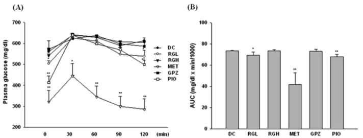
Fig. 1 −
Effects of RG, MET, GPZ & PIO on oral glucose tolerance test (A). Animals fasted overnight were given on oral glucose load of 1.5 g/
kg after oral administration of either vehicle or RG, MET, GPZ, PIO. (B) Area under the blood-glucose concentration curve was
measured over 120 min (AUC-120 min). Values represent as mean±S.E. (n=4). *p<0.05, **p<0.01 vs. DC.
실험결과 및 고찰
경구당부하시험
Fig. 1
은마우스에10
주간약물을경구투여한후측정한경구당부하시험결과를나타낸그림이다
. Fig. 1A
는120
분동안의혈당변화를나타낸 것이며
B
는각그룹의곡선하면적(area
under the curve, AUC)
을비교한그림으로당뇨대조군과비교시
RGL, MET, PIO
그룹에서곡선하면적이감소함을알수 있었다.
혈액지표분석
Table I
은10
주동안약물을경구투여한후, 12
시간절식시켜공복상태의실험동물에서전혈을채취한후측정한포도당
,
인슐린
,
당화혈색소,
인슐린저항성지수(HOMA-IR),
중성지방,
유리 지방산그리고지방세포에서분비되는adipokine
인adiponectin
과
leptin
의농도를나타낸것이다.
당뇨대조군에비해RGL
투여군은혈당이
19.8%(
p<0.05)
감소하였으며, RGH
또한당뇨대조군에비해혈당이
18.3%(
p<0.05)
감소하였다.
그러나인슐린수치는고려홍삼투여군에서유의적인차이를나타내지않았
으며
MET
투여군과PIO
투여군은당뇨대조군에비해각각67.9%(
p<0.001)
와56.9%(
p<0.001)
감소하였다.
인슐린저항성 지수(HOMA-IR)
는당뇨대조군에비해RGL
투여군에서27.7%
감소하였으며양성약물대조군에서각각
80.8%, 41.1%
그리고68.9%
감소하였다. HbA1c
는당뇨대조군에비해RGL
투여군, RGH
투여군, MET
투여군, GPZ
투여군, PIO
투여군에서각각11%, 6.4%, 18.9%, 16.1%, 27.9%
감소하였다.
지방세포에서지방분해
(lipolysis)
는인슐린에의해억제된다.
그러나인슐린저항성상태에서는지방분해가억제되지못한결 과혈중유리지방산
(non-esterified fatty acid, NEFA)
의수치는올라가게된다
.
이렇게상승된유리지방산은근육과지방조직으 로포도당이유입되는것을방해하며간에서당생성을촉진하게 된다.
21)또한장기적인유리지방산의상승은췌장에서인슐린분Table I −
Metabolic parameters in Korean red ginseng treated db/db mice
Parameter DC RGL RGH MET GPZ PIO
Glucose (mM) 0015.8±0.9 0012.7±1.3* 0012.9±0.4* 0005.1±0.4*** 0007.4±2.1*** 0006.8±0.6***
Insulin (
µU/ml) 0240.6±57.1 0216.6±18.2 0272.1±50.3 0143.0±37.0* 0302.6±28.3 0173.9±33.2*
HOMA-IR 0168.9±46.3 0122.3±22.4 0156.0±30.0 0032.4±13.0* 0099.5±41.3 0052.5±8.2*
HbA1c (%) 0007.0±0.2 0006.2±0.2* 0006.6±0.3* 0005.7±0.2** 0005.9±0.2** 0005.1±0.3***
Adiponectin (
µU/ml) 0031.5±1.6 0038.0±1.3* 0032.8±2.1 0029.5±2.3 0024.9±2.3 0061.9±5.5**
Leptin (ng/ml) 0054.5±1.3 0061.1±1.1** 0058.0±1.3 0057.6±1.1 0058.6±1.4* 0057.0±0.8
TG (mg/ml) 0094.9±9.4 0077.0±1.6 0095.9±5.2 0065.4±4.9* 0080.9±9.0 0089.0±5.8
NEFA (
µeqiv/l) 2179.3±78.0 1812.8±44.7*** 2278.6±173.3 1936.3±204.5 2022.8±442.0 1550.4±62.4***
Data are mean±standard error (n=4). Homeostasis model assessment was used to calculate an index of insulin resistance as insulin (
µU/ml)×glucose (mM)/22.5. *p<0.05, **p<0.01, ***p<0.001 compared to diabetic control group.
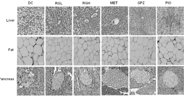
Fig. 2 −
Microscopic view of the liver, fat and pancreas sections obtained from DC, RGL, RGH, MET, GPZ and PIO treated groups. H & E,
magnification×200.
비도방해하는결과를낳는다
.
22)Table I
에서RGL
투여군은당 뇨대조군과비교시혈중유리지방산의수치가16.8%
감소하였다
.
비록고려홍삼은용량의존적으로혈중유리지방산의수치를낮추어주지는못하였지만저용량에서
17%
가까이유리지방 산수치를낮추어주었고이는포도당항상성에유리한영향을 미칠수있음을시사하였다.
또한RGL
투여군은통계적으로유의하지는않았지만혈중중성지방역시
DC
군에비해18.9%
감 소시켰다.
포도당항성성에다양한영향을미치는지방세포에서유리되 는
adipokine
인adiponectin
과leptin
호르몬의수치를그룹간비 교해보았다. apM1, GBP28, AdipoQ
혹은ACRP30
으로불리우기도하는
adiponectin
은분자량30 kDa
의유리단백질로비만당 뇨쥐에투여할경우간과근육조직에서AMPK activity
를촉진 시키고결과지방산산화와인슐린반응성을증가시키는활성이있음이알려지고있는물질이다
.
23,24)비만,
인슐린저항성, 2
형당뇨
,
대사증후군,
고지혈증,
고혈압,
산화적스트레스를보이는사람이나탄수화물고함유식이를섭취하는경우혈중
adiponectin
은감소하는경향을보이는반면체중을줄이거나콩단백질
,
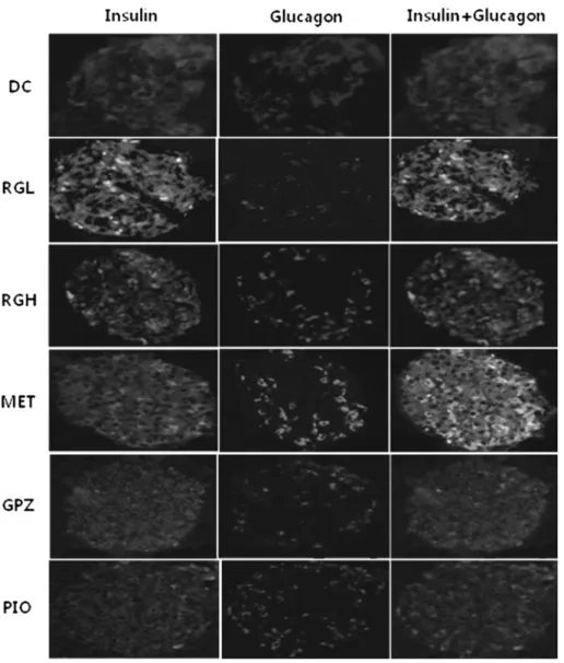
피 Fig. 3 −Double-immunofluorescence staining of insulin and glucagon in a db mouse pancreatic islet. Insulin and glucagon were coded in green
and red color, respectively. magnification×200.
Table II −
Number of the significant up- or down-regulated genes in hepatocytes and adipocytes treated with red ginseng or oral hypoglycemics
Up-regulated (>1.5-fold) Down-regulated (<1.5-fold)
Liver Fat Liver Fat
RGL 1453 3496 1540 3658
RGH 2387 4045 1513 4725
RGL+RGH 0461 1410 0261 1266
MET 3383 3352 2333 4006
GPZ 4250 4837 3025 7397
PIO 4942 3973 4511 4944
오글리타존 같은
PPAR-
γagonist
약물을 투여하면 혈중adiponectin level
이증가한다.
25)따라서혈중adiponectin
수치의증가는인슐린의작용성이증가되었다는근거가될수있다
. Table I
에서보듯이RGL
군에서는DC
군에비해adiponectin
및leptin
수치가20.6%, 12.1%
증가되었다.
이결과로부터고려홍삼은인슐린작용성을증가시키고지방 산산화를촉진하는물질들인
adiponectin
과leptin
의혈중농도를증가시키는활성이있음을알수있었다
.
조직의형태학적관찰
Fig. 2
는홍삼이간,
부고환지방및췌장형태에미치는영향을살펴본결과이다
.
간조직의hematoxylin-eosin
염색결과에서 홍삼투여군은DC
군에서많이보이는지방구(lipid droplets)
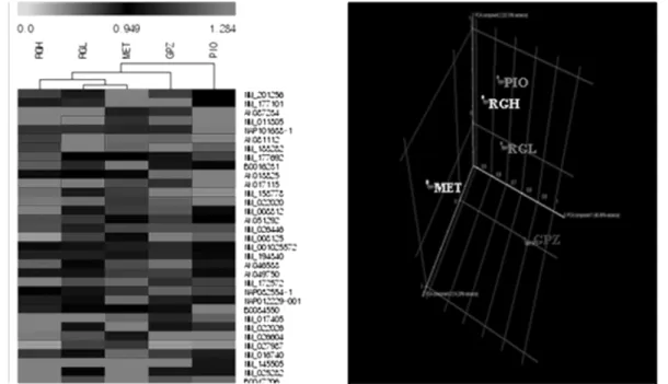
들 이거의사라진것을볼수있었고지방조직의염색결과에서는Fig. 4 −
Hierarchical clustering analysis and principal component analyses of gene expression data. Expression profile data were derived from pooled RNA of the livers of db mice treated with Korean red ginseng and oral hypoglycemics.
Fig. 5 −
Hierarchical clustering and principal component analyses of gene expression data. Expression profile data were derived from pooled
RNA of the fats of animals treated with red ginseng and oral hypoglycemics.
홍삼투여군의지방
size
가DC
군에비하여현저히작아졌음을알수가있었다
.
췌장조직의형태학적관찰결과에서DC
군마우스의
islets
은모양이불규칙적이고구조가파괴된양상을보였지만홍삼투여군의
islets
은DC
군마우스에비하여모양이나구조가뚜렸하였다
. Fig. 3
은췌장islets
에서insulin
과glucagon
의발현을면역형광염색하여관찰한결과이다
. DC
에비하여홍 삼투여군에서insulin
의발현은증가하였으나glucagon
의발현은감소하였다
.
이결과는홍삼추출물이비만성당뇨의β-cell
에서
insulin
의분비를촉진시킴과동시에 α-cell
에서glucagon
의 분비를억제함으로혈당을떨어뜨림을알수있었다.
Microarray
분석간에서
36,910
개의유전자를분석한결과DC
군에비해1.5
배이상높게나타난유전자는홍삼투여군에서
461
개였고반대로1.5
배이상낮게나타난유전자는261
개였다(Table II).
지방에서는
34,713
개의유전자를분석한결과홍삼투여군에서DC
군에비해
1.5
배이상높게나타난유전자는1410
개였고1.5
배이상 낮게나타난유전자는1266
개였다(Table II).
그리고간과지방에서각각
36,910
개, 34,713
개의유전자를사용하여hierarchical clustering analysis
와principal component analysis
를 하였다. Hierarchical clustering
은전체유전자의발현양상을분석하여그패턴이비슷한유전자또는투여군을묶어가는방법으로홍삼에 의한유전자발현을기존의항당뇨약물과비교할수있다
.
발현 이증가된것은붉은색으로발현이감소한것은녹색으로나타 내었다.
간에서는RGL
투여군과RGH
투여군의발현양상이비슷한것으로나타났고항당뇨약물중에서는
MET
투여군과가장가까운것으로나타났다
(Fig. 4).
지방에서는간의발현패턴과는달리
RGL
투여군이RGH
투여군보다MET
투여군과더Table III −
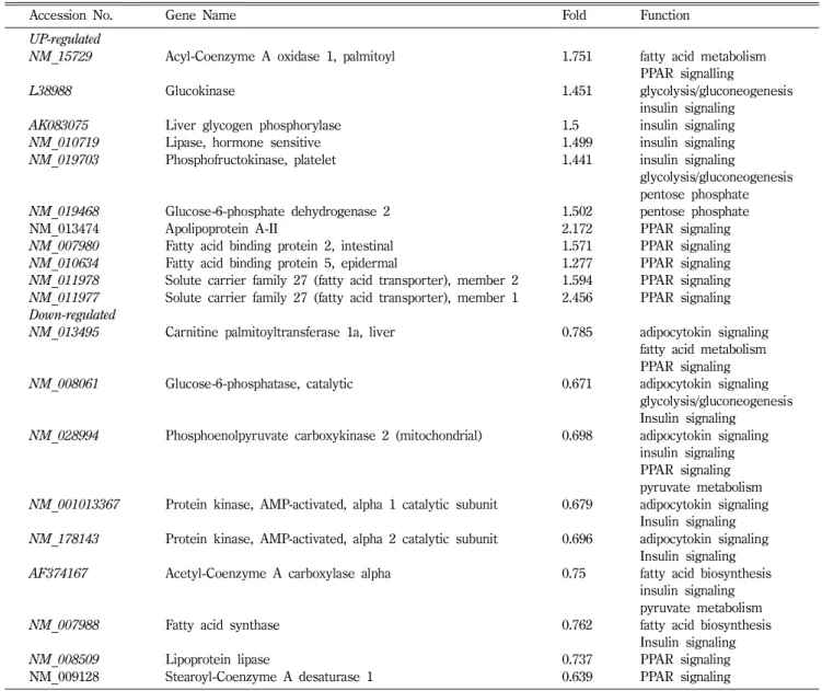
Differentially expressed genes in the liver of RG-treated db/db mice
Accession No. Gene Name Fold Function
UP-regulated
NM_15729 Acyl-Coenzyme A oxidase 1, palmitoyl 1.751 fatty acid metabolism
PPAR signalling
L38988 Glucokinase 1.451 glycolysis/gluconeogenesis
insulin signaling
AK083075 Liver glycogen phosphorylase 1.5 insulin signaling
NM_010719 Lipase, hormone sensitive 1.499 insulin signaling
NM_019703 Phosphofructokinase, platelet 1.441 insulin signaling
glycolysis/gluconeogenesis pentose phosphate
NM_019468 Glucose-6-phosphate dehydrogenase 2 1.502 pentose phosphate
NM_013474 Apolipoprotein A-II 2.172 PPAR signaling
NM_007980 Fatty acid binding protein 2, intestinal 1.571 PPAR signaling
NM_010634 Fatty acid binding protein 5, epidermal 1.277 PPAR signaling
NM_011978 Solute carrier family 27 (fatty acid transporter), member 2 1.594 PPAR signaling NM_011977 Solute carrier family 27 (fatty acid transporter), member 1 2.456 PPAR signaling Down-regulated
NM_013495 Carnitine palmitoyltransferase 1a, liver 0.785 adipocytokin signaling fatty acid metabolism PPAR signaling
NM_008061 Glucose-6-phosphatase, catalytic 0.671 adipocytokin signaling
glycolysis/gluconeogenesis Insulin signaling
NM_028994 Phosphoenolpyruvate carboxykinase 2 (mitochondrial) 0.698 adipocytokin signaling insulin signaling PPAR signaling pyruvate metabolism NM_001013367 Protein kinase, AMP-activated, alpha 1 catalytic subunit 0.679 adipocytokin signaling
Insulin signaling NM_178143 Protein kinase, AMP-activated, alpha 2 catalytic subunit 0.696 adipocytokin signaling
Insulin signaling
AF374167 Acetyl-Coenzyme A carboxylase alpha 0.75 fatty acid biosynthesis
insulin signaling pyruvate metabolism
NM_007988 Fatty acid synthase 0.762 fatty acid biosynthesis
Insulin signaling
NM_008509 Lipoprotein lipase 0.737 PPAR signaling
NM_009128 Stearoyl-Coenzyme A desaturase 1 0.639 PPAR signaling
비슷한양상을띄는것으로나타났다
(Fig. 5).
간에서유전자의발현
Table III
은간전체유전자중에서DC
군에비하여1.2
배이상으로나타났거나
0.8
배이하로나타난당뇨관련유전자를선 별한결과이다.
이결과에서고려홍삼은간에서탄수화물대사 에영향을주는것으로나타났다. Glucokinase(GK), phosphoenol- pyruvate caboxykinase(PEPCK), glucose-6-phosphatase(G6pase)
그리고
glucose-6-phosphate dehydrogenase(G6PD)
는glucose
가
glycogen
으로 저장되는key enzyme
들이다.
26)그중에서PEPCK
와G6pase
는gluconeogenesis
과정에촉매작용을하고G6PD
는pentose phosphate pathway
에작용하는enzyme
으로glucose metabolism
에결정적인작용을한다.
27,28)당뇨병환자는간에서
gluconeogenesis
에관여하는G6pase
나PEPCK
같은gene
들의발현이증가하고GK
나G6PD
같은gene
들의발현은 감소한다.
29,30)본연구에서glycolysis
관련gene
인glucokinase
는발현이
1.451
배증가하였고pentose phosphate pathway
에관련gene
인glucose-6-phosphate dehydrogenase 2
는1.502
배증가하였다
.
반면gluconeogenesis
관련gene
인PEPCK2
는발현이0.698
배감소하였고glucose-6-phosphatase
는0.671
배감소하였 다. Lipogenic gene
인fatty acid synthase(FAS)
와steroyl- Coenzyme A desaturase 1(SCD 1)
은각각0.762
배와0.639
배감소하였다
.
이결과를종합하면고려홍삼은간조직에서탄수화 물과지질의대사에영향을미쳐인슐린작용성을증가시키고포도당생성을억제하는활성이있음을알수있었다
.
선별된
marker
들의validation
홍삼의항당뇨활성작용기전이
metformin
과가장유사하다는 결론을 근거로
AMPK
를 중심으로marker
들의발현을Western blot
혹은RT-PCR
로검증하였다.
간조직에서AMPK
발현은
Western blot
방법으로 측정하였다. Fig. 6
에서보는 바와 같이홍삼은 용량의존적으로
AMPK
를현저하게phosphorylation
시켰으며ACC
역시현저하게phosphorylation
시켰다
. ACC
효소가인산화되면활성은떨어지고결과반응생성물인
malonyl CoA
의농도가떨어져지방산산화를위해미토 콘드리아로지방산을전달하는역할을수행하는효소인carnitine palmitoyltransferase-1(CPT1)
의활성이증가하여결과지방산의산화가촉진된다
.
31)따라서홍삼이간에서AMPK
를인산화시킨 다는실험결과는지방산산화를촉진하여TG
축적을저해하여 인슐린반응성을높인다는기전으로설명이가능하다.
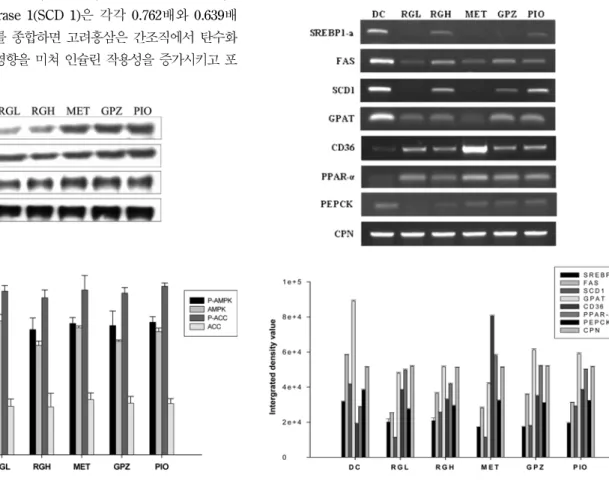
이어서홍삼의
AMPK
활성화 결과를 검증하기 위해target gene
들(lipogenesis, lipolysis, gluconeogenesis
관련유전자들)
의발현을RT-PCR
로측정한결과(Fig. 7)
홍삼은SREBP1a, FAS, SCD1,
GPAT
등지방합성에관련되는gene
들의발현을현저하게억Fig. 6 −
Western blot analysis of phospho-AMPK, AMPK, phospho-
ACC, ACC levels in liver.
Fig. 7 −RT-PCR analyses of SREBP1-
α,FAS, SCD1, GPAT, PPAR-
α,
CD36 and PEPCK genes in liver.
제한반면
CD36, PPAR-
α 등lipolysis gene
들의발현은현저하게증가시켰다
.
또한당신생과정에서rate-limiting enzyme
인PEPCK gene
의발현도DC
에비하여감소시켰다.
결 론
이상의결과들을요약하면홍삼은췌장에서
insulin
분비를촉진하는반면
glucagon
의분비를억제하여혈당을강하하였고hierarchical clustering
및principal component analysis
결과홍삼의당뇨관련유전자발현양상은
metformin
과가장유사하였으며
,
간에서AMPK activation
과ACC inactivation
을통하 여fatty acid
β-oxidation
을촉진하였고SREBP1a, FAS, SCD1, GPAT
등lipogenesis gene
들의발현을억제하여간에서지방산의합성을억제하며
CD36, PPAR-
α등lipolysis gene
들의발현 을증가하여인슐린저항성을개선시키는것으로나타났다.
결론적으로홍삼은간조직에서
AMPK signaling pathway
를활성화시켜
(
지방세포에서분비되는adipokine
인adiponectin
유 리에의해)
지방산의산화를촉진한결과중성지방의축적을차단하여 인슐린저항성을 개선하고 또한 당신생을 억제하여
(PEPCK, G6Pase
발현을억제함으로)
공복시혈당을감소시키 는 것으로사료된다.
고려홍삼은 비만한2
형 당뇨환자에서metformin
과같이인슐린저항성을개선하여공복시혈당을낮추어주는그러나
metformin
이나타낼수있는lactic acidosis
와 같은부작용은없는안전한혈당강하제로사용될수있을것으 로기대한다.
감사의 말씀
본연구는
2008
년도자생식물이용기술개발사업단(
과제번호M106KD010018-08K0401-01810)
의지원을받아수행되었습니다.
문 헌
1) Ugochukwu, N. H. and Figgers, C. L. : Modulation of the flux patterns in carbohydrate metabolism in the livers of streptozoticin-induced diabetic rats by dietary caloric restriction. Pharmacol. Res.
54, 172 (2006).
2) Cho, W. C., Chung, W. S., Lee, S. K., Leung, A. W., Cheng, C. H. and Yue, K. K. : Ginsenoside Re of Panax ginseng possesses significant antioxidant and antihyperlipidemic efficacies in streptozotocin-induced diabetic rats. Eur. J.
Pharmacol.
550, 173 (2006).
3) 2005
당뇨병기초통계연구TFT (
대한당뇨병학회,
건강보험심 사평가원)
자료.
4) Kim, Y. M., Cha, B. S., Kim, D. J., Choi, S. H., Kim, S. K., Ahn,
C. W., Lim, S. K., Kim, K. R., Huh, K. B. and Lee, H. C. : Predictive clinical parameters for therapeutic efficacy of rosiglitazone in Korean type 2 diabetes mellitus. Diabetes. Res.
Clin. Pract.
67, 43 (2005).
5) Zhang, B. B. and Moller, D. E. : New approaches in the treatment of type 2 diabetes. Curr. Opin. Chem. Biol.
4, 461 (2000).
6) Bailey, C. J. : Insulin resistance and antidiabetic drugs.
Biochem. Pharmacol.
58, 1511 (1999).
7) Scheen, A. J. : Thiazolidinediones and liver toxicity Diabetes.
Metab.
27, 305 (2001).
8) Belcher, G., Lambert, C., Edwards, G., Urquhart, R. and Matthews, D. R. : Safety and tolerability of pioglitazone, metformin, and gliclazide in the treatment of type 2 diabetes.
Diabetes Res. Clin. Pract.
70, 53 (2005).
9) Stades, A. M., Heikens, J. T., Erkelens, D. W., Holleman, F. and Hoekstra, J. B. : Metformin and lactic acidosis: cause or coincidence? A review of case reports. J. Intern. Med.
255, 179 (2004).
10) Kobayashi, M., Iwata, M. and Haruta, T. : Clinical evaluation of pioglitazone. Nippon. Rinsho.
58, 395 (2000).
11) Coleman, C. I., Hebert, J. H. and Reddy, P. : The effects of Panax ginseng on quality of life. J. Clin. Pharm. Ther.
28, 5 (2003).
12) Bucci, L. R. : Selected herbals and human exercise performance. Am. J. Clin. Nutr.
72, 624S (2000).
13) Tokuyama, S. and Takahashi, M. : Pharmacological and physiological effects of ginseng on actions induced by opioids and psychostimulants. Nippon Yakurigaku Zasshi.
117, 195 (2001).
14) Ryu, J. K., Lee, T., Kim, D. J., Park, I. S., Yoon, S. M., Lee, H. S., Song, S. U. and Suh, J. K. : Free radical-scavenging activity of Korean red ginseng for erectile dysfunction in non- insulin-dependent diabetes mellitus rats. Urology,
65, 611 (2003).
15) Shin, H. J., Kim, Y. S., Kwak, Y. S., Song, Y. B., Kim, Y. S. and Park, J. D. : Enhancement of antitumor effects of paclitaxel (taxol) in combination with red ginseng acidic polysaccharide (RGAP). Planta. Med.
70, 1033 (2004).
16) Wargovich, M. J. : Colon cancer chemoprevention with ginseng and other botanicals. J. Korean Med. Sci.,
16, S81 (2001).
17) Sung, J., Han, K. H., Zo, J. H., Park, H. J., Kim, C. H. and Oh, B. H. : Effects of red ginseng upon vascular endothelial function in patients with essential hypertension. Am. J. Chin.
Med.
28, 205 (2000).
18) Vuksan, V., Sung, M. K., Sievenpiper, J. L., Stavro, P. M.,
Jenkins, A. L., Di Buono, M., Lee, K. S., Leiter, L. A., Nam,
K. Y., Arnason, J. T., Choi, M. and Naeem, A. : Korean red
ginseng (Panax ginseng) improves glucose and insulin
regulation in well-controlled, type 2 diabetes: results of a randomized, double-blind, placebo-controlled study of efficacy and safety. Nutr. Metab. Cardiovasc. Dis.
18, 46 (2008).
19) Han, G. C., Ko, S. K., Sung, J. H. and Chung, S. H. : Compound K enhances insulin secretion with beneficial metabolic effects in db/db mice. J. Agric. Food. Chem.
55, 1064 (2007).
20) Chomczynski, P. and Sacchi, N. : Single-step method of RNA isolation by acid guanidium thiocyanate-phenol chloroform extraction. Anal. Biochem.
162, 156 (1987).
21) Stich, V. and Berlan, M. : Physiological regulation of NEFA availability: lipolysis pathway. Proc. Nutr. Soc.
63, 369 (2004).
22) Raz, I., Eldor, R., Cernea, S. and Shafrir, E. : Diabetes: insulin resistance and derangements in lipid metabolism. Cure through intervention in fat transport and storage. Diabetes Metab. Res. Rev.
21, 3 (2005).
23) Wang, Y., Lam, K. S., Yau, M. H. and Xu, A. : Post-translational modifications of adiponectin: mechanisms and functional implications. Biochem. J.
409, 623 (2008).
24) Whitehead, J. P., Richards, A. A., Hickman, I. J., Macdonald, G. A. and Prins, J. B. : Adiponectin--a key adipokine in the metabolic syndrome. Diabetes. Obes. Metab.
8, 264 (2006).
25) Aguilera, C. M., Gil-Campos, M., Cañete, R. and Gil, A. : Alterations in plasma and tissue lipids associated with obesity and metabolic syndrome. Clin. Sci. (Lond)
114, 183 (2008).
26) Scott, D. K., O'Doherty, R. M., Stafford, J. M., Newgard, C. B.
and Granner, D. K. : The repression of hormone-activated PEPCK gene expression by glucose is insulin-independent but requires glucose metabolism. J. Biol. Chem.
273, 24145 (1998).
27) Lochhead, P. A., Salt, I. P., Walker, K. S., Hardie, D. G. and Sutherland, C. : 5-aminoimidazole-4-carboxamide riboside mimics the effects of insulin on the expression of the 2 key gluconeogenic genes PEPCK and glucose-6-phosphatase.
Diabetes.
49, 896 (2000).
28) Ho, H. Y., Cheng, M. L. and Chiu, D. : T.Glucose-6-phosphate dehydrogenase--from oxidative stress to cellular functions and degenerative diseases. Redox. Rep.
12, 109 (2007).
29) Aoyama, H., Daitoku, H. and Fukamizu, A. : Nutrient control of phosphorylation and translocation of Foxo1 in C57BL/6 and db/
db mice. Int. J. Mol. Med.
18, 433 (2006).
30) Ugochukwu, N. H. and Babady, N. E. : Antihyperglycemic effect of aqueous and ethanolic extracts of Gongronema latifolium leaves on glucose and glycogen metabolism in livers of normal and streptozotocin-induced diabetic rats. Life. Sci.
73