애엽과 음양곽 혼합 발효물이 대식세포 활성에 미치는 영향
경원대학교 한의과대학 부인과학교실 류한우, 김윤상, 임은미
ABSTRACT
Fermented Artemisiae Argyi Folium and Epimedii Herba Mixture Effect on Macrophage’ Activity
Hahn-Woo Ryu, Yoon-Sang Kim, Eun-Mee Lee
Dept. of Gynecology, College of Oriental Medicine, Kyungwon University Purpose: This research aimed to study the effect of FAE(Ferment Artemisiae Argyi Folium and Epimedii Herba) on the mouse macrophage cell activity.
Methods: Effect of FAE, which was fermented by Sacchromyces cerevisiae STV89, on cell viability, amount of H2O2 within cells, amount of NO was measured and compaperd by using mouse macrophage cells.
Results:
1. Result of MTT assay conducted to observe the effect of FAE on the survival rate of mouse macrophage cells illustrated that, when FAE was proccessed for each concentration, there was no significant decrease of the survival rate.
2. FAE increased the amount of H2O2 within macrophage cells and increased inhibition of amount of H2O2 in macrophage induced by LPS.
3. FAE inhibited amount of NO in macrophage cells, and significantly inhibited increase of amount of NO in mcacrophage induced by LPS.
Conclusion: FAE produced by Artemisiae Argyi Folium and Epimedii Herba did not induce the decrease of macrophage cell survival rate, increased amount of H2O2 within cells, and reduced amount of NO. FAE significantly increase by LPS, reduced the increase of amount of NO in macrophage induced by LPS.
These results signify FAE has significant effect on immuno modulating activity of macrophage.
Key Words: Artemisiae Argyi Folium, Epimedii Herba, Fermented Herbal Medicine, Macrophage, Immuno activity.
5)
교신저자(김윤상) : 인천광역시 중구 용동 117번지 경원대학교 부속 길한방병원 부인과 전화 : 032-770-1227 이메일 : [email protected]
Ⅰ. 서 론
발효한약은 전통발효공법을 통해 한약 재를 미생물이 잘 이용할 수 있게 찌거 나 삶은 다음, 공기 중의 미생물 또는 유 산균과 같은 순수 분리 미생물을 이용하 여 발효한 한약을 말한다. 이는 한약성 분의 체내흡수율과 생체 이용률을 극대 화시킨 일종의 가공방법으로 약리적 기 능성뿐만 아니라 한약의 제형을 개선시 킬 수 있다1).
애엽은 국화과에 속한 다년생 초본으 로 性은 溫하고 味는 辛苦하여 散寒止痛 하고 溫經止血하여 吐血, 衄血, 崩漏, 姙 娠下血, 月經不調, 痛經, 胎動不安, 泄瀉 久痢, 帶下, 濕疹, 癰瘍 등에 응용한다2).
음양곽은 매자나무과에 속하는 다년생 초본인 삼지구엽초 및 기타 동속 식물의 줄기와 잎으로 性은 溫하고 味는 辛甘하 여 補腎壯陽, 祛風除濕하여 陽萎不擧, 小 便淋瀝, 筋骨攣急, 腰膝無力, 風濕痺痛, 四肢不仁 등에 사용한다2).
애엽과 음양곽은 모두 면역반응을 증 강 시켜 염증을 억제하는 효능이 보고되 었고3,4), 한방부인과적 임상질환에 빈용 되고 있으므로 이들을 혼합 발효하여 대 식세포 활성에 미치는 영향을 확인해 보 고자 하였다.
이에 저자는 애엽과 음양곽을 효모로 발효시켜 추출하여 얻은 시료 FAE를 마 우스 대식세포에 농도별로 처리하여 cell viability, 세포내 H2O2 및 NO 생성에 미 치는 영향을 측정, 비교한 결과 대식세 포의 면역관련 활성에 대한 유의한 효능 이 있었기에 보고하는 바이다.
Ⅱ. 실 험
1. 재 료 1) 약 재
실험에 사용된 애엽과 음양곽은 경방 제약에서 구입하였고, 모든 약재는 초음 파 세척기를 이용하여 불순물을 제거하 고 실험에 사용하였으며, 발효에 사용된 균주로는 Sacchromyces cerevisiae STV89 를 사용하였다.
2) Cell line
실험에 사용된 대식세포는 mouse macrophage(Raw 264.7 cell line)이고, 한 국 세포주 은행에서 구입하였다.
3) 시약 및 기기 (1) 시 약
본 실험을 위해서 ethyl alcohol(Samchun Chemical, Korea), methyl alcohol(Samchun Chemical, Korea), dimethyl sulfoxide (DMSO, Sigma, USA), Dulbecco's modied essential medium(DMEM, Sigma, USA), 1×PBS (Sigma, USA), dihydrorhodamine 123(DHR 123, Sigma, USA), ethylenediaminetetraacetic acid(EDTA, Sigma, USA), acetic acid(Sigma, USA), isopropanol(Sigma, USA), Trypsin -EDTA(Sigma, USA), sulfanilamide(Sigma, USA), H2SO4(Sigma, USA), potassium phosphate(Sigma, USA), NO assay kit (Oxford Biomedical Research, USA) 등 이 사용 되었다.
(2) 기 기
본 실험에 사용된 기기는 CO2 incuvator (NUAIRE, USA), pulverizer(Rong tsong, Taiwan), rotary vacuum evaporator(Eyela, Japan), air compressor(Tamiya, Japan), homogenizer(Omni, USA), research microscope
(Becton dickinson, USA), centrifuge(Hanil, Korea), fume hood(Hanil, Korea), clean bench(Jeio thec, Korea), ultrasonic cleaner (Branson, USA), microplate reader(Bio -Rad, USA), thermo aluminum bath(Fine PCR, USA), vortex mixer(Vision Scientific Co, Korea), water bath(Intron biotech, Korea), ice-maker(Vision Scientific Co, Korea) 등이다.
2. 방 법
1) 시료의 제조
(1) 애엽과 음양곽 추출물 제조 정확히 중량을 측정한 애엽 50 g을 환 류추출기에 1차 증류수 2,000 mL와 함께 넣은 뒤 탕액이 끓는 시점으로부터 2시 간 동안 가열하여 나온 추출액을 filter paper(Advantec No.2, Japan)로 감압 여과 한 후 여과액을 rotary vacuum evaporator (회전식 감압 농축기)를 이용하여 농축 하였다. 이 농축액을 동결건조기를 이용 하여 건조시켰고, 동결건조 추출물은 3.7 g을 얻었으며, 수율은 7.4%였다. 음양곽 도 같은 방법을 시행하여 동결건조 추출 물 17.6 g을 얻었고, 수율은 35.2%이었 다.
(2) FAE 발효 추출물 제조
위에서 제조된 애엽과 음양곽 추출물 을 이용하여 각각 다음과 같은 방법을 통하여 발효 애엽과 발효 음양곽을 얻었 다.
① 조효소 조제 : 조효소제인 3g의 Herbzyme-α(한국효소)에 증류수 100mL 를 가하고 37℃에서 30분간 침출하여 여 과시킨 후 그 여액을 조효소액으로 사용 하였다.
② 열수추출하여 건조한 애엽(3.0g, pH:5.44)
과 음양곽(3.0g, pH:4.82)을 screw cap tube에 각각 0.95g을 담고 미리 추출된 조효소액을 2.2mL을 첨가하여 37℃에서 2시간 효소반응 하였다.
③ 효소반응 후 95℃에서 10분간 살균 하였다.
④ Sacchromyces cerevisiae STV89를 애엽과 음양곽에 4%씩 접종한 후 30℃
에서 4일간 배양하였다.
⑤ 배양 후 60℃에서 20분간 열처리하 였다.
⑥ Sacchromyces cerevisiae STV89에 서 배양 후 애엽 발효물의 pH는 5.55였 고 음양곽 발효물의 pH는 4.24였다.
위에서 얻은 애엽 발효물과 음양곽 발 효물을 1:1로 혼합하여 시료 FAE를 만 들어 사용하였다. 발효물의 ph가 발효 전후와 비교하여 변화가 거의 없이 유사 하므로 발효가 잘 되었다는 것을 의미한 다. 만약 변화가 크다면 부패의 의미로 볼 수 있다.
2) 세포 배양
Raw 264.7 cell은 37℃, 5% CO2 조건 에서 10% FBS, penicillin(100IU/mL), streptomycin(100㎍/mL)이 첨가된 DMEM 배지에서 배양하였다. Cell은 75cm2 flask (Falcon, USA)에서 충분히 증식된 후 배양 3일 간격으로 배양세포 표면을 phosphate buffered saline(PBS) 용액으 로 씻어준 후 50mL flask 당 1mL의 0.25% trypsin-EDTA용액을 넣고 실온 에서 1분간 처리한 다음 trypsin용액을 버리고 37℃에서 5분간 보관하여 세포를 탈착하여 계대 배양하였다. 탈착된 세포 는 10% FBS가 첨가된 DMEM 배양액 10mL에 부유시킨 다음 새로운 배양용기 (50mL culture flask)에 옮겨 1:2의 split
ratio로 CO2 배양기(37℃, 5% CO2)에서 배양하였다.
3) 세포독성 검사
준비된 시료 FAE가 Raw 264.7 cells에 나타내는 세포독성 유발 효과를 알아보 기 위하여 Mosmann 등5-7)의 방법을 응 용하여 MTT assay를 실시하였다. 96 well plate에 1×10⁴ cells/well의 cell을 100㎕씩 넣고 37℃, 5% CO2 incubator에 서 24시간동안 배양한 후 배지를 버리고 배양세포 표면을 phosphate buffered saline (1×PBS) 용액으로 씻어주었다. 같은 양 의 배지와 PBS에 녹인 시료(25, 50, 100, 200㎍/mL)를 각 well에 처리하고 24시 간 동안 배양하였다. 배양이 끝난 후 PBS에 녹인 1㎎/mL MTT(Sigma, USA) 를 100㎕씩 각 well에 처리하여 알루미늄 호일로 차광시킨 후 2시간 동안 같은 조 건에서 배양하였다. 배양액을 모두 제거 한 후 DMSO를 100㎕ 처리하고 37℃에 서 2시간 방치 후 microplate reader (Molecular Devices, USA)를 이용하여 540nm에서 흡광도를 측정하였다. Cell viability는 다음 공식으로 계산되었다.
Cell viability(%)=AT/AC×100 AC-absorbance of control
AT-absorbance of tested extract solution.
4) Hydrogen peroxide(H2O2) assay 세포내 H2O2 생성은 Roesler 등8-12)의 방법을 응용, DHR 123 assay를 실시하 여 측정하였다. DHR은 비형광이지만 세포 내에서 세포 내 H2O2에 의하여 산 화되어 녹색의 형광을 발현하는 물질인 rhodamine 123(R123)로 바뀌게 된다. 그 러므로 여러 가지 산화적 반응을 일으키 는 물질들로 인해 mouse의 대식세포내 에서 발생하는 hydrogen proxide의 수준
을 DHR 123 assay을 이용하여 측정할 수 있다. 96 well plate 1×10⁴ cells/well의 cell을 100㎕씩 넣고 37℃, 5% CO2
incubator에서 24시간동안 배양한 후 배 지를 버리고 배양세포 표면을 phosphate buffered saline(1×PBS) 용액으로 씻어 주었다. 시료 단독 혹은 LPS와 시료를 동 시에 처리하기 전에 우선 DHR(10uM)이 담긴 배지를 30분간 각 well에 처리한 뒤 배지를 제거하였다. 시료 단독(25, 50, 100, 200, 400㎍/mL) 혹은 LPS를 처리 (2㎍/mL)하거나 시료와 LPS를 함께 배 지에 담아 각 well에 처리하고 2, 4, 22, 24, 46, 48시간동안 37℃, 5% CO2 incubator 에서 배양한 후 microplate reader(Bio- Rad, USA)를 이용하여 490nm에서 흡광 도를 측정하였다. 세포내 H2O2 생성은 다음 공식으로 계산되었다.
Intracellular Productions of H2O2(%)
=AT/AC×100
AC-absorbance of control
AT-absorbance of tested extract solution.
5) Nitric oxide(NO) 생성 측정 세포로부터 생성되는 NO의 양은 Weissman
등13-16)의 방법을 응용, 세포배양액 중에
존재하는 NO2-를 Griess 시약을 이용, 측 정하였다. NO의 기질인 L-arginine은 L-citruline과 NO로 변하는데, 이는 빠르 게 안정된 NO2, MINO2, M(NO3)n으로 변한다. Griess 시약(Griess reagent : 0.5%의 sulfanilamide, 2.5%의 phosphoric acid 및 0.5%의 naphthyl ethylene diamine) 은 NO2와 화학 반응하여 보라색의 아조 염을 형성하고 이것은 NO의 농도와 일 치하기 때문에, 아조염의 농도로부터 NO2의 농도를 추정하기 위해 microplate reader를 이용, 540nm에서 흡광도를 측
정하여 NO의 생성정도를 비교하였다.
이를 위해 다음과 같이 실험하였다. 시 료 단독(50, 100, 200㎍/mL) 혹은 LPS 를 처리(2㎍/mL)하거나 시료와 LPS를 함께 배지에 담아 각 well에 처리하고 24 시간 동안 37℃, 5% CO2 incubator에서 배양한 후 세포배양 상등액 60㎕을 채취 하여 여기에 Griess 시약 60㎕을 혼합하 여 15분 동안 반응시킨 후 microplate reader(Bio-Rad, USA)를 이용하여 540nm에서 흡광도를 측정하였다. 세포 의 NO 생성은 다음 공식으로 계산되었 다.
Productions of NO(%)=AT/AC×100 AC-absorbance of control
AT-absorbance of tested extract solution.
3. 통계처리
본 실험에서 얻은 결과에 대해서는 평 균치±표준편차(mean±SD)로 나타내었으 며, 대조군과 각 실험군과의 평균의 차 이는 Student's t-test와 ANOVA test로 분석하여 p-value값이 0.05 미만일 때 통 계적으로 유의성이 있는 것으로 판정하 였다.
Ⅲ. 결 과
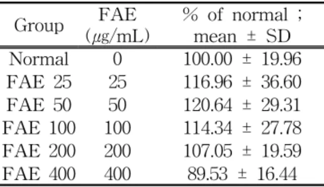
1. FAE가 대식세포의 세포생존율에 미치는 영향
FAE가 대식세포의 생존율에 미치는 영향을 비교한 결과 24시간 처리한 후 25㎍/mL 이상의 모든 농도에서 유의한 감소는 없었다(Table 1).
Group FAE (㎍/mL)
% of normal ; mean ± SD Normal 0 100.00 ± 19.96 FAE 25 25 116.96 ± 36.60 FAE 50 50 120.64 ± 29.31 FAE 100 100 114.34 ± 27.78 FAE 200 200 107.05 ± 19.59 FAE 400 400 89.53 ± 16.44 FAE : Mixture of Fermented Artemisiae Argyi Folium and Fermented Epimedii Herba.
Fermentation was done with Sacchromyces cerevisiae STV89.
Normal : Non-treated with FAE.
Results are represented as mean±SD.
Table 1. Cell Viabilities of Raw 264.7 Cell Incubated with FAE for 24hr.
2. FAE가 대식세포내 H2O2의 생성에 미치는 영향
1) FAE의 투약 시간에 따른 H2O2의 생성량 변화
FAE가 포함된 배지에 Raw 264.7 세 포들을 2, 4, 22, 24, 46, 48시간동안 배양 한 후 세포 내의 H2O2의 생성에 미치는 영향을 비교한 결과 50㎍/mL 이상에서 모두 유의한 증가를 나타내었다(Table 2, 3, 4, 5, 6, 7).
Group FAE (㎍/mL)
% of normal ; mean ± SD Normal 0 100.00 ± 4.85 FAE 25 25 98.84 ± 15.84 FAE 50 50 115.41 ± 23.40*
FAE 100 100 123.11 ± 14.44*
FAE 200 200 128.44 ± 17.36*
FAE 400 400 128.59 ± 8.35*
FAE : Mixture of Fermented Artemisiae Argyi Folium and Fermented Epimedii Herba.
Fermentation was done with Sacchromyces cerevisiae STV89.
Normal : Non-treated with FAE.
Results are represented as mean±SD.
* : represents P < 0.05 compared to the normal.
Table 2. Intracellular Production of H2O2 of Raw 264.7 Cell Incubated with FAE for 2hr.
Group FAE (㎍/mL)
% of normal ; mean ± SD Normal 0 100.00 ± 4.56 FAE 25 25 101.20 ± 11.84 FAE 50 50 115.72 ± 19.20*
FAE 100 100 124.36 ± 9.95*
FAE 200 200 127.80 ± 14.40*
FAE 400 400 129.46 ± 5.05*
FAE : Mixture of Fermented Artemisiae Argyi Folium and Fermented Epimedii Herba.
Fermentation was done with Sacchromyces cerevisiae STV89.
Normal : Non-treated with FAE.
Results are represented as mean±SD.
* : represents P < 0.05 compared to the normal.
Table 3. Intracellular Production of H2O2 of Raw 264.7 Cell Incubated with FAE for 4hr.
Group FAE (㎍/mL)
% of normal ; mean ± SD Normal 0 100.00 ± 3.80 FAE 25 25 96.52 ± 2.48*
FAE 50 50 111.50 ± 4.78*
FAE 100 100 123.68 ± 3.46*
FAE 200 200 131.01 ± 3.92*
FAE 400 400 138.66 ± 4.90*
FAE : Mixture of Fermented Artemisiae Argyi Folium and Fermented Epimedii Herba.
Fermentation was done with Sacchromyces cerevisiae STV89.
Normal : Non-treated with FAE.
Results are represented as mean±SD.
* : represents P < 0.05 compared to the normal.
Table 4. Intracellular Production of H2O2 of Raw 264.7 Cell Incubated with FAE for 22hr.
Group FAE (㎍/mL)
% of normal ; mean ± SD Normal 0 100.00 ± 3.89 FAE 25 25 95.05 ± 2.42*
FAE 50 50 109.91 ± 3.85*
FAE 100 100 122.48 ± 3.09*
FAE 200 200 129.09 ± 3.71*
FAE 400 400 138.84 ± 4.64*
FAE : Mixture of Fermented Artemisiae Argyi Folium and Fermented Epimedii Herba.
Fermentation was done with Sacchromyces cerevisiae STV89.
Normal : Non-treated with FAE.
Results are represented as mean±SD.
* : represents P < 0.05 compared to the normal.
Table 5. Intracellular Production of H2O2 of Raw 264.7 Cell Incubated with FAE for 24hr.
Group FAE (㎍/mL)
% of normal ; mean ± SD Normal 0 99.99 ± 4.33 FAE 25 25 96.90 ± 4.13 FAE 50 50 111.27 ± 3.63*
FAE 100 100 125.31 ± 3.25*
FAE 200 200 133.05 ± 3.28*
FAE 400 400 142.71 ± 5.28*
FAE : Mixture of Fermented Artemisiae Argyi Folium and Fermented Epimedii Herba.
Fermentation was done with Sacchromyces cerevisiae STV89.
Normal : Non-treated with FAE.
Results are represented as mean±SD.
* : represents P < 0.05 compared to the normal.
Table 6. Intracellular Production of H2O2 of Raw 264.7 Cell Incubated with FAE for 46hr.
Group FAE
(㎍/mL) % of normal ; mean ± SD Normal 0 100.00 ± 4.29 FAE 25 25 97.89 ± 4.39 FAE 50 50 112.74 ± 3.71*
FAE 100 100 126.61 ± 3.17*
FAE 200 200 133.82 ± 3.58*
FAE 400 400 142.98 ± 5.21*
FAE : Mixture of Fermented Artemisiae Argyi Folium and Fermented Epimedii Herba.
Fermentation was done with Sacchromyces cerevisiae STV89.
Normal : Non-treated with FAE.
Results are represented as mean±SD.
* : represents P < 0.05 compared to the normal.
Table 7. Intracellular Production of H2O2 of Raw 264.7 Cell Incubated with FAE for 48hr.
2) FAE의 LPS로 유발된 H2O2 생성 감소에 대한 영향
LPS 단독으로 1, 22, 24시간동안 배양 한 경우 Normal 군보다 유의하게 H2O2
생성이 감소하였고, LPS와 FAE를 함께 배양한 경우 FAE가 25㎍/mL이상일 때 LPS에 의한 감소를 모두 유의하게 회복, 증가시켰다(Table 8, 9, 10).
Group FAE (㎍/mL)
LPS (㎍/mL)
% of control
; mean ± SD
Normal 0 0 125.20±8.47
Control 0 2 100.00±43.72# FAE 25 25 2 182.45±56.06*
FAE 50 50 2 199.91±52.92*
FAE 100 100 2 219.53±72.17*
FAE 200 200 2 204.32±28.93*
FAE 400 400 2 217.64±12.16*
FAE : Mixture of Fermented Artemisiae Argyi Folium and Fermented Epimedii Herba.
Fermentation was done with Sacchromyces cerevisiae STV89.
Normal : Non-treated with LPS.
Control : Treated with LPS only.
Results are represented as mean±SD.
# : represents P < 0.05 compared to the normal.
* : represents P < 0.05 compared to the control.
Table 8. Intracellular Production of H2O2 of Raw 264.7 Cell Incubated with FAE and LPS for 1hr.
Group FAE (㎍/mL)
LPS (㎍/mL)
% of control
; mean ± SD
Normal 0 0 223.23±14.42
Control 0 2 100.00±5.60#
FAE 25 25 2 189.90± 11.07*
FAE 50 50 2 194.44±8.44*
FAE 100 100 2 230.47±7.25*
FAE 200 200 2 276.26±7.43*
FAE 400 400 2 353.70±13.75*
FAE : Mixture of Fermented Artemisiae Argyi Folium and Fermented Epimedii Herba.
Fermentation was done with Sacchromyces cerevisiae STV89.
Normal : Non-treated with LPS.
Control : Treated with LPS only.
Results are represented as mean±SD.
# : represents P < 0.05 compared to the normal.
* : represents P < 0.05 compared to the control.
Table 9. Intracellular Production of H2O2 of Raw 264.7 Cell Incubated with FAE and LPS for 22hr.
Group FAE (㎍/mL)
LPS (㎍/mL)
% of control ; mean ± SD
Normal 0 0 208.90±13.33
Control 0 2 99.99±14.24# FAE 25 25 2 313.88±111.10*
FAE 50 50 2 334.54±116.70*
FAE 100 100 2 340.27±115.90*
FAE 200 200 2 253.25±6.34*
FAE 400 400 2 324.29±17.78*
FAE : Mixture of Fermented Artemisiae Argyi Folium and Fermented Epimedii Herba.
Fermentation was done with Sacchromyces cerevisiae STV89.
Normal : Non-treated with LPS.
Control : Treated with LPS only. # represents P < 0.05 compared to the normal.
Results are represented as mean±SD.
* : represents P < 0.05 compared to the control.
Table 10. Intracellular Production of H2O2 of Raw 264.7 Cell Incubated with FAE and LPS for 24hr.
3. FAE가 대식세포내 NO의 생성에 미치는 영향
1) FAE의 의한 NO 생성량 변화 24시간 배양에서 50, 100㎍/mL일 때 유의한 감소를 나타내었다(Table 11).
Group FAE (㎍/mL)
% of normal ; mean ± SD Normal 0 100.00 ± 29.93 FAE 50 50 56.47 ± 28.27*
FAE 100 100 69.70 ± 39.46*
FAE 200 200 97.65 ± 64.09*
FAE : Mixture of Fermented Artemisiae Argyi Folium and Fermented Epimedii Herba.
Fermentation was done with Sacchromyces cerevisiae STV89.
Normal : Non-treated with FAE.
Results are represented as mean±SD.
* : represents P < 0.05 compared to the normal.
Table 11. NO Production of Raw 264.7 Cell Incubated with FAE for 24hr.
2) FAE의 LPS로 유발된 NO 생성 증 가에 대한 영향
24시간동안 LPS 단독으로 배양한 경 우 Normal 군보다 유의하게 NO생성이 증가하였고, LPS와 FAE를 함께 배양한 경우 FAE가 25㎍/mL이상일 때 농도에 비례하여 LPS에 의한 증가를 유의하게 억제시켰다(Table 12).
Group FAE (㎍/mL)
LPS (㎍/mL)
% of control ; mean ± SD
Normal 0 0 6.04 ± 1.42
Control 0 2 100.00 ± 3.14# FAE 25 25 2 79.11 ± 8.35*
FAE 50 50 2 73.17 ± 6.82*
FAE 100 100 2 64.23 ± 7.47*
FAE 200 200 2 43.77 ± 5.03*
FAE 400 400 2 16.38 ± 2.00*
FAE : Mixture of Fermented Artemisiae Argyi Folium and Fermented Epimedii Herba.
Fermentation was done with Sacchromyces cerevisiae STV89.
Normal : Non-treated with LPS.
Control : Treated with LPS only.
Results are represented as mean±SD.
# : represents P < 0.05 compared to the normal.
* : represents P < 0.05 compared to the control.
Table 12. NO Production of Raw 264.7 Cell Incubated with FAE and LPS for 24hr.
Ⅳ. 고 찰
발효한약은 우리 조상들의 지혜가 담 긴 전통발효공법을 통해 한약재를 찌거 나 삶은 다음, 공기 중의 미생물이나 유 산균과 같은 순수 분리 미생물을 이용하 여 발효한 한약재를 말하는데, 이는 기 존 전탕, 과립제 및 환약 등의 제형에 비 해 한약성분의 체내흡수율과 생체 이용 률을 극대화시킨 방법으로 약리적 기능 성과 안전성, 한약의 제형개량 및 포제 방법 등을 향상시킴으로써 한약의 수요 창출과 고부가가치의 새로운 한약제품을 개발할 수 있다는데 그 의의가 있다1). 최근 발효 한약에 대한 관심이 높아지고 있는 반면에 개별 한약재나 한약처방을 발효시킨 시료에 대한 연구가 아직은 미 흡한 실정이다.
발효한약에 관한 선행 연구로는 유산 균과 한국산 겨우살이 추출물의 항종양 활성 및 면역 자극 효과17), 버섯균사체 로 발효시킨 복령과 후박의 항산화 및 항암효과18), 인진호, 금은화, 지구자 등 을 주원료로 제조된 발효한약 추출물로 숙취해소 효능1) 및 차가버섯과 어성초 함유 발효 조성물로 암 예방 및 항암 식․의약품의 소개 개발 가능성 등이 있 다19).
애엽은 국화과에 속한 다년생 초본인 황해쑥(Artemisia argyi), 쑥(A. princeps), 또는 산쑥(A. montana)의 葉 및 어린 줄기를 여름에 꽃이 피기 전에 따서 曬 乾한 것인데2), 氣味가 辛苦溫하고, 肝, 脾, 腎經에 歸經하며, 溫經止血, 散寒止 痛, 去濕止痒의 효능이 있어 일반적으로 吐血, 衄血, 喀血, 便血, 心腹冷痛, 泄瀉
久痢, 霍亂轉筋, 濕疹, 疥癬, 痔瘡 및 癰 瘍 등의 질환에 응용한다20). 또한, 한방 부인과에서는 溫經固衝任止血의 효능으 로 胎動不安에 艾葉炭을 安胎藥과 함께 사용하거나 下元虛寒으로 인한 月經過多 와 崩漏下血에 艾葉炭을 사용하고, 溫經 止痛하는 효능으로 虛寒性 子宮寒冷, 久 不受孕 및 經前經行少腹冷痛에 사용하며
21), 외용으로 帶下, 陰痒, 冷症, 骨盤痛 및 月經痛 등의 질환에 사용한다22).
애엽의 주성분은 monoterpenoid 화합 물, aromatic 화합물, aliphatic acohol, sesquiterpene 화합물, arteminolide A-D, surfated polysaccaride, phenylpropanoid 화합물, olyoxyflavone 화합물, adenine, choline, vitamine A, B, C, D와 amylase
등으로23-31) 약리 작용으로는 항암작용
32-34), 항백혈병작용35), 항균작용25,36,37), 항 염증 및 항산화작용38), 혈소판 응집 억 제작용39), 혈관이완작용40), Trp-P-2의 비활성화41), 항천식작용42) 및 소화성궤 양에 대한 세포보호작용 등이 보고되었 다43,44).
음양곽은 매자나무과(berberidaceae)에 속 하는 다년생 초본인 삼지구엽초(Epimedium koreanum Nakai) 및 기타 동속 식물의 줄기와 잎을 여름과 가을 사이에 채취하 여 건조한 것인데, 氣味가 辛甘溫하고, 肝, 腎經에 歸經하며, 補腎壯陽, 祛風除 濕의 효능이 있어 일반적으로 陽萎不擧, 小便淋瀝, 筋骨攣急, 半身不遂, 腰膝無力, 風濕痺痛 및 四肢不仁 등의 질환에 응용 한다2). 또한 한방부인과적으로 腎虛로 인한 月經不調, 男女不姙, 産後身痛, 골 다공증 및 갱년기의 고혈압 등에 사용한 다2,22,45).
음양곽의 flavonoid들은 항염, 항알러
지, 항바이러스 및 항종양 작용 등의 다 양한 약리작용을 있어 세포성 면역반응 과 대식세포의 활성 및 말초순환 백혈구 수의 증가46), 생식기능에 대한 작용, 미 생물에 대한 작용, 진해거담 및 혈압에 대한 강압 작용 등이 밝혀져 있고47), 항 산화 성분에 대한 연구48,49), 신경세포의 산소자유기독성에 대한 항산화 작용 연 구50), 항고혈압작용51), 간의 대사계에 대 한 연구52) 및 음양곽의 flavonoid 성분에 대한 연구53,54) 등이 보고되었으며, 그 중 quercetin 등은 mast cell로부터의 histamine 유리억제 작용55,56), icariin은 고혈압으로 인한 합병증에 대한 효과57) 등이 보고되 었다.
이에 저자는 발효 한약의 애엽과 음양 곽을 각각 효모 Sacchromyces cerevisiae STV89로 발효 추출한 뒤 혼합하여 얻은 시료 FAE로 마우스 대식세포를 이용한 cell viability, 세포내 H2O2 생성, NO 생 성에 미치는 영향을 측정하여 FAE가 대 식세포의 면역관련 활성에 대한 변화를 알아보았는데, 애엽과 음양곽을 혼합하 여 사용한 것은 두 가지 약물 모두 면역 반응을 증강 시켜 염증을 억제하는 효능 이 있고3,4), 한방부인과 임상에서 빈용하 고 있으므로 함께 혼합 발효하여 실험하 였다.
발효미생물은 빠른 增殖性, 물질의 資 化性, 다양한 화학활성, 반응 特異性, 인 공변이의 容易性, 높은 均一性 및 고온 고압의 不必要 등의 특징이 있고58), 그 중 Sacchromyces계통은 일정한 생화학 적 성질, 물에서의 높은 분산도, 자기소 화에 대한 내성에 기인한 높은 보존성, 당밀배지에서의 빠른 증식속도 및 높은 수득률 등의 장점이 있어 본 실험에서
Sacchromyces cerevisiae STV89(효모)를 균주로 이용하여 발효하였다59).
산화적 스트레스를 일으키는 물질인 lipopolysaccharide(LPS)는 그람음성균의 세포벽에 존재하는 구성물질의 일종으로 대 식세포나 단핵구의 활성으로 인한 cytokine 을 분비하게 하고, 시상하부와 부신피질 의 활성을 유도하여 감염과 염증을 유발
시키므로60,61) 본 실험에서는 LPS를 이용
하여 마우스 대식세포의 세포내 H2O2
생성 감소와 NO 생성 증가를 유도한 후 시료가 미치는 영향을 살펴보았다.
H2O2는 세포내에서 발생하는 reactive oxygen species(ROS)의 일종으로 세포 의 산화적 stress를 유발하고62), 염증반응 에서 neutrophils 등의 세포는 ROS를 많 이 생성함으로써 immunologic reaction을 유발하며63), 최근연구에서는 macrophage 의 H2O2 생성 증가가 T-cell과 관련된 arthritis를 억제하는 함으로써 자가면역 질환 발생을 방어하는 작용이 있음이 보 고되었다64).
NO는 세포기능 발현 과정에서 signalling molecule로 활동하는데, 산화질소 합성효 소(nitric oxide synthase; NOS)에 의해 체내에서 생산되고, 이는 혈관확장을 통한 혈압감소, 혈소판 활동(platelet activity) 의 조절, 면역반응(immune response)에 의 기여, 신경전달(neurotransmission) 조절 및 세포분화(cell differentiation) 등 에 관여한다65-71).
본 실험에서 FAE가 대식 세포의 생존 율에 미치는 영향을 관찰하여 독성유무 를 살펴보았는데, 24시간 처리한 결과 FAE 25㎍/mL 이상의 모든 농도에서 유 의한 감소는 없는 것으로 나타나 FAE가 특별한 독성을 나타내지 않는 것으로 사
료되며, 독성기준은 Mosmann 등5-7)의 방법을 응용하여 MTT assay를 실시하 였다.
FAE가 포함된 배지에 Raw 264.7 세 포들을 2, 4, 22, 24, 46, 48시간동안 배양 한 후 세포 내의 H2O2의 생성에 미치는 영향을 비교한 결과 50㎍/mL 이상에서 모두 농도 의존적으로 유의하게 증가하 였고, FAE가 LPS로 염증이 유발된 대 식세포의 H2O2의 생성에 미치는 영향을 비교한 결과에서도 1, 22, 24시간동안 함 께 배양한 경우 FAE의 농도 25㎍/mL이 상일 때 LPS에 의한 H2O2의 감소를 모 두 유의하게 회복, 증가시키는 것으로 나타났으며, 이러한 실험결과는 FAE가 대식세포의 H2O2생성감소와 연관되는 류마티스성 관절염 증상악화 등에 대한 대응약물로서 개발될 수 있음을 의미한 다.
FAE가 대식세포의 NO의 생성에 미 치는 영향을 비교하였는데, 24시간 처리 한 결과 50, 100㎍/mL일 때 유의한 감소 를 나타내었고, LPS와 FAE를 함께 24 시간동안 배양하여 염증을 유발한 경우 FAE의 농도 25, 50, 100, 200, 40㎍/mL 의 모든 농도에서 LPS에 의한 NO의 증 가를 농도 의존적으로 유의하게 억제시 켰으며, 이 결과는 애엽의 단독 실험 보 고72)와 유사하게 FAE도 대식세포의 NO 생성증가와 관련된 염증반응을 완화할 수 있음을 의미한다.
이상의 결과, FAE는 대식세포의 생존 율의 감소를 유발하지 않으면서 대식세 포내의 H2O2의 생성을 증가시켰고, NO 의 생성은 감소시켰으며, LPS로 유발된 염증에서 대식세포내의 H2O2의 생성감 소를 유의하게 증가시켰고, 증가된 NO
생성을 유의하게 감소시키는 등 FAE가 대식세포의 면역작용과 관련하여 유의한 활성이 있는 것으로 나타났다.
이를 바탕으로 애엽과 음양곽 혼합 발 효물을 이용한 면역 치료제 개발을 위해 서 추후 많은 연구가 더 필요할 것으로 사료된다.
Ⅴ. 결 론
애엽과 음양곽을 효모 Sacchromyces cerevisiae STV89로 발효시킨 후 혼합한 시료 FAE로 마우스 대식세포를 이용하 여 cell viability를 관찰하고 세포내 H2O2생성과 NO 생성을 측정하였으며, LPS로 염증을 유발시킨 후 H2O2와 NO 의 생성에 미치는 영향을 측정하여 다음 과 같은 결론을 얻었다.
1. FAE의 대식세포의 생존율에 미치는 영향을 확인하기 위해 MTT assay를 수행한 결과 FAE를 25, 50, 100, 200, 400㎍/mL 농도로 처리했을 때 각각 대식세포에 유의한 생존율 감소를 유 발하지 않았다.
2. FAE를 2, 4, 22, 24, 46, 48hr의 시간 경과별로 측정한 결과 대식세포의 세 포내 H2O2의 생성을 증가시켰고, LPS 로 유발된 염증으로 대식세포내 억제 된 H2O2의 생성을 1, 22, 24hr의 시간 경과별로 유의하게 증가시켰다.
3. FAE는 대식세포의 NO의 생성을 억 제시켰으며 LPS로 유발된 염증으로 대식세포내 생성 증가된 NO를 FAE 가 유의하게 억제시켰다.
이상의 결과, 애엽과 음양곽의 혼합 발효물은 대식세포의 면역작용과 관련하 여 유의한 활성이 있는 것으로 사료된 다.
⃞ 투 고 일 : 2009년 4월 19일
⃞ 심 사 일 : 2009년 4월 29일
⃞ 심사완료일 : 2009년 5월 10일
참고문헌
1. 정용준 등. 발효한약추출물 HP-1이 알코올을 투여한 쥐의 알코올 대사에 미치는 영향. 동의생리병리학회지. 2007
;21(2):387-391.
2. 韓醫科大學 本草學 編纂委員會. 本草 學. 서울:영림사. 1991;405-406, 553-554.
3. 한효상. 艾葉 발효 추출물의 세포독성 과 면역활성에 관한 연구. 경원대학교 대학원 박사학위논문. 2008.
4. 최용덕 등. 음양곽 수추출물이 생쥐의 면역글로불린 및 Cytokine 생성에 미 치는 영향. 동의생리병리학회지. 2001
;15(2):300-310.
5. Ferrari M, Fornasiero MC, Isetta AM.
MTT colorimetric assay for testing macrophage cytotoxic activity in vitro.
J Immunol Methods. 1990;131(2):165-172.
6. Oez S, Platzer E, Welte K. A quantitative colorimetric method to evaluate the functional state of human polymorphonuclear leukocytes. Blut. 1990;60(2):97-102.
7. Gerlier D, Thomasset N. Use of MTT colorimetric assay to measure cell activation. J Immunol Methods. 1986
;94(1-2):57-63.
8. Roesler J et al. Diagnosis of chronic granulomatous disease and of its mode of inheritance by dihydrorhodamine 123 and flow microcytofluorometry.
Eur J Pediatr. 1991;150(3):161-165.
9. Crow JP. Dichlorodihydrofluorescein and dihydrorhodamine 123 are sensitive indicators of peroxynitrite in vitro:
implications for intracellular measurement of reactive nitrogen and oxygen species.
Nitric Oxide. 1997;1(2):145-157.
10. Van Pelt LJ et al. Limitations on the use of dihydrorhodamine 123 for flow cytometric analysis of the neutrophil respiratory burst. J Immunol Methods. 1996;191(2):187-196.
11. Jirapongsananuruk O et al. Diagnostic paradigm for evaluation of male patients with chronic granulomatous disease, based on the dihydrorhodamine 123 assay. J Allergy Clin Immunol.
2003;111(2):374-379.
12. Richardson MP et al. A simple flow cytometry assay using dihydrorhodamine for the measurement of the neutrophil respiratory burst in whole blood:
comparison with the quantitative nitrobluetetrazolium test. J Immunol Methods. 1998;219(1-2):187-193.
13. Weissman BA, Gross SS. Measurement of NO and NO synthase. Curr Protoc Neurosci. 2001;7(7):13.
14. Giustarini D et al. Nitrite and nitrate measurement by Griess reagent in human plasma: evaluation of interferences and standardization. Methods Enzymol.
2008;440:361-380.
15. Reher VG et al. Nitric oxide levels in saliva increase with severity of chronic periodontitis. J Oral Sci.
2007;49(4):271-276.
16. Casanova JA et al. Use of Griess reagent containing vanadium(III) for post-column derivatization and simultaneous determination of nitrite and nitrate in baby food. J AOAC Int. 2006;89(2):447-451.
17. 윤택준 등. 유산균으로 발효된 한국 산 겨우살이 추출물의 Macrophage 자극에 의한 면역적 활성화와 종양 전이 억제효과. 한국식품과학 학회 지. 1999;31(3):838-847.
18. 손미예. 버섯균사체로 발효시킨 복령 과 후박의 항산화 및 항암효과. 식품 산업과영양. 2007;12(2):51-57.
19. 차재영 등. 차가버섯과 어성초 함유 발효 조성물이 인체 위암 AGS 및 대장암 HCT-15 세포 생육에 미치는 영향. 한국응용생명화학지. 2004;47(2) :202-207.
20. 동의보감 국역위원회. 對譯 東醫寶 鑑. 서울:법인문화사. 1999;1941.
21. 임은미. 여성본초학. 경기:전국의학 사. 2005;45-46.
22. 김동일, 곽금화, 이태균. 애엽 분말환 을 이용한 질강내 훈증․훈연 및 온 열치료기(허브컴)의 한방부인과 임 상응용에 관한 연구 1. 대한한방부인 과학회지. 2002;15(1):77-88.
23. 한방약리학교재편찬위원회. 한방약리학.
서울:도서출판 신일북스. 2006;354-357.
24. Umano K et al. Volatile chemicals identified in extracts from leaves of Japanese mugwort(Artemisia princeps
pamp,). J Agric Food Chern. 2000
;48:3463-3469.
25. 조연희, 장매희. 인진쑥, 황해쑥, 사자발 쑥의 정유성분 및 항균 효과. 한국 국 제농업개발학회지. 2001;13(4):313-320.
26. 함인혜 등. 강화산애엽의 보관기간에 따른 정유성분의 변화. 대한본초학회 지. 2007;22(4):271-277.
27. Lee SH et al. Arteminolides B, C, and D, new inhibitors of farnesyl protein transferase from Artemisia argyi. J Org Chern. 2002;67:7670-7675.
28. Hayashi T et al. Sulfated polysaccharide from the leaves of Artemisia Princeps activates heparin cofactor Ⅱ independently of the Lys173 and Arg189 residues of heparin cofactor Ⅱ. Thromb Res.
1997;87:105-112.
29. Yoshikawa M et al. Bioactive constituents of Chinese natural medicines. I. New sesquiterpene ketones with vasorelaxant effect from Chinese Moxa, the processed leaves of Artemisia argyi Levl. et Vant.: moxartenone and moxartenolide.
Chern Pharm Bull(tokyo). 1996;44
;1656-1662.
30. 류수노 등. 수집약쑥의 유효성분 함 량변이. 한국작물학회지. 2006;51(S) :220-223.
31. 김호절. 한약약리학. 서울:집문당. 2001
;309-311.
32. Jeong MA et al. Jaceosidin, a pharmacologically active flavone drived from Artemisia argyi, inhibits phorbol -ester-induced upregulation of COX-2 and MMP-9 by blocking phosphorylation of ERK-1 and -2 in cultured human
mammary epithelial cell. Ann NY Acad Sci. 2007;1095:458-466.
33. Lee HG et al. Inhibitory effect of jaceosidin isolated from Artemisia argyi on the function of E6 and E7 oncoproteins of HPV 16. J Ethnopharmacol. 2005
;98(3):339-343.
34. Lee SH et al. Arteminolides B, C, and D, new inhibitors of farnesyl protein transferase from Artemisia argyi. J Org Chem. 2002;67(22) :7670-7675.
35. Adams M, Efferth T, Bauer R. Activity -guided isolation of scopoletin and isoscopoletin, the inhibitory active principles towards CCRF-CEM leukaemia cells and multi-drug resistant CEM /ADR5000 cells, from Artemisia argyi.
Planta Meed. 2006;72(9):862-864.
36. Wenqiang G et al. Comparison of composition and antifungal activity of Artemisia argyi Lévl. et Vant inflorescence essential oil extracted by hydrodistillation and supercritical carbon dioxide. Nat Prod Res.
2006;20(11):992-998.
37. 이영숙, 염정록, 장일무. 애엽(Artemisia argyi Levl, et Vant., Compositae, 황해쑥)의 항균력에 대한 연구. 생약 학회지. 1990;20(3):249.
38. Kim MJ et al. Jaceosidin induces apoptosis in ras-transformed human breast epithelial cells through generation of reactive oxygen species. Ann NY Acad Sci. 2007;1095:483-485.
39. Zhong Y, Cui S. Effective chemical constituent of Artemisia argyi Levl.
et Vant for inhibition of platelet aggregation. Zhongguo Zhong Yao Za Zhi. 1992;17(6):353-354, 383.
40. Yoshikawa M et al. Bioactive constituents of Chinese natural medicines. I. New sesquiterpene ketones with vasorelaxant effect from Chinese moxa, the processed leaves of Artemisia argyi Levl. et Vant.: moxartenone and moxartenolide.
Chem Pharm Bull. 1996;44(9):1656-1662.
41. Nakasugi T, Nakashima M, Komai K. Antimutagens in gaiyou(Artemisia argyi Levl. et Vant.). J Agric Food Chem. 2000;48(8):3256-3266.
42. Hwang YC, Jenkins EM. Effect of acupuncture on young pigs with induced enteropathogenic Escherichia cili diarrhea. Am J Vet Res. 1988
;49(9):1641-1643.
43. Srivastava R, Kulshreshtha K. Bioreactive polysaccharides from plants. Phytochem.
1989;28:2877-2883.
44. Ignarro LJ et al. Proc Natl Acad Sci USA. 1987;84(24):9265-9269.
45. 國家中醫藥管理局中華本草編委會. 中 華本草. 上海:上海科學技術出版社. 1999
;308-314, 624-625, 684, 716.
46. 김인훈. 음양곽 추출분획물이 마우스 의 면역 반응에 미치는 영향. 원광대 학교 석사학위논문. 1990.
47. 김창민 등. 中藥大辭典. 서울:도서출 판 정담. 1998;4400-4406.
48. 김성렬, 김진화, 김승겸. 음양곽추출 물 중의 항산화성분 분리 밀 성질.
Korean J Food Sci Technol. 1992
;24(6):535-540.
49. 이종원, 도재호, 이성계. 음양곽의 항
산화활성. J Korean Soc Food Sci Nutr. 2000;29(4):732-736.
50. 박승택, 전병훈, 박병림. 배양 희소돌 기아교세포에 있어서 산소자유기 神 經毒性에 대한 淫羊藿의 效果. 대한 동의병리학회지. 1997;11(2):58-62.
51. 최형일 등. 淫羊藿의 항고혈압작용에 관한 연구. 大韓本草學會誌. 1997;12(1) :35-44.
52. 이영구 등. 음양곽의 추출물이 노화 에 따른 흰쥐 간의 이물질대사 효소 계에 미치는 영향. Korean J Med Corp Sci. 2002;10(1):29-36.
53. Kim JH et al. Effects of aqueous extract of Epimedii Herba on the induction of oral tolerance in mice.
Biol Pharm Bull. 2002;25(8):1000-1005.
54. 김삼식 등. 음양곽의 Flavonoid 성분 에 관한 연구. 생약학회지. 1988;19(2) :93-96.
55. Amellal M et al. Inhibition of mast cell histamine release by flavonoids and biflavonoids planta Medica.
1985;1:16.
56. Elliot M, Gray D. Flavonoid inhibition of human basophil histamine release stimulated by various agents. Biochem Pharmacol. 1984;33:3333.
57. Tang W, Eisenbrand G. Chinese drugs of plant origin. NY:Springer-Verlag.
1992;491.
58. 성낙계 등. 신판 발효공학. 서울:형설 출판사. 1995;14-15.
59. 하덕모. 신편 발효공학. 서울:문운당.
1994;288.
60. Lindemann RA, Economou JS, Rothrmel H. Production of interleukin-1 and
tumor necrosis factor by human peripheral blood monocytes activated by periodontal bacteria and extrocted lipopolysaccharide. J. Dent Res. 1988
;67(8):1131-1135.
61. Lehnardt S et al. Activation of innate immunity in the CNS triggers neurodegeneration through a Toll-like receptor 4-dependent pathway. Proc Natl Acad Sci USA. 2003;100(14) :8514-8519.
62. Gelderman KA et al. Macrophages suppress T cell responses and arthritis development in mice by producing reactive oxygen species. J Clin Invest. 2007;117(10):3020-3028.
63. Hultqvist M et al. Lack of reactive oxygen species breaks T cell tolerance to collagen type II and allows development of arthritis in mice. J Immunol. 2007;179(3):1431-1437.
64. Hultqvist M et al. Enhanced autoimmunity, arthritis, and encephalomyelitis in mice with a reduced oxidative burst due to a mutation in the Ncf1 gene. Proc Natl Acad Sci USA. 2004;101(34) :12646-12651.
65. Garthwaite J, Charles SL, Chess- Williams R. Endothelium-derived
relaxing factor release on activation of NMDA receptors suggests a role as intracellular messenger in the brain. Nature. 1988;336:385-388.
66. Furchgott RF, Zawadzki JV. The obligatory role of endothelial cells in the relaxation of arterial smooth muscle by acetylcholine. Nature.
1980;288:373-376.
67. Albina JE et al. Nitric oxide-mediated apoptosis in murine peritoneal macrophage. J Immunol. 1993;150 :5080-5085.
68. Fehsel K et al. Nitric oxide induces apoptosis in mouse thymocytes. J Immunol. 1995;155:2858-2865.
69. Michael R et al. Essential function of nitric oxide synthase in Drosophila.
Current Biology. 2004;14:881-882.
70. McKnight GM et al. Dietary nitrate in man: friend or foe. Br. J. Nutr.
1999;81(5):349358.
71. Busse R, Mulsch A. Induction of nitric oxide synthase by cytokines in vascular smooth muscle cells.
FEBS Lett. 1990;275:87-90.
72. 이혜성. 애엽으로 부터 분리한 수용 성 추출물의 염증 억제 효과에 관한 연구. 인제대학교 대학원 석사학위논 문. 2007.