玄兎丹의 RAW 264.7 대식 세포에서의 항염증 효과에 관한 연구
김마룡
1#
, 강옥화1#
, 공룡1
, 서윤수1
, 주전1
, 김상아1
, 김은수1
, 신민아1
, 이영섭2
, 권동렬1*
1 : 원광대학교 약학대학 한약학과, 원광한약연구소 2 : 농촌진흥청 국립원예특작과학원 인삼특작부 인삼특작이용팀
The study of anti-inflammatory effect of
Hyeonto-dan extract
in RAW 264.7 macrophageMa-Ryong Kim 1# , Ok-Hua Kang 1# , Ryong Kong 1 , Yun-Soo Seo 1 , Tian Zhou 1 , Sang-A Kim 1 , Eun-Su Kim 1 , Min-A Sin 1 , Young-Seob Lee 2 , Dong-Yeul Kwon 1*
1 : Department of Oriental Pharmacy, College of Pharmacy, Wonkwang Oriental Medicines Research Institute, Wonkwang University, Iksan, Jeonbuk 570-749
2 : Department of Herbal Crop Research, National Institute of Horticultural & Herbal Science, RDA, 92 Bisanro, Eumsung, Chungbuk 369-873, Republic of Korea
ABSTRACT
Objectives : This study aimed to investigate the unknown mechanisms behind the anti- inflammatory activity of
Hyeonto-dan(HT) 70% ethanol extract on LPS-stimulated RAW 264.7 cells.
Methods : Cells were treated with
Hyeonto-dan1 h prior to addition of 200 ng/mL of LPS. Cell viability was measured by the MTS assay. Nitric oxide levels were determined by the Griess assay. PGE
2were measured using EIA kit. Pro- inflammatory cytokine production was measured by the enzyme-linked immunosorbent assay (ELISA). The expression of COX-2, iNOS, and MAPKs was investigated by Western blot, qRT-PCR. NF-κ B/p65 localization and interaction of the TLR-4 receptor with LPS was examined by immunofluorescence assays.
Results :
Hyeonto-danhad no cytotoxicity at the measured concentration.
Hyeonto-daninhibited NO production and pro-inflammatory cytokines such as IL-6, TNF-α, and PGE2 as well as the protein and mRNA expression of iNOS and COX-2. Moreover,
Hyeonto-daninhibited the interaction between LPS and TLR-4 in murine macrophages. It suppressed phosphorylation of extracellular signal-regulated kinase (ERK 1/2), c-jun N-terminal kinase (JNK 1/2) and p38. Finally, it inhibited translocation of NF-κB in response to competitive LPS.
Conclusions : Based on the results of this study,
Hyeonto-daninhibited the binding of TLR-4 receptor to LPS and inhibited the phosphorylation of extracellular signaling pathway MAPKs. These inhibitory effects are thought that the amount of NF-κ B delivered to the nucleus was decreased and the inflammatory reaction was prevented by decreasing the production of LPS-induced PGE
2,NO, IL-6 and TNF-α .
1)
Key words :
Hyeonto-dan, Anti-inflammatory effect, RAW 264.7 cells, TLR-4.
*Corresponding author : Professor Dong-Yeul Kwon, Department of Oriental Pharmacy, College of Pharmacy and Wonkwang- Oriental Medicines Research Institute, Iksan-daero 460, Iksan, Jeonbuk 570-749, Republic of Korea.
·Tel:+82-63-850-6802 ·E-mail : [email protected]
#First author : Dr Ma-Ryong Kim, Department of Oriental Pharmacy, College of Pharmacy and Wonkwang-Oriental Medicines Research Institute, Iksan-daero 460, Iksan, Jeonbuk 570-749, Republic of Korea.
·Tel:+82-63-850-6802 ·E-mail : [email protected]
#First author : Dr Ok-Hua Kang, Department of Oriental Pharmacy, College of Pharmacy and Wonkwang-Oriental Medicines Research Institute, Iksan-daero 460, Iksan, Jeonbuk 570-749, Republic of Korea.
·Tel:+82-63-850-6802 ·E-mail : [email protected]
·Received:19 January 2017 ·Revised:2 March 2017 ·Accepted:15 March 2017
Ⅰ. 서 론
염증은 생체의 세포나 조직이 어떠한 원인에 의하여 손상 을 받으면 면역세포가 이를 인지하여 다양한 염증매개물질을 분비함으로써 몸을 보호해주는 보호기전이다
1,2). 염증반응에 관여하는 주요 세포는 macrophage로 알려져 있으며, macrophage는 여러 자극이나 면역세포들이 분비하는 cytokines, hydrogen peroxide, viruses, antigen 등에 의 해 활성화되어 proinflammatory cytokines, nitric oxide (NO)와 prostaglandin E
2(PGE
2)를 생성함으로써 통증, 열, 부종 등의 염증 반응을 유발한다
2).
염증반응을 유발하는 cytokines, iNOS, COX-2의 발현은 전사인자인 nuclear factor-κ B (NF-κ B)에 의해 조절이 된 다고 보고되고 있다
3). NF-κB의 활성은 inhibitor of κ B (Iκ B) 와의 결합에 의해 조절되는데 활성화되지 않은 상태에서는 cytosol에서 NF-κB가 IκB 와 결합하여 불활성된 형태로 존 재한다
4). 그러나 macrophage가 LPS와 같은 자극원에 의해 자극을 받게 되면, Iκ B kinase (IKK)는 Iκ B의 serine 32와 serine 36을 인산화 시키고, 인산화된 Iκ B는 NF-κ B와 분리 되어 proteasome에 의해 분해된다
5,6). Iκ B와 분리된 NF-κB는 핵내로 이동하여 TNF-α , IL-6, IL-1β , COX-2, iNOS의 promoter region에 결합함으로써 다양한 cytokines의 분비를 조절한다
7). 특히, 만성염증 질환에서는 지속적으로 NF-κ B가 활성화되어 염증매개물질이 꾸준히 발현되는 것으로 나타나
6), NF-κ B의 활성을 억제함으로서 만성염증질환을 치유하고자 하는 연구가 지속적으로 이루어지고 있다
3).
Toll-like receptors (TLRs)는 세포의 표면에 존재하는 병 원체 감지 수용체로 인체에 침입한 병원체들을 가장 먼저 감 지한다. TLRs은 하위 전염증성 활성들을 개시함으로서 초기 면역반응을 유도한다. 외래의 병원체와 TLRs간의 상호작용에 의해 촉발된 면역반응은 초기의 급성 염증반응의 매개자들을 작동시킨다. 현재까지 발견된 TLR family의 유전자들은 13 종류로 그중, TLR-4는 LPS와 결합하고 그 자극으로 인해 하위 염증반응이 나타나는 것으로 보고되어 있다
8). TLR-4에 의하여 유발되는 염증 발병 기전은 기존에 알려진 것들 이외 에도 다양한 질환들에서 중추적인 역할을 하는 것으로 알려져 있다
9). TLR-4 신호전달 체계의 차단은 상기와 같은 다양한 질환에 응용할 수 있으며, 다수의 생약이 이와 유사한 기전을 통하여 항염증 효과를 보이고 임상적 적용이 가능하다는 보고 가 있다.
玄兎丹은 1345년 저술된『世醫得效方 卷第七 痟渴』 에 수 록된 처방으로 禁精하는 효능을 가지고 있으며, 三消渴病을 치료한다고 되어있다. 또한 禁遺精하는 처방으로『東醫寶鑑 雜病篇 卷六 消渴』 에 수록되어있다. 구성 약재는 같으나 배합 비율에서 약간의 차이가 있으며『世醫得效方』에 수록된 玄兎 丹은 茯苓, 山藥, 蓮子肉, 五味子, 菟絲子를 각각 3:6:3:7:10의 비율로 사용하도록 되어있으며『東醫寶鑑』에서는 3:3:3:7:10의
비율로 사용하도록 되어있다
10,11). 본 연구에서는 동의보감의 비율에 따라 시료를 조제하여 실험에 사용하였다.
玄兎丹에 대한 연구 논문이 거의 없기에 저자는 LPS로 유 발시킨 RAW 264.7 cell 염증 모델에서 현토단의 항염증 효 과를 구명하고자 실험을 진행하였고, 유의한 결과를 얻었기에 보고 하는 바이다.
Ⅱ. 재료 및 방법
1. 재료
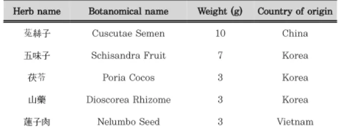
1) 약재 및 추출건조된 약재를 table 1.의 조성에 따라 혼합한 후 분쇄하여 약재 총량의 5배의 70% EtOH로 2회 환류 추출하였으며, 감압 농축 및 동결 건조하여 추출물 3.538 g (수득율 13.61%)을 얻었다. 현토단에 들어가는 약재인 菟絲子, 五味子, 茯苓, 山藥, 蓮子肉은 ***에서 구입하였다(Table 1).
Herb name Botanomical name Weight (g) Country of origin
菟絲子 Cuscutae Semen 10 China
五味子 Schisandra Fruit 7 Korea
茯苓 Poria Cocos 3 Korea
山藥 Dioscorea Rhizome 3 Korea
蓮子肉 Nelumbo Seed 3 Vietnam
Table 1. The composite of Hyeonto-dan
2) 시약
Roswell Park Memorial Institute (RPMI) 1640 과 Bovine serum albumin과streptomycin, penicillin은 Hyclone에서 구입하였다 (Logan, UT, USA). LPS, MTS, 4′, 6-diamidino -2-phenylindole (DAPI)는 Sigma에서 구입하였다 (St. Louis, MO, USA). iNOS와 TLR4, COX-2, β -actin, peroxidase- conjugated secondary antibody는 Santa Cruz Biotechnology Inc.에서 구입하였다 (Santa Cruz, CA, USA).
anti-mouse TNF-α antibody와 biotinylated anti-mouse TNF-α antibodies, Anti-mouse IL-6 antibody biotinylated anti-mouse IL-6 antibodies는 BD Biosciences에서 구입 하였다 (San Jose, CA, USA). 추가적으로, QuantiTect Reverse Transcription kit와 RNeasy Mini kit는 Qiagen 에서 구입하였다 (Hilden, Germany). 마지막으로, fluorochrome-conjugated LPS와 fluorochrome-conjugated secondary antibodies는 Life Technologies Corp.에서 구 입하였다 (Seoul, Republic of Korea).
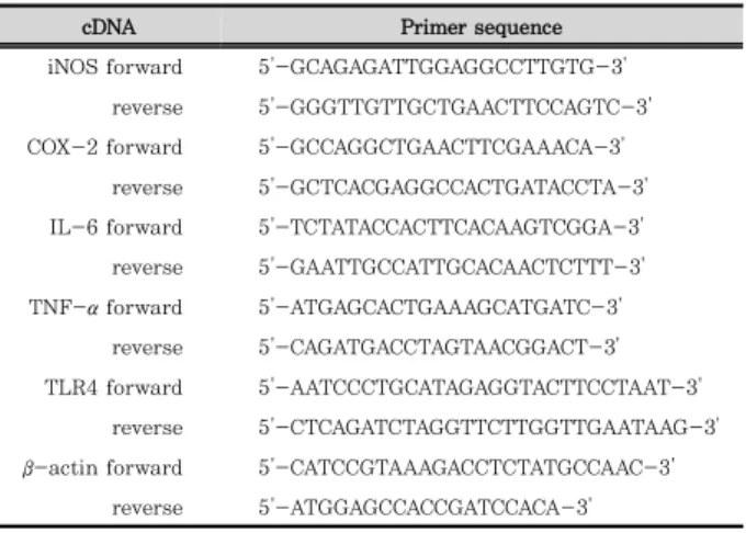
3) Primer 준비
qRT-PCR을 위해 사용한 primer의 구성은 다음과 같다
(Table 2).
cDNA Primer sequence iNOS forward 5'-GCAGAGATTGGAGGCCTTGTG-3'
reverse 5'-GGGTTGTTGCTGAACTTCCAGTC-3' COX-2 forward 5'-GCCAGGCTGAACTTCGAAACA-3'
reverse 5'-GCTCACGAGGCCACTGATACCTA-3' IL-6 forward 5'-TCTATACCACTTCACAAGTCGGA-3' reverse 5'-GAATTGCCATTGCACAACTCTTT-3' TNF-α forward 5'-ATGAGCACTGAAAGCATGATC-3'
reverse 5'-CAGATGACCTAGTAACGGACT-3' TLR4 forward 5'-AATCCCTGCATAGAGGTACTTCCTAAT-3'
reverse 5'-CTCAGATCTAGGTTCTTGGTTGAATAAG-3' β -actin forward 5'-CATCCGTAAAGACCTCTATGCCAAC-3'
reverse 5'-ATGGAGCCACCGATCCACA-3'
Table 2. Primer sequences for qRT-PCR
2. 방법
1) 세포 배양설치류 대식 세포주는 한국세포주 은행으로부터 분양받았다.
RAW 264.7세포는 100 U/㎖의 페니실린과 100 ㎎/㎖의 스 트렙토마이신을 첨가하고 10% FBS을 첨가한 RPMI 1640 배 지에서 37℃의 온도, 5% CO
2의 습한 대기 조건으로 배양하 였다.
2) 세포 생존율 측정
RAW 263.7 세포 생존율은 MTS 분석법으로 측정하였다.
RAW 263.7 세포 (5×10
5cells/㎖)를 96 well plate에 100 ㎕ 의 RPMI 1640배지에서 18시간 배양한 다음, 현토단을 농도 별로 처리하여 24시간 배양하였다. 각 실험군에 5 ㎎/㎖ 농 도의 MTS를 50 ㎕ 씩 넣은 후 2시간 동안 배양한 후 fomazon 생성량을 microplate reader를 이용하여 550 ㎚에서 흡광도를 측정하였으며, 세포 독성은 세포만을 배양한 비 처 리군을 생존률 100%로 하여 약물처리군의 상대적인 세포 생 존율을 이용해 계산하였다.
3) NO 생성량 측정
현토단을 여러 농도로 처리하고 여기에 200 ng/㎖의 LPS 를 각각 주입한 후 대식 세포주를 24시간 배양하였다. Griess reagent(1% sulfanilamide, 0.1% N -(1-naphthyl)- ethylene diamine dihydrochloride in 2.5% phosphoric acid solution)와 수집한 배양한 상층액을 동량으로 혼합한 후 10분간 실온에서 방치하였다. Epoch Microplate Spectrophotometer (Biotek, Winooski, VT, USA)를 이용 하여 570 ㎚에서 흡광도를 측정하여 아질산의 농도는 결정하 였다. 세포가 없는 배양액에 아질산 0-30 ㎛을 넣어 측정하 였으며, 이 값을 표준으로 하여 여러 실험군의 아질산 값의 흡광도를 측정하였다.
4) PGE2생성량 측정
RAW 264.7 세포를 RPMI 1640 배지를 이용하여 1.5×10
5cells/㎖로 조절한 후 24 well plate 에 접종하고, 5% CO
2항온기에서 18시간 배양 하였다. 이후 배지를 제거하고
현토단을 각각 다양한 농도로 처리한 후, LPS (200 ng·㎖
-1) 로 대식 세포주를 자극하였다. 세포 부유액을 원심 분리하여 세포들을 제거한 상층액을 수집하고 상층액에 함유된 PGE
2생성량을 사용자 매뉴얼에 기재된 방법대로 EIA kit(R&D Systems Inc., Minneapolis, MN, USA)를 이용하여 정량해 분석하였다.
5) Cytokine 생성량 측정
전처리가 끝난 RAW 264.7 세포의 부유액을 원심분리한 후 세포들을 침전시켜 상층액을 수집하였으며, 상층액에 포함 된 IL-6와 TNF-α 의 생성량을 사용자 매뉴얼에 기재된 방법 대로 ELISA kit(R&D Systems Inc., Minneapolis, MN, USA)를 이용하여 정량해 분석하였다.
6) Western blot analysis
RAW 263.7 세포에 현토단을 농도별로 처리하여 30분간 배양한 후 LPS (200 ng·㎖
-1)를 처리하였다. MAPK는 LPS 자극 1시간 후에 인산화 정도를 확인하였으며 COX-2와 INOS는 자극 후 24시간 후에 발현량을 확인하였다.
7) RNA 분리 및 quantitative reverse-transcription PCR (qRT-PCR)
전 처리된 세포를 모아 PBS로 세척 후 이지 블루(easy blue, 인트론사) 1 ㎖를 가하여 교반하였다. 클로로포름 200 ㎕를 넣고 교반하여 13,000rpm, 4℃에서 10분간 원심분리 하였다.
상등액 400 ㎕에 이소프로판올을 동량을 가하여 원심분리하여 RNA를 얻었다. 여기서 얻어진 RNA에 역전사효소(reverse transcriptase), 1 mM dNTP 0.5 ㎍을 섞어 cDNA를 만들 었다. qRT-PCR는 사용자 매뉴얼에 기재된 방법대로 power SYBR® Green PCR master mix를 이용하여 진행하였다.
결과물은 StepOnePlus Real-Time RT-PCR System과 StepOne software v2.3 (Applied Biosystems, Foster City, CA, USA)을 사용하여 β-actin mRNA의 발현량을 기준으로 상대적 발현 값을 정량하였다.
8) 면역형광염색법 (Immunofluorescence staining)
RAW 264.7는 chambered cover glasses (Nunc)에서 18 시간 동안 배양한 후, 현토단과 LPS를 처리하였다. 세포는 4% formaldehyde를 사용하여 실온에서 15분 동안 고정하였 으며, 100% MeOH을 10분 동안 −20℃에서 처리하여 투과 성을 높였다. 표본은 blocking buffer (PBS with 5% serum and 0.3% Triton X-100)를 사용하여 1시간 동안 blocking 하였으며 4℃에서 1차 항체를 하룻밤 동안 처리하였다. 형광 물질이 축합된 2차 항체는 실온, 암소 조건에서 1시간 처리하 였다. PBS로 세척 후, 세포핵을 DAPI를 이용하여 대비 염색 하였다. 표본의 형광정도는 형광 현미경을 사용하여 관찰하였 다(Carl Ziess, Oberkochen, Germany). 세포막에 결합된 LPS는 AlexaFluor 488와 축합된 LPS를 사용하여 관찰하였다.
9) 통계분석
통계분석은 Scheffe’s test법을 따라 ANOVA를 실행하였다.
모든 실험은 3회 이상 반복적으로 이루어졌으며, 실험결과는 각 항목에 따라 SPSS statistics 22 software (SPSS Inc.
Chicago, IL, USA)를 사용하여 평균치 ± 표준편차 (SD)를 구하여 신뢰 수준 P < 0.05에서 통계적 유의성을 평가하였다.
Ⅲ. 결 과
1. 세포생존율에 대한 현토단의 효과
현토단의 대식 세포의 생존율에 대한 영향을 확인하기 위해 MTS 시험법을 시행하였다. 세포는 다양한 농도로 24시간 동안 처리하였다. 실험 결과 현토단 500 ㎍/㎖내에서 RAW 264.7 세포에 대해 독성이 나타나지 않았으며, 이후의 항염증 활성 실험을 진행하였다(Figure 1.).
Figure 1. Effects of Hyeonto-dan on RAW 264.7 cells viability.
Cell viability was evaluated with the MTS assay. Data are means
± standard deviations of duplicate determinations from three separate experiments.
2. LPS로 유도된 NO 생성에 대한 현토단의 효과
NO의 생성에 현토단이 미치는 영향을 알아보기 위해 배지로 분비된 질산염의 농도를 Griess시약을 사용하여 측정하였다.
LPS 비처리군에 비하여 LPS 처리 시 NO의 생성이 급격히 증가하였다. 현토단을 LPS와 함께 처리한 경우 농도 의존적 으로 감소가 일어남을 확인하였다(Figure 2).
3. LPS로 유도된 PGE 2 생성에 대한 현토단의 효과
PGE
2의 생성에 현토단이 미치는 영향을 알아보기 위해 EIA kit를 이용하여 정량해 분석하였다. LPS 비처리군에 비하여 LPS 처리 시 PGE
2의 생성이 급격히 증가하였다. 현토단을 전 처 리한 경우 생성량이 감소하는 것을 확인하였다(Figure 3).
4. LPS로 유도된 IL-6와 TNF-α 의 생성 및 mRNA 발현에 대한 현토단의 억제 효과
LPS로 자극된 대식세포에서 cytokines인 IL-6와 TNF-α 의 합성에 대한 현토단의 효과를 알아보았다. 그 결과, LPS 처리 시 IL-6와 TNF-α 의 분비량이 유의적으로 상승함을 확 인 할 수 있었다. 하지만 다양한 농도의 현토단을 동시에 처 리한 경우 생성량이 줄어들었다 (Fig. 4A). TNF-α 의 생성량
역시 현토단 처리 시 농도 의존적으로 줄어드는 것을 확인 할 수 있었다 (Fig. 4C). 더욱이 qRT-PCR의 결과를 보면 cytokines 의 mRNA수준에서의 발현량 역시 LPS 단독 처리군과 비교하여 줄어드는 것이 확인 되었다 (Figure 4B, D).
Figure 2. Effects of Hyeonto-dan on NO production in RAW 264.7 cells. The cells were treated with Hyeonto-dan at the indicated concentration for 30 min and then stimulated with 200 ng/ml LPS for 24 h. NO release was measured by the Griess assay. The culture supernatant was isolated and analyzed. *P <
0.05 compared to the LPS-treated group. Significant differences between treated groups were determined using analysis of variance followed by Scheffe’s test for multiple comparisons. Values are means ± standard deviations of duplicate determinations from three separate experiments.
Figure 3. Effects of Hyeonto-dan on PGE2 production in RAW 264.7 cells. These were determined according to the manufacturer’s protocol. *P < 0.05 compared to the LPS-treated group. Significant differences between treated groups were determined using analysis of variance followed by Scheffe’s test for multiple comparisons.
Values are means ± standard deviations of duplicate determinations from three separate experiments.
5. LPS로 유도된 iNOS와 COX-2의 발현에 대한 현토단의 억제 효과
iNOS와 COX-2의 발현을 단백질과 mRNA수준에서 현토
단의 영향을 알아보았다. 단백질과 mRNA수준에서 iNOS와
COX-2의 발현정도를 Western blot법과 qRT-PCR법을 활
용하여 정량하였다. iNOS와 COX-2는 비 처리군에서 단백질과
mRNA수준에서 측정되지 않았으며, LPS 처리 시 유의적으로
상승하였다. 현토단은 최고농도에서 LPS 단독 처리군에 비해
COX-2의 발현을 단백질 수준 (Figure 5A, B)과 mRNA수
준 (Figure 5C)에서 억제하였으며, iNOS 역시 단백질 수준
(Figure 5A, B)과 mRNA수준 (Figure 5C)에서 억제하였다.
Figure 4. Effects of Hyeonto-dan on LPS-induced IL-6, TNF-α production (A, B) and mRNA expression (C, D) in RAW 264.7 cells. RAW 264.7 cells were pretreated with the indicated concentration of Hyeonto-dan for 30 min before being incubated with LPS (200 ng/㎖) for 24 h. The culture supernatant was isolated and analyzed to measure IL-6 production. *P < 0.005 compared to the LPS-treated group.
Significant differences between treated groups were determined using Scheffe’s test. Values are means ± standard errors of duplicate determinations from three separate experiments.
Figure 5. Effects of Hyeonto-dan on protein (A,B) and mRNA (C) expression of iNOS and COX-2 in LPS-stimulated RAW 264.7 cells.
The cells were pretreated with Hyeonto-dan for 30 min, and then incubated with LPS (200 ng/㎖) for the indicated time. Representative Western blots of at least three separate experiments are shown. *P < 0.005 compared to the LPS-treated group. Significant differences between the treated groups were determined using analysis of variance followed by Scheffe’s test for multiple comparisons. Values are means ± standard deviations of duplicate determinations from three separate experiments.
6. MAPKs의 인산화에 대한 현토단의 효과
MAPKs는 세포의 성장과 분화 및 사이토카인과 스트레스 제어에 중요한 역할을 한다. 따라서 현토단의 억제 메커니즘이 MAPKs 경유하는지 알아보기 위해 MAPKs의 인산화를 Western blot을 통해 확인하였다. 그 결과 LPS에 의해 활성화 된 RAW 264.7 대식세포에 HT을 처리한 경우 JNK와 ERK, p38의 인산화를 억제하는 것을 확인하였다 (Figure 6).
Figure 6. Effects of Hyeonto-dan on phosphorylation of MAPK family in LPS-stimulated RAW 264.7 cells. The cells were pretreated with Hyeonto-dan for 30 min, and then incubated with LPS (200 ng/㎖) for 1 h. Representative Western blots of at least three separate experiments are shown.
7. NF-κB의 핵 내로의 이동에 대한 현토단의 효과
기존의 연구에서 iNOS, COX-2, cytokines과 같은 염증성 인자의 조절에 NF-κ B가 연관성이 보고되었다. LPS 비 처리군에 비해 LPS 처리군에서 NF-κ B의 핵 내로의 이동이 증가되었다. 현토단을 LPS와 같이 처리한 군의 경우 NF-κB의 이동이 현저하 게 감소되었다(Figure 7). 따라서 현토단의 염증 억제 활성에 있어서 NF-κ B의 이동을 막는 것이 주요 기전 중 하나로 보여진다.
Figure 7. Effects of Hyeonto-dan on NF-κB nuclear translocation. Cells were pretreated with Hyeonto-dan for 30 min and then incubated with LPS (200 ng/㎖) for 1 h. Localization of NFκB/p-65 was visualized with a fluorescence microscope after immunofluorescence staining.
Nuclei were counterstained with DAPI. Similar results were obtained in three independent experiments, and results of one representative experiment are shown.
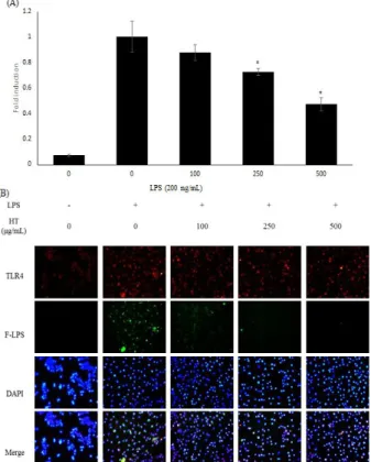
8. TLR-4 발현에 대한 현토단의 효과
LPS에 의해 유도되는 TLR-4 기전에 대한 현토단의 효과를 면역형광염색법과 qRT-PCR법을 통해 측정하였다. 수용체의 발현량은 24시간 동안 LPS로 자극한 후 확인하였으며, LPS 비처리군에 비해 증가하였다. 현토단과 LPS를 함께 처리한 군에서는 TLR-4의 발현이 mRNA 수준에서 유의적으로 감 소되었다 (Figure 8A). 또한 형광 시약이 부착된 LPS를 처리 하였을 때 세포 표면에 결합된 LPS의 양이 현토단 처리 시 감 소하였다 (Figure 8B). 이러한 결과로 보아 현토단은 TLR-4 발현을 감소시키며 LPS와 경쟁적으로 작용하여 LPS가 세포 표면에 결합되는 것을 방해하는 것으로 생각된다.
Figure 8. Effects of Hyeonto-dan on TLR-4 expression and the interaction between LPS and TLR-4. Panel (A) indicates expression of TLR-4 in mRNA levels. Cells used in the qRT-PCR were pretreated with Hyeonto-dan for 30 min and then incubated with LPS (200 ng/㎖) for 24 h. (B) Expression of TLR-4 on membrane of RAW 264.7 cells and interactions between LPS and TLR-4 were visualized with a fluorescence microscope after immunofluorescence staining. Cells for immunofluorescence were pretreated with Hyeonto-dan for 30 min and then incubated with fluorescence conjugated-LPS (200ng/㎖) for 24 h. Similar results were obtained in three independent experiments, and the results of one representative experiment are shown. *P < 0.005 compared to the LPS-treated group. Significant differences between the treated groups were determined using analysis of variance followed by Scheffe’s test for multiple comparisons. Values are means ± standard deviations of duplicate determinations from three separate experiments.
Ⅳ. 고 찰
현토단의 잠재적인 항염증 효과와 염증 반응 억제 기전을 LPS로 유도한 염증 모델에서 확인하였다. 활성 산소종의 일
종인 NO는 혈관과 근육조직의 이완 조절에 관여하는 면역계 방어물질로서 인체 생리에 중요한 역할을 한다. 그리고 생리 적인 효과뿐만 아니라 감염이나 조직 손상으로 인한 염증반응 을 매개하는 인자로서 병리과정에도 작용한다
12). NO는 L-arginine이 L-citrulline으로 전환되면서 형성되며, 그 과 정에서 nitic oxide synthase (NOS)라는 효소가 작용하게 된 다. 활성화된 대식세포에서 iNOS의 발현에 의해 과도하게 생 성된 NO는 세포독성이나 조직손상 등 염증반응을 촉진할 뿐만 아니라 IL-1β , IL-6, TNF-α 등과 같은 전염증성 cytokine 및 COX-2와 같은 염증 매개물질들의 생합성을 촉진하여 과 도한 면역반응으로 인한 다양한 질병을 초래하게 한다
13). COX-2는 정상적인 상태에서 발현하지 않고, 주로 염증반응 에 관여하는 PGE
2를 생성하여 발열, 혈관 확장을 유발하는 대표적인 염증 지표물질이다
14). 그러므로 NO와 PGE
2는 염증 반응 동안 iNOS와 COX-2의 하위 반응기로서 작용한다. 대식 세포에 LPS 자극을 주면 NO와 PGE
2의 생산을 증가한다. 하 지만 LPS와 현토단을 함께 처리 하였을 때 NO와 PGE
2의 생 성량이 감소하였다(Figure 2,3). 추가적으로 iNOS와 COX-2의 발현 역시 단백질수준과 mRNA수준 모두에서 감소하였다 (Figure 5).이러한 결과로 보아 현토단의 NO, PGE
2억제효 과는 iNOS와 COX-2발현 억제를 통한 작용으로 생각된다.
대식세포는 감염이나 조직의 손상으로 인한 염증 반응 동안 NO나 PGs와 같은 다른 염증성 인자와 마찬가지로 IL-6와 TNF-α 를 분비한며, 이는 다른 면역세포로부터 추가적인 반 응을 유도한다
15). LPS로 처리된 RAW 264.7세포에서 IL-6 와 TNF-α 의 분비가 증가된다(Figure 4). 하지만, 현토단을 전처리하면 IL-6와 TNF-α 의 생산량과 mRNA 발현량이 감 소한다. 이를 통해 현토단의 항염증 작용은 전염증성 매개자 의 발현을 억제함으로서 나타남을 알 수 있다.
MAPKs는 세포외 자극에 의해 반응하여 전사인자, 세포골격 단백질, 효소 등을 포함한 다수의 단백질들의 인산화를 통해 세포질 내에서 핵으로 정보를 전달하는데 중요한 역할을 하며, 세포 성장과 분화 및 생존에 관여한다
16). 이러한 MAPKs에는 extracellular signal regulated kinase(ERK), c-Jun N- terminal kinase (JNK), p38 kinase (p38)의 3가지 신호 전달경로가 존재하는데, 이들은 물리적, 화학적인 자극에 의해 활성화되고 inhibitory kappa B kinase kinase (IKK)의 활 성을 매개함으로서 NF-κ B를 핵으로 전위시켜 염증성 cytokine 및 iNOS, COX-2의 발현을 촉진한다
17). 현토단은 MAPK 신호 전달 경로를 차단함으로서 NF-κB 활성화의 억제하는 것으로 나타났다(Figure 6).
염증반응을 유발하는 cytokines, iNOS, COX-2의 발현은 전사인자인 nuclear factor-κB (NF-κB)에 의해 조절이 된 다고 보고되고 있다
3). LPS에 의해 세포가 자극되면 Iκ B가 인 산화 되며 NF-κ B가 유리되고 유리된 NF-κ B는 세포의 핵내로 이동한다. NF-κB활성의 주요 과정은 p65의 핵내로의 이동 하면서 나타나는 p65의 전사촉진 활성이라고 보고되었다
18). 염증매개자에 대한 억제 작용은 NF-κB유리를 억제하는 것 으로 나타났다(Figure 7).
Toll-like receptor는 선천성 면역계를 담당하는 세포에서
주로 발현되어 있는 transmembrane signaling molecule로
9)현재까지 발견된 TLR family의 유전자들은 13종류로 일반적
으로 extracellular domain을 통하여 pathogen-associated molecular patterns (PAMPs)나 micro-organsim- associated molecular patterns (MAMPs)를 인식하여 선천 성 면역 반응을 일으키고 염증세포의 활성화를 유발한다
19). LPS는 LPS-결합 단백질(lipopolysaccharide binding protein, LBP)과 결합하고 결합체는 대식세포나 가지세포의 CD40과 결합함으로서 Toll-like receptor (TLR)-4의 수용체를 활성 화시킨다
9).
LPS로 자극된 TLR-4는 TLR-4-NF-κ B기전을 통해 NO, TNF-α , IL-6의 발현 증가를 촉발시키고 매개자들의 자가 피해를 유발한다
20). 따라서 이번 연구에서는 RAW 264.7 세포주에서 현토단과 LPS의 TLR-4에 대한 상호작용 을 연구하였다. 현토단을 처리하였을 때 세포표면에 부착된 LPS의 양이 특이적으로 감소하였으며 (Figure 8), 이를 통해 현토단이 TLR-4의 결합에 간섭함을 알 수 있다. 추가적으로 현토단은 LPS로 자극된 RAW 264.7 세포주에서 TLR-4의 발현을 억제하였다. 그러므로 현토단의 항염증작용에 있어서 TLR-4에 대한 경쟁적인 작용이 주요 기전으로 생각된다.
이번 연구에서 LPS로 자극된 RAW 264.7 대식세포주에서 염증반응에 의해 NO, PGE
2,IL-6,TNF-α와 같은 염증매개 체의 발현이 증가되었다. 또한 NF-κ B의 핵 내로의 이동과 TLR-4의 분비 역시 LPS자극 이후 증가되었다. 과거의 연구 들에서 TLR4-NFκ B 기전은 LPS자극에 의한 염증반응의 주 요기전으로 보고되어있다. 실험 결과 현토단은 자극된 RAW 264.7세포에서 LPS와 TLR-4의 결합을 억제하였다. 또한 LPS자극에 의해 유도된 NO의 생성을 억제하고 NF-κ B 기전 을 조절함으로서 염증 반응을 약화시켰다. 현토단의 하위 염 증성 매개자(iNOS, COX-2)와 인자들(NO, PGE2, IL-6, TNF-α )에 대한 억제작용이 위의 기전을 통해 나타나는 것으 로 추측된다. 따라서 현토단이 염증성 질환의 예방 및 치료에 유효할 것으로 생각된다.
Ⅴ. 결 론
현토단의 염증반응 억제 효과를 알아보기 위해 LPS를 처 리한 RAW 264.7 세포에서 NO, PGE
2의 생성, iNOS, COX-2의 발현, 염증성 사이토카인인 IL-6와 TNF-α 의 분비, 신호전달 기전을 알아보기 위해 세포내 신호전달 단백질인 MAPK의 활 성 및 TLR-4 활성 정도를 측정하여 다음과 같은 결론을 얻 었다.
1. 현토단은 LPS로 유도된 대식세포에서 NO와 PGE
2생 성을 농도 의존적으로 억제하였다.
2. 현토단은 LPS로 유도된 iNOS와 COX-2의 발현을 단 백질 수준과 mRNA수준 모두 억제하였다.
3. 현토단은 LPS로 유도된 RAW264.7 세포에서 IL-6와 TNF-α 발현을 현저하게 억제하였다.
4. 현토단은 LPS로 유도된 RAW264.7 세포에서 JNK와
ERK, p38의 인산화를 억제하였다.
5. 현토단은 LPS로 유도된 NF-κ B의 핵 내로의 이동을 억제 하였으며 상위 기전인 TLR4와 LPS의 결합을 현 저하게 억제하였다.
이러한 결과로 미루어 보아 현토단은 대식세포에 작용하여 LPS와 경쟁적으로 작용함으로서 TLR4-NF-κB 기전을 저해 하여 MAPKs의 인산화 활성을 떨어트려 iNOS와 COX-2의 발현을 억제하고 NO와 PGE
2,IL-6,TNF-α 의 생성을 억제함 으로서 항염증 효과를 나타내었다.
감사의 글
이 논문은 한국연구재단의 이공분야기초연구사업(NRF- 2016R1D1A1B03934552)과 2017년도 정부[미래창조과학부]의 재원으로 한국연구재단의 지원을 받아 수행된 기초연구사업 (No. 2017-0062484)의 지원에 의해 수행된 결과로 이에 감 사드립니다.
참고문헌