Ethanol extract of Sinsun-yukza-hwan, a Korean medicinal prescription, promotes hair growth
in C57BL/6 mice, an alopecia animal model
Ji Yoon Kim
1#, Mi Ryeo Kim
2*1 : Course of Beutytechnology, Center for Continuing Education, Dongnam Health University 2 : Department of Herbal Pharmacology, School of Korean Medicine, Daegu Haany University
ABSTRACT
Objectives : In Korean medicine, a prescription of Sinsun-yukza-hwan (Shenxian-liuzi-wan, SSY) has been used in clinic for treatment of alopecia via oral. This study was performed to determine transdermal effects of the ethanol extract from SSY on hair growth and -related gene expressions in mice.
Methods : We analyzed index compound, 5-hydroxy-methyl-2-furaldehyde (HMF), in SSY extract by ultra performance liquid chromatography (UPLC). 6 weeks old C57BL/6 mice with removed hair were used as an alopecia animal model.
Mice were divided into 3 experimental groups including normal (3 water: 1 ethanol: 2 polyethylene glycol mixture as a vehicle), SSY extract and 5% minoxidil (as a positive control), treated groups. SSY was applied topically on the hair- shaved skin of C57BL/6 mice every day for 15 days. The color, thickness and density of hair were monitored every 5
thday by naked eye, photograph and phototrichogram using folliscope. Also hair growth-associated gene expressions were measured by immunoblotting assay.
Results : Hair density of minoxidil or SSY-treated group was significantly increased compared to that of vehicle application on the 15
thday, respectively. And hair thickness of minoxidil and SSY groups was increased compared to that of vehicle treated group on the 15
thday, respectively. Induction of insulin-like-growth factor 1(IGF-1) and vascular endothelial growth factor (VEGF) were also significantly accelerated by SSY extract compared to those of vehicle-applied group.
Conclusions : These results provide scientific evidence to support the potent multi-application of SSY as a cosmeceutical material for promoting hair growth.
1)
Key words : Sinsun-yukza-hwan (Shenxian-liuzi-wan, SSY), hair density, hair thickness, insulin-like-growth factor 1(IGF-1), vascular endothelial growth factor (VEGF), alopecia animal model
Ⅰ. Introduction
The number of people who suffer from hair loss is increasing every year
1). Therefore, there is a growing interest in research focused on development of useful cosmeceuticals for treatment of hair loss or alopecia. Also there are a number of factors which may contribute to hair loss at an earlier age, both in men and women.
Some of the factors found to be associated with increased hair loss are an increase in social activity for women, western-style dietary habits, nutritional unbalance based on wrong eating habits, heavy mental stress, and dieting for weight loss
2).
Hair plays an important role in protecting the scalp and the skull fracture from any external shock and in waste elimination from the body. It’s another role is to
*Corresponding author : Mi Ryeo Kim, Department of Herbal Pharmacology, College of Korean Medicine, Daegu Haany University, 136 Sincheondong-Ro, Suseong-Gu, Daegu, Korea.
·Tel : +82-53-770-2241 ·E-mail : E-mail : [email protected]
#First author : Ji Yoon Kim, Course of Beutytechnology, Center for Continuing Education, Dongnam Health University, 50-74 Cheoncheon-Ro, Jangan-Gu, Suwon, Korea.
·Tel : +82-31-249-6333 ·E-mail : [email protected]
·Received : 11 April 2018 ·Revised : 9 May 2018 ·Accepted : 25 May 2018
contribute to one’s beauty. Since the social interaction has increased, people have also paid attention to self- image. As a consequence, they are very interested in preventing hair loss
3,4).
From the medical perspective, hair loss has been associated autoimmune diseases, aging, topical bloodstream, stress, dyscrinism, seborrheic scalp, and environmental pollutants
5-7). On the other hand, Korean medicine indicates that hair loss is caused by Hyeoryeol (e.g., the hotness of blood), Hyeoleo (e.g., blood coagulation in the flesh), stress, hepatorenal failure, etc.
8,9).
From the Korean medicine perspective, hair loss increases the pore vulnerability after hair falls out, whereas in the clinical perspective, it is mainly identified by two types, alopecia areata and alopecia seborrheica
10). Alopecia areata generally appeared from a topical non-inflammatory hair loss of the scalp. The first stage of alopecia areata is called “Gwichedu”, the symptom of a fistful of hair fell out. The second stage is “Jeondok”, the symptom of whole hair felling out. The last stage is “Bodok”, alopecia universalis. On the other hand, alopecia seborrheica is hair loss caused by a seborrheic condition of the scalp or a heavy sebum secretion that affects a number of young adults and middle-aged men. The main symptom is dandruff and urtication. If it is serious, these symptoms may result in baldness
11).
Several treatments such as systemic steroid therapy, transplant, and hair follicle stimulants can be used to treat hair loss; however, none of these are very effective
12-14). Furthermore, there are two US FDA approved drugs (minoxidil and finasteride) that are used for hair growth, but they require treatment and may have some side effects. As such, there is an increased interest to develop new treatment on hair loss using medicinal herbs in Korean medicine.
Therefore, a better understanding is needed that Sinsun-yukza-hwan (Shenxian-liuzi-wan, SSY) has known preventing hair loss and gray hair, in
‘Eoyakwonbang’, old Korean medical book. In this study, we tested the effect of ethanol extract of SSY on hair growth to investigate it’s applicability as a safe ingredient in cosmetic.
Ⅱ. Materials and methods
1. Analysis of index compound from SSY extract
We used an Ultra Performance Liquid Chromatography (UPLC, Waters, USA) with ACQUITY
TMphotodiode array detector (PDA) and BEH C
18column(1.7 ㎛, 2.1×100) for analysis. Then, microwave extractor (Branson 3210,
USA) was used for sample extraction and standard preparation dissolution with 30% methanol. Index compound from Rehmanniae Radix Preparata in SSY, 5-hydroxy-methyl-2-furaldehyde (HMF), were detected in 280 n m at room temperature. A mobile phase, mixed liquid of the water and acetonitrile which contain 0.1%
Formic acids, was flowed at the rate of 0.4 ㎖/min.
2. Animals
We used 5-week age (average weight: 20 g) C57BL/6 male mice (Hyochang Science, Daegu). They were housed in a animal care facility where the following conditions maintained throughout the experimental period:
temperature 23±3℃, relative humidity 50±10%, 12 hours of lamp cycle. After having the adaption for a week, the experiment took place under permission of animal research ethics committee (DHU 2014-018).
3. Sample preparation
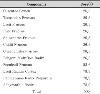
SSY is composed of 12 Korean medicinal herbs, Cuscutae Semen, Toosendan Fructus, Lycii Fructus, Rubi Fructus, Shizandrae Fructus, Cnidii Fructus, Chaenomelis Fructus, Poligoni Multiflori Raulus, Foeniculi Fructus, Lycii Radicis Cortex, Rehmanniae Radix Preparata, and Achyranthis Radix, which were purchased from market for Korean herbal medicine (Daewon-yakeopsa, Daegu, Korea). We mixed these components as described in Table 1 and extracted it for 10 hours at 100℃ with 10-fold amount of 50% ethanol. Thus, some freeze-dried powder was obtained by following the filtering and enrichment process which was kept in -20℃ with 16.2% yield.
Components Dose(g)
Cuscutae Semen 26.3
Toosendan Fructus 26.3
Lycii Fructus 26.3
Rubi Fructus 26.3
Shizandrae Fructus 26.3
Cnidii Fructus 26.3
Chaenomelis Fructus 26.3
Poligoni Multiflori Radix 26.3
Foeniculi Fructus 52.6
Lycii Radicis Cortex 78.9
Rehmanniae Radix Preparata 78.9
Achyranthis Radix 78.9
Total 500
Table 1. Composition of SSY
4. Treatment
Mice were anesthetized by intraperitoneal (i.p) injection of pentobarbital (50 ㎎/㎏, Hanlim Co.). After an anesthesia, we used a small hair-clipper to remove a patch of hair from the back of each mouse(6-weeks aged) w. Additionally, we used a depilatory (Niclin, Ildong Co.) to remove some hair follicles and micro-cells. The skin was washed with tepid water and left to stabilize the skin following a 24 hours convalescent. After the completion of this process, the mice were divided in three treatment groups, such as normal, minoxidil and SSY, with 6 mice in each group. After that, we applied each solution to dorsal dermis depilated (200 ㎕ per head) daily. As such, we used a mixed vehicle [d-H2O (3): PEG (1): Ethanol (1)] for the normal group, a 5%
minoxidil solution for the minoxidil group (purchased from Hyundai Pharm), and a 15% SSY for the SSY group for a certain period of time.
5. Macroscopic observation
Using a digital camera, photographs were taken on Day 0, and at intervals of 5 days. As spontaneous hair growth begins, both observation results and data were collected until Day 15 after the start of the treatment.
6. Phototrichogram analysis
After the completion of applying a sample, we extracted skin texture and fixed it in 4% formaldehyde and the stored for the sample analysis. We analyzed all collected samples at the same time. The extracted textures were unfolded using Whatman paper and were tested by folliscope (version. 2.8, Lead M, Korea). We set two parts in tomography images and calculated the hair density and thickness per unit area (㎠).
7. Western blotting
After adding lysis buffer (50 mM Tris pH 7.8, 120 mM NaCl, 2mM EDTA, 1% Triton X-100) the extracted skin samples were homogenized (Biospec, Korea). Soluble protein fraction was extracted using a centrifuge. Since we set the required protein by Bradford, samples passed the electrophoresis process in 12% SDS-PAGE gel.
Western blotting was done to isolate individual proteins from this crude mixture and was blotted using PVDF
membranes.
PVDF membrane was blocking with 5% skim milk solution for an hour. Membrane was incubated using the primary antibodies (1:1,000) for 12 hours in 4℃.
Membranes were washed using PBST (3 times washing per 10 minutes), followed by incubation in secondary antibodies (1:1,000) in the room temperature for one and half hours. The final step was to wash using 1x PBST (3 times washing per 10 minute). Membranes were developed using ECL substrate. We finally assessed intensity of each protein using an image analytic tool (Gel Documentation system, UVP, USA).
8. RNA separation and RT-PCR
In order to extract RNA from the collected skin texture, we used TRIzol (Invitrogen, Grand Island, NY) reagent. Samples were homogenized, and centrifuged at 3,250 x g, 4℃ for 10 minutes. The upper layer was collected, and added to chloroform (100 ㎕). The mixture was again centrifuged at 3,250 x g, 4℃ for 10 minutes, and the upper was collected. Furthermore, isopropanol (100 ㎕) was added to this mixture. After the centrifugation at 3,250 x g, 4℃ for 10 minutes, we got RNA pellet. The pellet was washed twice with 75%
Ethanol. After air-dry, it was diluted by 0.5~10 ㎍/㎕
using diethyl pyrocarbonate (DEPC) – treated water.
cDNA was prepared by using a Mastercycler gradient
(Eppendorf, Hamburg, Germany). Thermocycler mixture
consisted of 2.5 mM dNTP, 10X butter, DEPC water,
premixed primer (GenoTech, Korea) and Taq DNA
polymerase into RT product (template cDNA). QuantiTect
SYBR Green PCR kit (QIAGEN, Germany) was used for
the gene expression analysis. PCR reaction procedure
was as follows: 94℃, 3 minutes (1 cycle), 38 cycles (45
second at 94℃, 45 second at 59℃, and 45 second at
72℃) were used afterwards. We completed the reaction
after the extension at 72℃ for 10 minutes. The amplified
production was electrophoresed using 1.2% agarose
gel, and the DNA band was confirmed using Gel Doc
(Bio Lad, Italy). Primers used are as follows: GAPDH (fwd
5’-TGGAATCCTGRGGCATCCATGAAA–3’, rev 5’–TAAA
ACGCAGCTCAGTAACAGTCC–3’) as the internal
transcription marker. Both VEGF (fwd 5’- CAAGGCCA
GCACATAGGAGA-3’, rev 5’-GCAACGCGAGTCTGTGTT
TT-3’) and IGF-1 (fwd 5’-GAAGGTGAAGGTCGGAGTC
A-3’, rev 5’-AGTCCTT CCACGATACCAAAG-3’).
9. Liver function tests
We analyzed the activity of both aspartate transaminase (AST) and alanine transaminase (ALT) using a spectrophotometric analysis (Asan kit, Korea).
We took 1 ㎖ of GOT and GTP-based substrate solution and incubated it at 7℃ for 5 minutes. After that, we added to plasma 200 ㎕. AST was reacted at 37℃ for 60 minutes, whereas ALT was reacted at 37℃ for 30 minutes. Then, we added to 1 ㎖ of dinitrophenyl hydrazine (color formation solution), and incubated at room temperature for 20 minutes again. They were mixed with 10 ㎖ of 0.4N NaOH, and incubated for an additional 10 minutes. Finally, we measured the absorbance value.
10. Statistical analysis
Data were expressed as mean ± S.E.. We used SPSS 11.5 to test our experiments. We conducted one-way ANOVA and tested the significance of the mean value with Ad-hoc analysis at p< 0.05.
Ⅲ. Results
1. Analysis of index compound from SSY extract
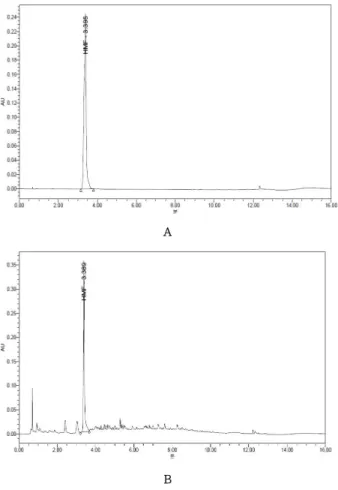
Standard solution was diluted with the methanol to be contained 25, 50, 100 ng per ㎖ and was injected into UPLC system. A R2 figure of standard curve was over 0.999 from all the standard solutions. We determined an index compound, HMF which is resulted from Rehmanniae Radix Preparata, one of the components of SSY extract and confirmed by retention time. Concentration of HMF was 37.10 ± 1.20 ppm in extract via peak area on chromatogram (Figure 1).
2. Observation of SSY efficacy on hair growth with the naked eye
When the patch of hair was removed from the back, the color of the skin was ting ed with light pink. The color of the skin changed to black when experiments were in progress. After removing hair at the surface of the skin, there was no observation of hair growth phenomenon until Day 5 following the treatment;
however, there were partial hair growth in all groups after 10 days of treatment. More specifically, for the vehicle-treated group, 4 in 6 mice showed partial hair
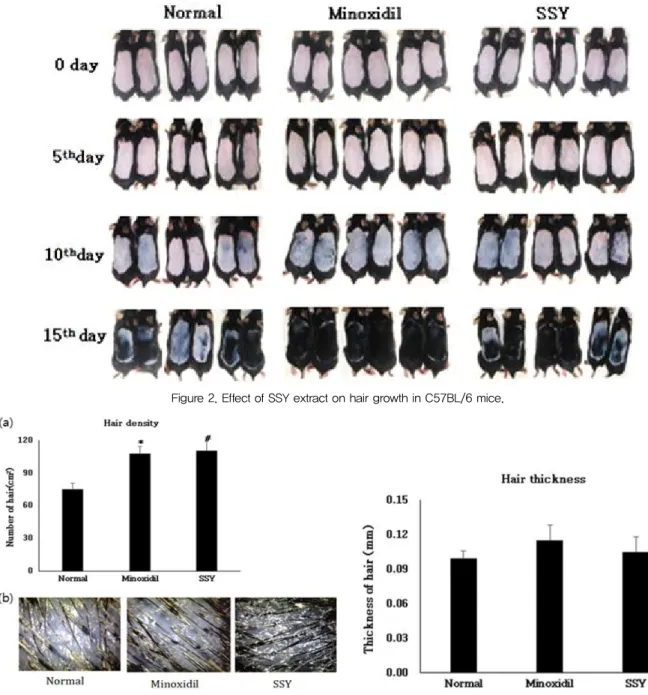
growth. For the SSY group, all mice showed partial hair growth with pink color of the skin underneath. In particular, pink color was much abundant compared to the minoxidil-treated mice. After 15 days of treatment, we confirmed that the overall progress of hair growth was normalized: 1 in 6 (the vehicle), 3 in 6 (the SSY), and 6 in 6 (the minoxidil group) (Figure 2).
A
B
Figure 1. Quantitative analysis of HMF in SSY extract by UPLC.
A : chromatogram of HMF standard solution, B : chromatogram of SSY extract
3. Effects of SSY extract on hair density and thickness
After the completion of applying extract to the skin,
we observed how SSY influences hair density. Skin
tissue collected from treated area was filmed by Follisope,
a high-resolution hair analysis system. The findings
showed that both SSY and minoxidil treatment increased
density of hair, compared to the vehicle treatment
(Figure 3). However, as shown in Figure 4, there were
no statistical differences in hair thickness among three
groups.
Figure 2. Effect of SSY extract on hair growth in C57BL/6 mice.
Figure 3. Effect of SSY extract on hair density in hair-removed C57BL/6 mice.
Data are mean ± S.E. of 6 mice per group. *p < 0.05 Minoxidil vs.
Normal, #p < 0.05 SSY vs Normal.
Figure 4. Effect of SSY extract on hair thickness in hair- removed C57BL/6 mice.
Data are mean ± S.E. of 6 mice per group.
4. Effects of SSY extract on IGF-1 protein expression
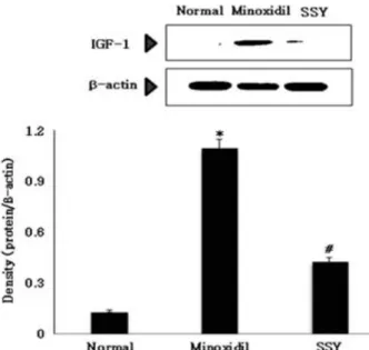
The expression of insulin-like growth factor-1 (IGF-1), a protein implicated in promoting hair growth, was measured. After 21 days of applying an SSY extract to the skin, we confirmed the expression of IGF-1. An increase in IGF-1 protein expression was observed in both minoxidil and SSY treated group compared to the vehicle control group (Figure 5).
5. Effect of SSY extract on VEGF protein expression
`We confirmed the relationship between SSY treatment and hair growth. In so doing, we measured the intensity of protein expression of vascular endothelial growth factor (VEGF), a protein that plays a role in normal hair growth by promoting hair root differentiation and improving blood circulation of the vascular endothelial.
After 15 days of treatments, VEGF expression was
significantly increased in both minoxidil and SSY-treated
groups compared to the vehicle-treated group (Figure 6).
Figure 5. Effect of SSY extract on IGF-1 protein expression in hair-removed C57BL/6 mice.
Data are mean ± S.E. of 6 mice per group. *p < 0.05 Minoxidil vs.
Normal, #p < 0.05 SSY vs. Normal.
Figure 6. Effect of SSY extract on VEGF protein expression in hair-removed C57BL/6 mice.
Data are mean ± S.E. of 6 mice per group. *p < 0.05 Minoxidil vs.
Normal, #p < 0.05 SSY vs. Normal.
6. Effect of SSY on IGF-1 and VEGF gene expression
At 15th day after treatment, IGF-1 gene expression was significantly increased in minoxidil and SSY-treated groups compared to that the vehicle-treated group, respectively (Figure 7). Also, in both minoxidil and SSY-treated groups VEGF gene expression was significantly increased compared to that the vehicle- treated group (Figure 8). These results were correlated with patterns of protein change.
Figure 7. Effect of SSY extract on IGF-1 mRNA expression in hair-removed C57BL/6 mice.
Data are mean ± S.E. of 6 mice per group. *p < 0.05 Minoxidil vs.
Normal, #p < 0.05 SSY vs. Normal.
Figure 8. Effect of SSY extract on VEGF mRNA expression in hair-removed C57BL/6 mice.
Data are mean ± S.E. of 6 mice per group. *p < 0.05 Minoxidil vs. Normal, #p < 0.05 SSY vs. Normal.
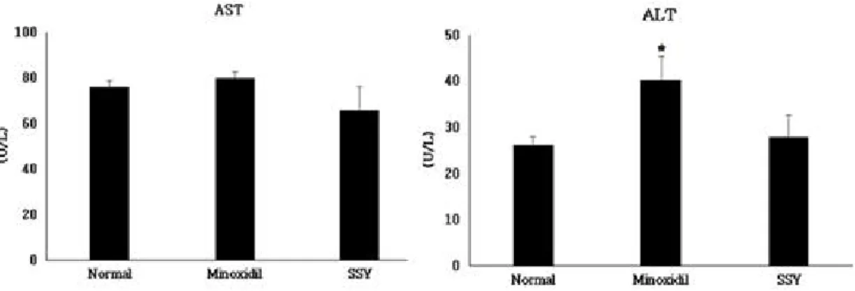
7. Effect of SSY extract on liver function
To test any possible liver toxicity associated with SSY treatment, we tested aspartate transaminase (AST) and alanine transaminase (ALT). These are enzymes routinely for measuring liver toxicity in serum samples.
There were no significant differences of two indexes (AST
and ALT) between SSY-treated versus vehicle-treatment
group. As shown in Figure 9, however, minoxidil treated
animals showed much higher ALT activity.
Figure 9. Effect of SSY extract on liver function in hair-removed C57BL/6 mice.
AST (aspartate transaminase), ALT (alanine transaminase). Data are mean±S.E. of 6 mice per group, *p < 0.05 Minoxidil vs. Normal