• 검색 결과가 없습니다.

Vitamin D Status in South Korean Military Personnel with Acute Eosinophilic Pneumonia: A Pilot Study

N/A
N/A
Protected

Academic year: 2021

Share "Vitamin D Status in South Korean Military Personnel with Acute Eosinophilic Pneumonia: A Pilot Study"

Copied!
7
0
0

로드 중.... (전체 텍스트 보기)

전체 글

(1)

Vitamin D Status in South Korean Military Personnel with Acute Eosinophilic

Pneumonia: A Pilot Study

Byung Woo Jhun, M.D.

1

, Se Jin Kim, M.D.

1

, Kang Kim, M.D.

1

, Ji Eun Lee, M.D.

1

and Duck Jin Hong, M.D.

2

1

Division of Pulmonary and Critical Care Medicine, Department of Medicine,

2

Department of Laboratory Medicine, The Armed Forces Capital Hospital, Seongnam, Korea

Background: A relationship between low vitamin D levels and the development or outcomes of respiratory diseases has been identified. However, there is no data on the vitamin D status in patients with acute eosinophilic pneumonia (AEP).

We evaluated the vitamin D status in patients with AEP among South Korean military personnel.

Methods: We prospectively compared the serum levels of total 25-hydroxyvitamin D [25(OH)D], 25(OH)D3, and 25(OH)D2 among patients with AEP, pulmonary tuberculosis (PTB), and community-acquired pneumonia (CAP).

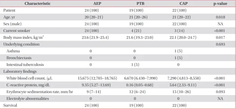
Results: In total, 65 patients with respiratory diseases, including AEP (n=24), PTB (n=19), and CAP (n=22), were identified. Of the 24 patients with AEP, 2 (8%) had deficient total 25(OH)D levels (<10 ng/mL), 17 (71%) had insufficient total 25(OH)D levels (≥10 to <30 ng/mL), and only 5 (21%) had sufficient total 25(OH)D levels (≥30 to <100 ng/mL). The difference in the total 25(OH)D levels among patients with AEP, PTB, and CAP was not statistically significant (p=0.230).

The median levels of total 25(OH)D, 25(OH)D3, and 25(OH)D2 were 22.84, 22.84, and 0.00 ng/mL, respectively, and no differences in the 25(OH)D level were present among patients with AEP, PTB, and CAP with the exception of the total 25(OH)D level between patients with AEP and PTB (p=0.042).

Conclusion: We have shown that low vitamin D levels are frequently found in patients with AEP and are comparable with those in patients with PTB and CAP.

Keywords: Deficiency; Pulmonary Eosinophilia; Pneumonia; Vitamin D

Introduction

Vitamin D is a prohormone important in bone mineraliza- tion and calcium homeostasis. Dermal synthesis after ultra- violet radiation exposure is the major source of vitamin D; a minority of vitamin D comes from dietary sources. Vitamin D3, or cholecalciferol, is formed when ultraviolet radiation converts 7-dehydrocholesterol in epidermal keratinocytes and dermal fibroblasts to previtamin D. Vitamin D2, or ergo- calciferol, is formed when ergosterol in plants is exposed to irradiation and is frequently found in plant dietary sources.

Both of these forms of vitamin D are converted in the liver to 25-hydroxyvitamin D [25(OH)D], the major circulating form of vitamin D and a good indicator of an individual’s vitamin D status. It is then converted in the kidney to 1,25(OH)D, the biologically active form

1

.

Copyright © 2015

The Korean Academy of Tuberculosis and Respiratory Diseases.

All rights reserved.

Address for correspondence: Byung Woo Jhun, M.D.

Division of Pulmonary and Critical Care Medicine, Department of Internal Medicine, The Armed Forces Capital Hospital, 177 Saemaeul-ro, Bundang-gu, Seongnam 463-040, Korea

Phone: 82-31-725-6298, Fax: 82-31-706-0987 E-mail: [email protected] Received: Nov. 27, 2014

Revised: Feb. 9, 2015 Accepted: Mar. 17, 2015

cc

It is identical to the Creative Commons Attribution Non-Commercial

License (http://creativecommons.org/licenses/by-nc/4.0/).

(2)

Recent studies on the role of vitamin D in human disease have consistently suggested the presence of an association between low vitamin D levels and the development of vari- ous respiratory diseases, including pulmonary tuberculosis (PTB)

2-4

, respiratory tract infections

5-8

, chronic obstructive lung disease

9,10

, and asthma

11

. In addition, molecular studies have also suggested that the vitamin D may be a potent regu- lator in the inflammatory process in various diseases such as autoimmune encephalomyelitis

12

and inflammatory bowel disease

13

, and it may repress production of inflammatory cyto- kines in T helper cells, including interferon-γ, interleukin (IL)-2, and IL-5

14

.

Acute eosinophilic pneumonia (AEP)

15-17

is an uncommon inflammatory lung disease of unknown etiology characterized by acute respiratory or systemic symptoms, diffuse radio- graphic pulmonary infiltrates, and infiltration of eosinophils into the lung

18

, and specific cytokines including IL-5 plays im- portant roles in the eosinophil recruitment and in maintaining active inflammatory processes

19

. However, no data on the vita- min D status in patients with AEP is currently available. There- fore, we prospectively compared the serum levels of vitamin D in South Korean military personnel with AEP with those of pa- tients with PTB and community-acquired pneumonia (CAP), both of which are known to be associated with low vitamin D levels. Our goal was to identify a possible association between low vitamin D levels and the development of AEP.

Materials and Methods

1. Study patients

Consecutive adult patients newly diagnosed with AEP, PTB, or CAP were prospectively screened and recruited from 1 May 2014 to 1 August 2014 at the Armed Forces Capital Hos- pital (874-bed military referral hospital) in Gyeonggi Province, South Korea.

A definitive diagnosis of AEP was based on a modification of criteria proposed by Philit et al.

18

as previously reported

16

: (1) acute onset of febrile respiratory manifestations <1 month in duration regardless of hypoxemia, (2) bilateral diffuse in- filtrates on chest radiography or computed tomography, (3)

>25% eosinophils on bronchoalveolar lavage or eosinophilic pneumonia on lung biopsy, and (4) absence of known causes of pulmonary eosinophilia, including drugs, toxins, and infec- tions.

The diagnosis of PTB was considered to be definitive if (1) microbiological testing (acid-fast bacillus staining, polymerase chain reaction, or culture) using lower respiratory tract speci- mens confirmed infection with Mycobacterium tuberculosis or (2) the patient’s clinical presentation met the criteria for clinical active PTB established by the World Health Organiza- tion

20,21

. CAP was defined as the presence of a new infiltrate on

chest radiography plus at least one of the following: (1) fever (body temperature of ≥38.0

o

C) or hypothermia (body temper- ature of <35.0

o

C); (2) altered breath sounds on auscultation; or (3) respiratory symptoms including cough, sputum, pleuritic chest pain, or dyspnea

22

.

Samples for measurement of the serum 25(OH)D level was performed after the study patients had provided written informed consent. The present study was approved by the Armed Forces Capital Hospital Institutional Review Board, which permitted review and publication of patient records.

This work was supported by the Korean Military Medical Research Project by ROK Ministry of National Defense (ROK- MND-2014-KMMRP-021).

2. Patient management and data collection

All study patients were treated appropriately according to their respiratory disease: a 2-week course of corticosteroids was given to patients with AEP

16,17

, anti-tuberculosis drugs were given to patients with PTB, and empirical antibiotics were given to patients with CAP. Peripheral blood samples for measurement of the 25(OH)D level were obtained from patients diagnosed with AEP, PTB, and CAP before starting treatment, and the total levels of 25(OH)D, 25(OH)D3, and 25(OH)D2 were evaluated. The degree of total 25(OH)D was classified as deficient (<10 ng/mL), insufficient (≥10 to <30 ng/

mL), or sufficient (≥30 to <100 ng/mL). The serum levels of total 25(OH)D, 25(OH)D3, and 25(OH)D2 were compared among patients with AEP, PTB, and CAP.

Data on the patients’ baseline characteristics, body mass in- dex, underlying conditions, and laboratory findings including the white blood cell count, C-reactive protein level, and eryth- rocyte sedimentation rate were also collected. Follow-up data were last obtained on 1 August 2014.

3. Measurement of serum 25(OH)D level

1) Reagents and calibration materials: We used a mul- tilevel serum calibrator set as a calibrator and MassCheck 25(OH)D3/D2 serum control as the internal quality control material (Chromsystems, Munich, Germany). 25(OH)D2/

D3 (IsoScience, Trevose, PA, USA) was used as the internal standard and was diluted with methanol: isopropanol (8:2, v:v). Other reagents and organic solvents were of high-perfor- mance liquid chromatography grade.

2) Sample processing: In total, 150 μL of blood samples,

quality control materials, or correction material and 20 μL of

internal standard were mixed; 150 μL of 0.2 M ZnSO

4

were

then added, and the mixture was shaken in a vortex mixer for

10 seconds. Next, 300 μL of methanol were added and the se-

rum proteins were precipitated. Hexane (750 μL) was added

and shaken in a vortex mixer for 30 seconds. After 5 minutes

(3)

of centrifugation, the supernatant was transferred to a Waters Maximum Recovery Vial (Waters, Manchester, UK) and dried at 50

o

C in nitrogen. Finally, 75 μL of 70% methanol were add- ed and dissolved, and 20 μL of the mixture were injected into a liquid chromatography–mass spectrometry (LC-MS/MS) system.

3) Ultra performance LC-MS/MS: An ACQUITY ultra- performance LC system (Waters, Milford, MA, USA) with a BEH phenyl column (2.1×50.0 mm, 1.7 μm) was used, and the test was performed under the following conditions: dose, 20 μL; analysis time, 5 minutes; flow rate, 0.45 mL/min; mobile phase A, 2 mM ammonium acetate with 0.1% formic acid solution (distilled water); and mobile phase B, 2 mM ammoni- um acetate with 0.1% formic acid (methanol). The ACQUITY TQD was used, and the test was performed under the follow- ing conditions: electrospray ionization, positive ion mode, and multiple reaction monitoring. All analyses of the measured data were performed using the QuanLynx 4.0 software.

4. Statistical analysis

Data are presented as medians (interquartile range, IQR) for continuous variables and as numbers (percentage) for categorical variables. Data were compared using the Mann- Whitney U test for continuous variables and the chi-squared or Fisher exact test for categorical variables. All statistical analyses were performed using the PASW software ver. 18.0

(SPSS Inc., Chicago, IL, USA). A two-sided p-value of <0.05 was considered to indicate statistical significance.

Results

1. Baseline patient characteristics

In total, 65 patients with respiratory diseases, including AEP (n=24), PTB (n=19), and CAP (n=22) were identified during the study period. The infectious etiologies of CAP and PTB were microbiologically identified in 14 (64%) and 11 (58%) patients, respectively. Baseline characteristics of the study patients are shown in Table 1. All patients were young males. The median ages were 20, 21, and 21 years in patients with AEP, PTB, and CAP, respectively, and the median age of patients with AEP tended to be higher than that of patients with PTB and CAP. All patients with AEP were current smok- ers, and their median body mass index was highest among all three respiratory disease groups. Among the patients with CAP, one patient had asthma and one had bronchiectasis; one patient with PTB had concurrent intestinal tuberculosis. The white blood cell count and C-reactive protein level were signif- icantly higher in patients with AEP, with a median of 15,675/

μL (IQR, 12,705–18,765/μL) and 9.35 mg/dL (5.27–13.69 mg/

dL), respectively. The results of the electrolyte tests, including the phosphate and calcium levels, in all study patients were within the reference ranges.

Table 1. Baseline characteristics of the study patients

Characteristic AEP PTB CAP p-value

Patient 24 (100) 19 (100) 22 (100)

Age, yr 20 (20–21) 21 (20–26) 21 (20–22) 0.018

Sex (male) 24 (100) 19 (100) 22 (100) NA

Current smoker 24 (100) 4 (21) 3 (14) <0.001

Body mass index, kg/m

2

23.6 (21.9–25.4) 21.6 (19.3–23.0) 22.1 (20.0–24.7) 0.017

Underlying condition 0.693

Asthma 0 0 1 (5)

Bronchiectasis 0 0 1 (5)

Intestinal tuberculosis 0 1 (5) 0

Laboratory findings

White blood cell count, /μL 15,675 (12,705–18,765) 6,670 (6,430–7,990) 7,290 (4,813–8,558) <0.001 C-reactive protein, mg/dL 9.35 (5.27–13.69) 0.16 (0.05–0.68) 5.64 (2.33–9.11) <0.001

Erythrocyte sedimentation rate, mm/hr 9 (7–14) 12 (6–24) 15 (10–26) 0.093

Electrolyte abnormalities 0 0 0 NA

Survival 24 (100) 19 (100) 22 (100)

Values are presented as median (interquartile range) or number (%).

AEP: acute eosinophilic pneumonia; PTB: pulmonary tuberculosis; CAP: community acquired pneumonia; NA: not applicable.

(4)

2. Comparisons of serum 25(OH)D levels in the study patients

Serum 25(OH)D levels according to the different respiratory diseases are shown in Table 2. Of the 24 patients with AEP, 2 (8%), 17 (71%), and five (21%) had deficient, insufficient, and sufficient total 25(OH)D levels, respectively. Of the 19 patients with PTB, one (5%) had deficient total 25(OH)D levels and the others (n=18, 95%) had insufficient levels. Of the 22 patients with CAP, one (4%), 18 (82%), and three (14%) had deficient, insufficient, and sufficient total 25(OH)D levels, respectively (Figure 1). The differences in the proportions of degree of the serum total 25(OH)D levels among patients with AEP, PTB, and CAP were not statistically significant (p=0.230).

The median serum levels of total 25(OH)D were 22.84 ng/

mL (IQR, 17.07–27.87), 16.08 ng/mL (IQR, 12.38–22.37), and 22.37 ng/mL (IQR, 14.85–26.89) in patients with AEP, PTB,

and CAP, respectively. The median serum levels of 25(OH) D3 were 22.84 ng/mL (IQR, 16.47–27.81), 15.95 ng/mL (IQR, 12.38–22.37), and 22.01 ng/mL (IQR, 14.07–26.67) in patients with AEP, PTB, and CAP, respectively. The median serum levels of 25(OH)D2 were 0.00 ng/mL (IQR, 0.00–0.21), 0.00 ng/mL (IQR, 0.00–0.00), and 0.00 ng/mL (IQR, 0.00–0.74) in patients with AEP, PTB, and CAP, respectively. The serum 25(OH)D2 levels were below the detection limit in 15 (63%), 15 (79%), and 13 (59%) of patients with AEP, PTB, and CAP, respectively.

We also compared the total 25(OH)D (Figure 2), 25(OH)D3 (Figure 3), and 25(OH)D2 (Figure 4) levels among the study patients with AEP, PTB, and CAP. There were no significant differences in the 25(OH)D levels among the study patients, with the exception of the total 25(OH)D level between pa- tients with AEP and PTB (p=0.042) (Figure 2).

Table 2. Comparisons of serum 25(OH)D levels in the study patients

AEP PTB CAP p-value

Patients 24 (100) 19 (100) 22 (100)

Degree of serum total 25(OH)D 0.230

Sufficiency (≥30 to <100 ng/mL) 5 (21) 0 3 (14)

Insufficiency (≥10 to <30 ng/mL) 17 (71) 18 (95) 18 (82)

Deficiency (<10 ng/mL) 2 (8) 1 (5) 1 (4)

Serum levels of 25(OH)D

Total 25(OH)D, ng/mL 22.84 (17.07–27.87) 16.08 (12.38–22.37) 22.37 (14.85–26.89) 0.096 25(OH)D3, ng/mL 22.84 (16.47–27.81) 15.95 (12.38–22.37) 22.01 (14.07–26.67) 0.098

25(OH)D2, ng/mL 0 (0–0.21) 0 0 (0–0.74) 0.236

Values are presented as median (interquartile range) or number (%).

AEP: acute eosinophilic pneumonia; PTB: pulmonary tuberculosis; CAP: community acquired pneumonia.

Figure 1. Degree of serum total 25(OH)D levels among patients with acute eosinophilic pneumonia (AEP), pulmonary tuberculosis (PTB), and community-acquired pneumonia (CAP).

Figure 2. Comparisons of serum total 25(OH)D levels among pa- tients with acute eosinophilic pneumonia (AEP), pulmonary tuber- culosis (PTB), and community-acquired pneumonia (CAP). NS:

not significant.

(5)

Discussion

In the present study, we compared the vitamin D status among South Korean military personnel with AEP, PTB, and CAP. Most patients with AEP (n=19, 79%) had insufficient or deficient serum total 25(OH)D levels, and the serum levels of both 25(OH)D3 and 25(OH)D2 in patients with AEP were as low as those of patients with PTB and CAP, without significant differences. This result was observed despite the fact that all patients with AEP were young, previously healthy males with high physical activity levels and that it was late spring and summer in South Korea, which would have allowed for ad- equate ultraviolet radiation exposure.

Studies on the role of vitamin D in respiratory diseases have consistently indicated that low vitamin D levels may be as- sociated with a higher incidence, greater severity, or poorer treatment responses in various respiratory diseases, including PTB

2-4,23

, respiratory tract infections

5-8,24

, chronic obstructive lung disease

9,10

, and asthma

11

. In PTB, for example, several can- didate polymorphisms of the vitamin D receptor and vitamin D binding protein that modulate the development of tubercu- losis have been identified. Another study of 259 patients with PTB showed that supplementation with high doses of vitamin D accelerated clinical and radiographic improvement

3

, sug- gesting that vitamin D supplementation enhances primarily innate responses to mycobacterial infection. Furthermore, an association between a higher risk of upper respiratory infec- tions and low vitamin D levels has been identified

5,8,25

, and a recent clinical investigation showed that high vitamin D levels are associated with better lung function, less airway hyper- responsiveness, and improved corticosteroid responses in pa- tients with asthma

11

. To our knowledge, however, no reported studies have focused on the serum vitamin D levels of patients with AEP. In this context, our data suggest a possible associa-

tion between low vitamin D levels and the development of or increased vulnerability to AEP.

AEP is an uncommon inflammatory lung disease of un- known etiology. All patients exhibit typical clinical features including acute-onset respiratory distress with fever, diffuse pulmonary infiltration, and elevated proportions of eosino- phils in bronchoalveolar lavage fluid, but the detailed patho- physiology of the disease remains unknown. Some previous studies suggested that a hypersensitivity reaction to an un- identified inhaled antigen could be a part of the inflammatory process

26-29

and demonstrated that several cytokines are as- sociated with eosinophil recruitment in AEP

30,31

. However, no risk factors for the development of, or increased susceptibility to, AEP have been identified. Given the possible roles of vita- min D in inflammation and immune modulation in patients with respiratory diseases

32,33

, our data on the high incidence of low vitamin D levels in patients with AEP suggest that low vi- tamin D levels are associated with AEP. However, data on the association between vitamin D levels and the inflammatory process of AEP are lacking; further studies are needed.

We also evaluated the serum levels of 25(OH)D3, 25(OH) D2, and total 25(OH)D to determine the proportions from di- etary sources; we then compared the 25(OH)D3 and 25(OH) D2 levels among the study patients with various respiratory diseases. Interestingly, the serum 25(OH)D2 levels were below the detection limit in most patients regardless of the specific respiratory disease (15 [63%], 15 [79%], and 13 [59%]

patients with AEP, PTB, and CAP, respectively), and there were no significant differences in the 25(OH)D3 and 25(OH)D2 levels among the patient groups (Figures 3, 4). Although these findings may be understandable given that dermal synthesis is the major natural source of vitamin D and <10% of vitamin D comes from dietary sources, limited data exist on the 25(OH) D3 and 25(OH)D2 levels or differences between the 25(OH) Figure 4. Comparisons of serum 25(OH)D2 levels among patients with acute eosinophilic pneumonia (AEP), pulmonary tuberculosis (PTB), and community-acquired pneumonia (CAP). NS: not sig- nificant.

Figure 3. Comparisons of serum 25(OH)D3 levels among patients

with acute eosinophilic pneumonia (AEP), pulmonary tuberculosis

(PTB), and community-acquired pneumonia (CAP). NS: not sig-

nificant.

(6)

D3 and 25(OH)D2 levels in patients with respiratory disease.

Thus, further studies on the clinical implications of 25(OH)D3 and 25(OH)D2 in patients with respiratory disease may help to understand the mechanisms of vitamin D in respiratory disease.

This study had important limitations. First, there was no healthy control group. Although recent one study have indi- cated vitamin D deficiency rate of 47.3% in 3,047 males and 65.0% in age of 20–29 in Korean populations

34

, which were lower than that of our pilot study patients, clinical character- istics of our study patients may be different in terms of age, physical activity levels, ultraviolet radiation exposure, and diet, because our study was conducted at a military hospital. Thus, based on the present study, further well-controlled studies are needed. Second, only a small number of patients were enrolled because of the rarity of the diseases studied and the nature of pilot studies.

In summary, we investigated the vitamin D status in pa- tients with AEP and showed that low vitamin D levels are frequently found in patients with AEP and are comparable with those in patients with PTB and CAP. Further large-scale studies on the relationship between vitamin D deficiency and AEP are needed.

Conflicts of Interest

No potential conflict of interest relevant to this article was reported.

Acknowledgements

This work was supported by the Korean Military Medical Research Project by ROK Ministry of National Defense (ROK- MND-2014-KMMRP-021).

References

1. Holick MF. Vitamin D deficiency. N Engl J Med 2007;357:266- 81.

2. Kim JH, Park JS, Cho YJ, Yoon HI, Song JH, Lee CT, et al. Low serum 25-hydroxyvitamin D level: an independent risk factor for tuberculosis? Clin Nutr 2014;33:1081-6.

3. Salahuddin N, Ali F, Hasan Z, Rao N, Aqeel M, Mahmood F.

Vitamin D accelerates clinical recovery from tuberculosis:

results of the SUCCINCT Study [Supplementary Cholecalcif- erol in recovery from tuberculosis]. A randomized, placebo- controlled, clinical trial of vitamin D supplementation in pa- tients with pulmonary tuberculosis’. BMC Infect Dis 2013;13:

22.

4. Hong JY, Kim SY, Chung KS, Kim EY, Jung JY, Park MS, et al.

Association between vitamin D deficiency and tuberculosis in a Korean population. Int J Tuberc Lung Dis 2014;18:73-8.

5. Jolliffe DA, Griffiths CJ, Martineau AR. Vitamin D in the pre- vention of acute respiratory infection: systematic review of clinical studies. J Steroid Biochem Mol Biol 2013;136:321-9.

6. Bergman P, Lindh AU, Bjorkhem-Bergman L, Lindh JD. Vita- min D and respiratory tract infections: a systematic review and meta-analysis of randomized controlled trials. PLoS One 2013;8:e65835.

7. Aregbesola A, Voutilainen S, Nurmi T, Virtanen JK, Ronkainen K, Tuomainen TP. Serum 25-hydroxyvitamin D3 and the risk of pneumonia in an ageing general population. J Epidemiol Community Health 2013;67:533-6.

8. Remmelts HH, van de Garde EM, Meijvis SC, Peelen EL, Damoiseaux JG, Grutters JC, et al. Addition of vitamin D sta- tus to prognostic scores improves the prediction of outcome in community-acquired pneumonia. Clin Infect Dis 2012;55:

1488-94.

9. Janssens W, Lehouck A, Carremans C, Bouillon R, Mathieu C, Decramer M. Vitamin D beyond bones in chronic obstructive pulmonary disease: time to act. Am J Respir Crit Care Med 2009;179:630-6.

10. Janssens W, Bouillon R, Claes B, Carremans C, Lehouck A, Buysschaert I, et al. Vitamin D deficiency is highly prevalent in COPD and correlates with variants in the vitamin D-bind- ing gene. Thorax 2010;65:215-20.

11. Brehm JM, Schuemann B, Fuhlbrigge AL, Hollis BW, Strunk RC, Zeiger RS, et al. Serum vitamin D levels and severe asth- ma exacerbations in the Childhood Asthma Management Program study. J Allergy Clin Immunol 2010;126:52-8.e5.

12. Cantorna MT, Hayes CE, DeLuca HF. 1,25-Dihydroxyvitamin D3 reversibly blocks the progression of relapsing encephalo- myelitis, a model of multiple sclerosis. Proc Natl Acad Sci U S A 1996;93:7861-4.

13. Cantorna MT, Munsick C, Bemiss C, Mahon BD. 1,25-Dihy- droxycholecalciferol prevents and ameliorates symptoms of experimental murine inflammatory bowel disease. J Nutr 2000;130:2648-52.

14. Mahon BD, Wittke A, Weaver V, Cantorna MT. The targets of vitamin D depend on the differentiation and activation status of CD4 positive T cells. J Cell Biochem 2003;89:922-32.

15. Lee JE, Rhee CK, Lim JH, Lee SM, Shim YS, Lee CT, et al. Frac- tion of exhaled nitric oxide in patients with acute eosinophilic pneumonia. Chest 2012;141:1267-72.

16. Rhee CK, Min KH, Yim NY, Lee JE, Lee NR, Chung MP, et al.

Clinical characteristics and corticosteroid treatment of acute eosinophilic pneumonia. Eur Respir J 2013;41:402-9.

17. Jhun BW, Kim SJ, Kim K, Lee JE. Clinical implications of initial peripheral eosinophilia in acute eosinophilic pneumonia.

Respirology 2014;19:1059-65.

18. Philit F, Etienne-Mastroianni B, Parrot A, Guerin C, Robert D,

Cordier JF. Idiopathic acute eosinophilic pneumonia: a study

of 22 patients. Am J Respir Crit Care Med 2002;166:1235-9.

(7)

19. Jhun BW, Kim SJ, Kim K, Lee JE, Hong DJ. Clinical implica- tions of correlation between peripheral eosinophil count and serum levels of IL-5 and tryptase in acute eosinophilic pneu- monia. Respir Med 2014;108:1655-62.

20. World Health Organization. Treatment of tuberculosis: guide- lines for national programmes. 3rd ed. Geneva: World Health Organization; 2003.

21. Lee SW, Jang YS, Park CM, Kang HY, Koh WJ, Yim JJ, et al. The role of chest CT scanning in TB outbreak investigation. Chest 2010;137:1057-64.

22. Carratala J, Fernandez-Sabe N, Ortega L, Castellsague X, Ro- son B, Dorca J, et al. Outpatient care compared with hospital- ization for community-acquired pneumonia: a randomized trial in low-risk patients. Ann Intern Med 2005;142:165-72.

23. Ganmaa D, Giovannucci E, Bloom BR, Fawzi W, Burr W, Batbaatar D, et al. Vitamin D, tuberculin skin test conversion, and latent tuberculosis in Mongolian school-age children: a randomized, double-blind, placebo-controlled feasibility trial.

Am J Clin Nutr 2012;96:391-6.

24. Bergman P, Norlin AC, Hansen S, Rekha RS, Agerberth B, Bjorkhem-Bergman L, et al. Vitamin D3 supplementation in patients with frequent respiratory tract infections: a ran- domised and double-blind intervention study. BMJ Open 2012;2:e001663.

25. Quraishi SA, Bittner EA, Christopher KB, Camargo CA Jr. Vi- tamin D status and community-acquired pneumonia: results from the third National Health and Nutrition Examination Survey. PLoS One 2013;8:e81120.

26. Badesch DB, King TE Jr, Schwarz MI. Acute eosinophilic pneumonia: a hypersensitivity phenomenon? Am Rev Respir

Dis 1989;139:249-52.

27. Imokawa S, Sato A, Hayakawa H, Toyoshima M, Taniguchi M, Chida K. Possible involvement of an environmental agent in the development of acute eosinophilic pneumonia. Ann Al- lergy Asthma Immunol 1996;76:419-22.

28. Rom WN, Weiden M, Garcia R, Yie TA, Vathesatogkit P, Tse DB, et al. Acute eosinophilic pneumonia in a New York City firefighter exposed to World Trade Center dust. Am J Respir Crit Care Med 2002;166:797-800.

29. Uchiyama H, Suda T, Nakamura Y, Shirai M, Gemma H, Shi- rai T, et al. Alterations in smoking habits are associated with acute eosinophilic pneumonia. Chest 2008;133:1174-80.

30. Yoshimoto T, Mizutani H, Tsutsui H, Noben-Trauth N, Ya- manaka K, Tanaka M, et al. IL-18 induction of IgE: depen- dence on CD4+ T cells, IL-4 and STAT6. Nat Immunol 2000;1:

132-7.

31. Mato N, Bando M, Kusano A, Hirano T, Nakayama M, Uto T, et al. Clinical significance of interleukin 33 (IL-33) in patients with eosinophilic pneumonia. Allergol Int 2013;62:45-52.

32. Adams JS, Liu PT, Chun R, Modlin RL, Hewison M. Vitamin D in defense of the human immune response. Ann N Y Acad Sci 2007;1117:94-105.

33. Bartley J. Vitamin D, innate immunity and upper respiratory tract infection. J Laryngol Otol 2010;124:465-9.

34. Choi HS, Oh HJ, Choi H, Choi WH, Kim JG, Kim KM, et al.

Vitamin D insufficiency in Korea--a greater threat to younger

generation: the Korea National Health and Nutrition Exami-

nation Survey (KNHANES) 2008. J Clin Endocrinol Metab

2011;96:643-51.

수치

Table 1. Baseline characteristics of the study patients
Figure 1. Degree of serum total 25(OH)D levels among patients  with acute eosinophilic pneumonia (AEP), pulmonary tuberculosis  (PTB), and community-acquired pneumonia (CAP).
Figure 3. Comparisons of serum 25(OH)D3 levels among patients  with acute eosinophilic pneumonia (AEP), pulmonary tuberculosis  (PTB), and community-acquired pneumonia (CAP)

참조

관련 문서

때 문에 본 프로그램에서는 나노 기술의 원리를 깊이 있게 탐구하기 보다는 다양한 나노 소재와 나노 기술 소 개하고 학생들로 하여금 나노 기술을 우리 생활에 적용해

χ is the fraction of the emitted light flux absorbed in the water Ф OH is the quantum yield for generation of ·OH radicals... &lt;Example 1&gt;. We assume that for

미국에서 설날에 대해 정식으로 배울 기회가 많지 않아 설날이 중국 명절로 잘못 인식되기도 하는 현 상황 속에서 이번 행사를 통해 시애틀 통합 한국학교 학생들은

The Relationship between Physical Activities and Health-related Quality of Life in Korean Adults with Diabetes

지금까지의 과정은 기존에 있는 사용자 계정에 대한 처리였다.. 여기까지 성공하였다면 이제 외부로 메일을 보내 보자. 방법은t o: 뒤에 외부메일 주소를 입력하면

 North Korea's submarine can essentially perform one task: launch nuclear missiles at close targets like South Korea or Japan.. This is a suicide

상대방의 짧은 답변을 유도하며, ‘어떻게 생각하십니까?’와 같은 개방형 질문과 비논리적인 질문은 피한 다.. 상대방 답변을

순천시 금산마을 영광군 상정마을 영암군 학림마을 완도군 청학마을. 장성군 괴정마을 진도군 인천마을 함평군