DOI: http://dx.doi.org/10.3339/chikd.2015.19.2.71 ISSN 2384-0250 (online)
Prevention of Pediatric Acute Kidney Injury
The incidence of acute kidney injury (AKI) in critically ill pediatric patients has been reported as increasing to 25 %, depending on population characteristics. The etiology of AKI has changed over the last 10-20 years from primary renal disease to the renal conditions associated with systemic illness. The AKI in pediatric population is associated with increased mortality and morbidity, and preven tion is needed to reduce the consequence of AKI. It is known that the most important risk factors for AKI in critically ill pediatric patients are clinical conditions to be associated with decreased renal blood flow, direct renal injury, and illness severity. Renal hypoperfusion leads to neurohormonal activation including renin-angiotensin- aldosterone system, sympathetic nervous system, antidiuretic hormone, and prostaglandins. Prolonged renal hypoperfusion can result in acute tubular necrosis. The direct renal injury can be predisposed under the condition of renal hypoperfusion, and appropriate treatment of volume depletion is important to prevent AKI. The preventable causes of AKI include contrast-induced nephro pathy, hemodynamic instability, inappropriate mediation use, and multiple nephrotoxic insults. Given the evidence of preventable factors for AKI, several actions such as the use of protocol for prevention of contrast-induced nephropathy, appropriate treatment of volume depletion, vigorous treatment of sepsis, avoidance of combi- nations of nephrotoxic medications, and monitoring of levels of drugs should be recommended.
Key words: Acute kidney injury, Biomarker, Children
Heeyeon Cho, M.D., Ph.D.
Departments of Pediatrics, Samsung Medical Center, Sungkyunkwan University School of Medicine, Seoul, Korea
Corresponding author:
Heeyeon Cho, M.D., Ph.D.
Department of Pediatrics, Samsung Medical Center, Sungkyunkwan University School of Medicine, 81 Irwon-ro,
Gangnam-gu, Seoul 135-710, Korea Tel: +82-2-3410-3539
Fax: +82-2-3410-0043
E-mail: [email protected] Received: 15 September 2015 Revised: 13 October 2015 Accepted: 25 October 2015
This is an open-access article distributed under the terms of the Creative Commons Attribu tion Non-Commercial License (http://
crea tivecom mons.org/licenses/bync/3.0/) which permits unrestricted non-commercial use, distribution, and reproduction in any medium, provided the original work is properly cited.
Copyright © 2015 The Korean Society of Pediatric Nephrology
Introduction
The definitions and characterization of acute kidney injury (AKI) in children have advanced significantly over the past 2 decades. Acute kidney injury is common in critically ill children and is associated with increased morbidity and mortality1). Acute kidney injury in association with sepsis, multiple organ involvement, and fluid overload carries heightened risk1). Gene probes and urinary biomarkers represent intriguing tools for predicting and moni
toring pediatric AKI.
To prevent AKI, the stepbystep approach such as coronary angina is needed. First step is risk factor and symptom assessment1). Risk factor for coronary angina such as smoking or hypercholesterolemia has been known and the patients usually manifest the typical symptom such as chest pain.
However, AKI dose not hurt. It means that there is no typical symptom in
AKI. In AKI, the risk factor evaluation is more important.
Second step is to measure biomarker such as troponin I and assess AKI1). In the past, serum creatinine (Cr) has been used as a biomarker of AKI. It has been known that even as small elevation of serum Cr may reflect significant kidney damage and be associated with poor outcome1). It means that serum Cr is a late marker of AKI. The numerous biomar
kers have been addressed as an early marker of AKI. Third step is intervention such as thrombolysis in coronary angina. It is difficult to prevent AKI because AKI does not show typical symptoms and there was little data for the earlier biomarker of AKI. In the past decade, research has been expanded to discover risk factor, biomarkers and intervention in AKI. In the literature, the etiology, risk fac
tors, assessment modalities, and prevention in pediatric AKI patients will be addressed.
Etiology of AKI
The etiology of AKI has changed over the last 1020 years from primary renal disease (hemolytic uremic syndrome, glomerulonephritis) to the renal complications of systemic illness or associated treatment (sepsis, cardiac disease,
oncolo gic disease). Common causes of AKI in critically ill pediatric patients were renal ischemia, nephrotoxic medi
cation and sepsis, and these conditions lead to acute tubular necrosis. The etiology of AKI is classified to prerenal, renal (intrinsic), or postrenal disease (Table 1). Causes of pre
renal azotemia are absolute loss of effective blood volume such as hemorrhage and relative loss due to capillary leak in sepsis. Extracorporeal membrane oxygenation and heart failure can be prerenal causes. Pharmacologic agents such as indomethacin, tolazoline, angiotensinconverting
enzyme inhibitor, and angiotensin receptor blockers can cause AKI by decreasing renal perfusion. When renal blood flow decreases, renal autoregulation preserves glomerular filtration rate (GFR) by increasing renal sympathetic tone, activation of the reninangiotensinaldosterone system, and increased activation of hormones such as vasopressin and endothelin. If tubular function is intact, sodium and urinary urea reabsorption can increase in response to decreased renal blood flow. These renal hemodynamic changes maintain systemic volume expansion and blood pressure. This situation is reflected by low urine sodium concentrations, low urine urea concentration and increased blood urea to creatinine ratio. This period of renal hypoper
fusion, so called “renal angina” is critical to recognize and Table 1. Causes of pediatric acute kidney injury
Renal origin Systemic illness associated conditions
Renal hypoperfusion Low intravascular volume
Urinary losses, diuretics Hemorrhage/bleeding
Third-space loss: sepsis and capillary leak Decreased effective circulating volume
Renal-artery obstruction Cardiac dysfunction
Sepsis-associated vasodilation Intrinsic
Glomerular Glomerulonephritis
Vascular
Hemolytic uremic syndrome Disseminated intravascular coagulation
Interstitial Acute interstitial nephritis: drug-induced
Tubular
Acute tubular necrosis: ischemic, drug-induced Tumor lysis syndrome
Obstruction of urinary tract
Congenital anomalies Mass, stones
treat to prevent cellular damage. Prompt recognition and correction for the cause of renal hypoperfusion can restore GFR.
In contrast to prerenal AKI, renal function abnorma lities in intrinsic AKI have been supposed not to be immedia
tely reversible. The severity of intrinsic AKI ranges from mild tubular dysfunction to acute tubular necrosis and corticomedullary necrosis with irreversible renal damage.
The intrinsic kidney injury can be divided into glomerular, vascular, tubular, and interstitial disease. Glomerulone
phritis/vascular diseases should be suspected when children present with AKI without an identifiable cause. The exami
nation of the urine sediment can differentiate glomerular from tubular injury, and the laboratory evaluation for autoimmune antibodies and a diagnostic kidney biopsy may be helpful to differentiate the cause. Pharmacologic agents are one of the most common causes of nephrotoxic AKI, and these toxins can cause AKI by decreasing renal perfusion (nonsteroidal antiinflammatory drug, diure
tics, angiotensinconvertingenzyme inhibitor), by direct tubular injury (aminglycocides, cephalosporins, ampho
tericin B, rifampin, vancomycin, nonsteroidal antiinfla
mmatory drug, contrast media, myoglobin/hemoglobin), by interstitial nephritis, or tubular obstruction (acyclovir, uric acid).
The common causes of postrenal AKI are congenital malfor mations including imperforate prepuce, urethral stricture, prunebelly syndrome, and posterior urethral valves.
Other causes of acute obstruction include neurogenic bladder, extrinsic compression, and intrinsic obstruction from renal calculi or fungal balls. Depending on the cause and associated damage to the kidneys, the relief of the obstruction is essential to restore renal function.
In pediatric AKI series, the incidence of sepsis
associated AKI has ranged from 9% to 34%, and sepsis
associated AKI is also associated with lower survival.
The pathogenic mechanism of AKI in sepsis is complex and multifactorial, and has been known to be related to combination of blood flow alterations and cytokine
mediated injury.
The “peak concentration hypothesis” has been proposed that the elevations and imbalances of both proinflamma
tory and antiinflammatory mediators coupled with endo
thelial dysfunction and altered coagulation cascade can synergistically induce AKI.
AKI frequently occurs with other organ failure in criti
cally ill children. The complex pathophysiologic interac
tion of heart and kidney dysfunction is described as cardio renal syndrome. Type 1 cardiorenal syndrome is primarily caused by acute cardiac disease with secondary acute renal impact, and common in the perioperative setting with acute decompensated heart failure. The interaction of liver and kidney known as hepatorenal syndrome is well reco
gnized, but the pathophysiologic mechanisms remain unknown. AKI can occur in undergoing surgery for con
genital heart disease, and the mechanisms are multifac
torial. The ischemia results in ATP depletion and tubular cell death. The contact of RBC with the bypass circuit during cardiopulmonary bypass causes hemolysis, and in the presence of free radicals, the free iron participates in oxygen radical reactions. Loss of brush borders and disrup
tion of cell polarity and cytoskeleton extend to apoptosis, and the desquamation of tubule cells into the lumen leads to the cast obstruction.
Risk factor assessment
In critically ill adult patients, multiple risk factors for AKI such as old age, diabetes, liver cirrhosis, congestive heart failure, chronic kidney disease, or cardiopulmonary bypass have been identified1). However, children typically do not have the comorbid conditions noted for adult patients1). There is a little data to evaluate the risk factor for AKI in children. Bailey D et al. reported the risk factors of AKI in critically ill children in a singlecenter, prospective obser
vational study over 1 year2). The enrolled patients were the pediatric intensive care unit population with 3 days to 18 years of age2). Acute kidney injury was defined as doubling of baseline serum creatinine2). In chronic kidney disease patients, AKI was defined as 25% increase in serum Cr2). The incidence rate of AKI was 4.5%2). Significant risk factors for AKI were thrombocytopenia, age > 12 years, hypoxemia, hypotension, and coagulopathy2). A few studies for risk factors for developing AKI in children were per
formed and the risk factors included invasive mechanical ventilation, vasoactive medications, nephrotoxic medica
tions, sepsis, multiple organ failure, volume depletion, thrombocytopenia, hypoxemia, neurologic dysfunction, and stem cell transplantation1). It is recommended that all children with any of these risk factors should be monitored for the development of AKI.
Acute kidney injury assessment
1. Definition of Acute kidney injury
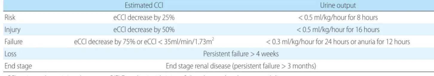
It has been known that children with small changes in serum Cr or with high degree of fluid overload are at risk for poor outcomes1). The widely used AKI criteria in children were based on the change of serum Cr, and AKI can be assessed by criteria. In 2004, the Acute Dialysis Quality Initiative group developed the first consensus multidimensional AKI definition, termed the RIFLE criteria. In 2007, AckanArikan developed and validated a pediatric modified version of the RIFLE criteria (pRIFLE) in critically ill children (Table 2)3). RIFLE has 3 AKI staging strata, Risk, Injury, Failure, and 2 outcome criteria, Loss and EndStage Kidney Disease3). AKI develo pment by RIFLE (RIFLER) is defined as a 50% rise in serum Cr (or 25%
decrease in estimated Cr clearance) over baseline or by urine output of less than 0.5 mL/kg/hour for 6 hours3). RIFLEI and RIFLEF are each defined by greater changes in Cr or
urine output3). And the RIFLE was modified by the Acute Kidney Injury Network (AKIN) in 2007 to include a 0.3 mg/dL serum Cr rise in less than 48 hours for the AKI defi nition4). In 2012, an international guideline was dev
eloped by the Kidney Disease Improving Global Outcomes (KDIGO) (Table 3)5). Acute Kidney Injury Work Group harmonized RIFLE, AKIN, and pRIFLE into a single standardized definition5). The KDIGO AKI definition and staging criteria should be used at the current time unless further modifications are warranted by prospective study.
2. Fluid overload
Acute kidney injury can be assessed by fluid overload.
Decreased renal function and oliguria in AKI result in fluid overload1, 6). However, fluid overload itself may con tri
bute to AKI1, 6). In patients with AKI, outcomes progressively worsened with increasing degree of fluid overload1, 6). Fluid balance was an independent risk factor for mortality, and the early institution of renal replacement therapy was asso
ciated with better outcome1, 6). Therefore, the aggressive use of diuretics and early initiation of hemofiltration is suggested. Goldstein SL et al. reported that lesser percent fluid overload at continuous venoveno hemofiltration (CVVH) initiation was associated with improved outcome when sample was adjusted for severity of illness6). Foland et al. reported that percent fluid overload was significantly Table 2. Pediatric-modified RIFLE (pRIFLE) criteria
Estimated CCl Urine output
Risk eCCl decrease by 25% < 0.5 ml/kg/hour for 8 hours
Injury eCCl decrease by 50% < 0.5 ml/kg/hour for 16 hours
Failure eCCl decrease by 75% or eCCl < 35ml/min/1.73m2 < 0.3 ml/kg/hour for 24 hours or anuria for 12 hours
Loss Persistent failure > 4 weeks
End stage End stage renal disease (persistent failure > 3 months)
eCCl, estimated creatinine clearance; pRIFLE, pediatric risk, injury, failure, loss and end-stage renal disease.
Table 3. Kidney Disease Improving Global Outcome (KDIGO) criteria
Stage Serum creatinine criteria Urine output criteria
1 Increase > 0.3mg/dL in 48 hours
or 1.5-1.9 times < 0.5ml/kg/hour for 6-12 hours
2 Increase 2-2.99 times < 0.5ml/kg/hour for 12 hours
3
>3.0 Increase or > 4.0 mg/dL or If <18 years of age then eCCl <35 mL/min/1.73m2
< 0.5ml/kg/hour for 24 hours or
< 0.3mL/kg/hour for 12 hours
eCCl, estimated creatinine clearance
lower in survivors compared with nonsurvivors in children with multiple organ dysfunction syndrome and percent fluid overload was independently associated with survival when severity of illness is adjusted7). Gillespie R et al.
reported that pediatric continuous renal replacement therapy (CRRT) survival outcomes progressively worsened with increasing percent fluid overload and children with high fluid overload (>10%) at CVVH initiation were at 3 times greater risk of mortality8).
3. Biomarker
Acute kidney injury can be assessed by biomarker, but serum Cr has a few limitations. Because, serum Cr is a late functional marker of AKI, serum Cr may not change until loss of 2550% kidney function1, 9, 10). It may take days after an injury before a rise in serum Cr. At lower glomerular filtra
tion rate, serum Cr will overestimate renal function1, 9, 10). Serum Cr varies by muscle mass, hydration status, sex, age, and method of measurement1, 9, 10). Novel biomarker to detect development and severity of kidney injury earlier than serum Cr in AKI is needed and the utilities of biomarkers are as follow9); (1) Early diagnosis, (2) Define severity of injury, monitor AKI course, (3) Define AKI subtypes & etiology (prerenal, septic, nephrotoxic), (4) Monitor response to AKI interventions, (5) Risk stratify for poor outcomes (dialysis need, CKD, mortality), (6) Identify location of renal tubular injury. The ideal qualities of a biomarker are as follow10); (1) Accurate, reliable, (2)
Relatively noninvasive/acceptable to patients, (3) Rapidly measurable, standardized assay, (4) Sensitive/specific with reproducible cutoff values.
Urine neutrophil gelatinaseassociated lipocalin (NGAL), kidney injury molecule 1 (KIM1), interleukin 18 (IL18), interleukin 6 (IL6), interleukin 8 (IL8), β2micro
glo bulin, CystatinC, and Livertype fatty acidbinding protein (LFABP) are urinary and serum biomarkers for the diagnosis of AKI in pediatric patients (Table 4)1119). NGAL is expressed in proximal and distal nephron and binds and transports ironcarrying molecules10, 11). NGAL plays an important role in injury and repair and rises very early (hours) after injury in animals, confirmed in children with cardiopulmonary bypass, critically ill children, and children with contrastinduced nephropathy1113). In a pro
spective cohort study of critically ill children, mean and peak urine NGAL concentrations increased with worsening pRIFLE status, and urine NGAL concentrations rose in AKI, 2 days before and after a 50% or greater rise in serum creatinine, without change in control urine NGAL12). This study suggested that urine NGAL might be a useful early AKI marker that predicted development of severe AKI in a heterogeneous group of patients with unknown timing of kidney injury. IL18 plays a role in inflammation, activat
ing macrophages and mediates ischemic renal injury13). IL
18 antiserum to animals protects against ischemic AKI10). Urinary IL18 was reported to be an AKI biomarker in critically ill children, children with portcardiac surgery, Table 4. Pediatric acute kidney injury biomarkers
Biomarker Sample Clinical setting Author Year
NGAL S/U CIN Hirsch et al.11) 2007
NGAL U Critically ill patients Zappitelli et al.12) 2007
NGAL U Post-cardiac surgery Parikh et al.13) 2011
IL-18 U Critically ill patients Washburn et al.14) 2008
IL-18 U Emergency department Du et al.15) 2011
IL-18 U Post-cardiac surgery Parikh et al.13) 2011
IL-6 U Post-cardiac surgery Dennen et al.16) 2010
IL-8 S Post-cardiac surgery Liu et al.17) 2009
KIM-1 U Emergency department Du et al.15) 2011
β2-MG U Emergency department Du et al.15) 2011
Cystatin-C S Post-cardiac surgery Krawczeski et al.18) 2010
LFABP U Post-cardiac surgery Portilla et al.19) 2008
NGAL, neutrophil gelatinase-associated lipocalin; S, serum; U, urine; CIN, contrast-induced nephropathy; IL-18, interleukein-18; IL-6, interleukin-6; IL-8, interleukin-8; KIM-1, kidney injury molecule-1; β2-MG, β2-microglobulin; LFABP, Liver-type fatty acid-binding protein.
and pediatric patients in emergency department1315). Washburn K et al. reported that urine IL18 might be used as a useful early AKI marker in critically ill children14). Urinary IL6 and serum IL8 was confirmed in children with portcardiac surgery16, 17). KIM1 is an epithelial trans
membrane protein, and is involved in cellcell interaction10). KIM1 appears to have strong relationship with severity of renal injury10). Du Y et al. reported that KIM1 and β2
microglobulin could be used as urinary biomarkers to detect AKI in pediatric emergency center15). Serum cystatin C and urinary LFABP was reported as a biomarker of AKI after pediatric cardiac surgery [18, 19]. In the pediatric setting, many studies were conducted postcardiac surgery, and the collaborations among pediatric nephrology, cardio
logy, emergency medicine, and critical care medicine are needed for the research of AKI biomarkers.
Therapeutic intervention
In the timecourse of AKI, initiation of AKI can involve hemodynamic changes in glomerular filtration rate, sub
clinical tubular injury or both processes occurring simulta
neously. A short time window may exist where specific therapy might reverse AKI. Established AKI requires days to weeks for recovery, and during this period supportive therapy and the avoidance of secondary renal injury is very important. Secondary injury may result in nonrecovery of renal function or chronic kidney disease and endstage renal disease.
Intervention of AKI is composed of diagnostic evalua
tion to uncover the underlying etiology, optimization of hemodynamics, minimize exposure to nephrotoxic medica
tions, conservative fluid management after an initial resus
cita tion, and maximize nutrition.
1. Fluid management
Attention to the patient’s volume status is essential throug
hout the hospital stay. Intake and output measurement does not take into account insensible losses. Therefore, obtaining daily weight measurement on the same scale is very important and, in combination with vital signs, heart rate, and blood pressure, it will give the clinician a better sense of the patient’s volume status.
2. Avoidance of further renal injury 1) Drug dosing in AKI
Toxicity of drugs can be avoided by careful monitoring and adjusting the dose in accordance with the patient’s chan ging renal function status. Antibiotics such as amino
glycosides or vancomycin can be used safely in patients with AKI with careful attention to serum drug levels.
Antibacterials usually need a high peak and lower trough level, and have to be given with longer dosing intervals20). Antiviral such as acyclovir might induce crystal nephro
pathy and acute tubular necrosis, and adequate hydration is necessary to prevent renal injury. Amphotericin B can cause renal vasoconstriction associated AKI and toxic effect to distal tubular epithelium. To prevent these adverse effects, sodium and volume loading prior to administra
tion of amphotericinB is needed. In the use of oral vori
conazole, dosage adjustment in AKI is not necessary.
Histamine antagonist can induce thrombocytopenia in AKI and dosage adjustment is needed. Proton pump inhibitor can be given with no dosage adjustment in AKI patients. Sucralfate is supposed to have a risk of aluminum accumulation in AKI. Phenytoin and levetiracetam can be used with dosage adjustment in AKI.
2) Contrastinduced nephropathy
Contrastinduced nephropathy has been known to be the third most common cause of hospitalacquired AKI in adults. The pathophysiology has been supposed to be renal vasoconstriction through endothepin1 or inhibition of nitric oxide and toxic effect to the renal tubular epithelium11). Prevention include the fluid volume loading with sodium bicarbonate, avoiding nephrotoxic drug prior to contrast, the use of lower osmolarity contrast agent, and Nacetylcystein11).
3. Specific intervention 1) Diuretics
Diuretics can be used to augment urine output but will not enhance solute clearance. Accordingly, increased urine output does not correlate with improvement of renal func
tion or with improvement of solute clearance. Comparison of loop diuretics given as an intermittent bolus or by con
tinuous infusion shows that the use of continuous infusion involves less exposure to these potentially toxic agents with
the same amount of urine output21). Thiazidelike diuretics (e.g., oral metolazone or intravenous chloro th iazide) can be given to enhance the effectiveness of loop diuretics.
2) Potential therapies
There is no evidence to support the use of renal dose
dopamine. However, fenoldopam, a selective dopaminergic1 Rc agonist has been reported to reduce the risk of AKI in critically ill adults and improve urine output in critically ill neonates following cardiopulmonary bypass22). Nacetyl
cystein has been known to be effective in the prevention of contrastinduced nephropathy, but there is no evidence for prevention of AKI in critically ill patients23). It has been reported that human natriuretic peptide nesiritide might be favorable for renal hemodynamic effects and increase urine output after cardiac surgery24). Additionally, growth factor, erythropoietin, or freeradical scavengers have been tried as a potential therapy in AKI2527).
4. Nutrition
Nutrition is important in patients with AKI as part of ongoing care. Specialized formulas to maximize nutrition and minimize both solute and fluid excess can be delivered to patients with AKI28, 29). Enteral nutrition has an advan
tage over parenteral nutrition and these formulas can be given either orally or by feeding tube to provide adequate caloric intake28, 29). These formulas deliver 2 cal/mL, with either no or low electrolytes (specifically, potassium and phosphorus)28, 29).
Conclusion
The AKI in pediatric population is associated with increased mortality and morbidity, and prevention is needed to reduce the consequence of AKI. To prevent AKI, the close monitoring of the pediatric patients with risk factor and the AKI assessment by AKI definition, fluid overload, and biomarker are necessary. Intervention should include the use of protocol for prevention of contrastinduced nephropathy, appropriate treatment of volume depletion, vigorous treatment of sepsis, avoidance of combinations of nephrotoxic medications, and monitoring of levels of drugs.
Critically ill children tend to be medically complex patients
with multiple organ dysfunction, and the comprehensive team approach for early recognition of AKI and appro
priate interventions are necessary to promote renal recovery and overall survival. The collaborative research should be conducted to find more accurate diagnostic methods such as biomarkers to detect AKI in pediatric patients under the complex situations such as intensive care unit, emer
gency department, preterm birth, postcardiac surgery, and trauma.
References
1. Stuart L. Goldstein, Lakhmir S. Chawla. Renal angina. CJASN 2010;5:943-9.
2. Bailey D, Phan V, Litalien C, Ducruet T, Mérouani A, Lacroix J, et al.
Risk factors of acute renal failure in critically ill children: A prospective descriptive epidemiological study. Pediatr Crit Care Med 2007;8:29-35.
3. Akcan-Arikan A, Zappitelli M, Loftis LL, Washburn KK, Jefferson LS, Goldstein SL. Modified RIFLE criteria in critically ill children with acute kidney injury. Kidney Int 2007;71:1028-35.
4. Mehta RL, Kellum JA, Shah SV, Molitoris BA, Ronco C, Warnock DG, et al; Acute Kidney Injury Network. Acute Kidney Injury Network: report of an initiative to improve outcomes in acute kidney injury. Crit Care 2007;11:R31.
5. Kellum JA, Lameire N; KDIGO AKI Guideline Work Group.
Diagnosis, evaluation, and management of acute kidney injury:
a KDIGO summary (Part 1). Crit Care 2013;17:204.
6. Goldstein SL, Currier H, Graf Cd, Cosio CC, Brewer ED, Sachdeva R. Outcome in children receiving continuous venovenous hemofiltration. Pediatrics 2001;107:1309-12.
7. Foland JA, Fortenberry JD, Warshaw BL, Pettignano R, Merritt RK, Heard ML, et al. Fluid overload before continuous hemo- filtra tion and survival in critically ill children: a retrospective analysis. Crit Care Med 2004;32:1771-6.
8. Gillespie RS, Seidel K, Symons JM. Effect of fluid overload and dose of replacement fluid on survival in hemofiltration. Pediatr Nephrol 2004;19:1394-9.
9. Devarajan P. Proteomics for biomarker discovery in acute kidney injury. Semin Nephrol 2007;27:637-51.
10. Nguyen MT, Devarajan P. Biomarkers for the early detection of acute kidney injury. Pediatr Nephrol 2008;23:2151-7.
11. Hirsch R, Dent C, Pfriem H, Allen J, Beekman RH 3rd, Ma Q, et al.
NGAL is an early predictive biomarker of contrast-induced nephropathy in children. Pediatr Nephrol 2007;22:2089-95.
12. Zappitelli M, Washburn KK, Arikan AA, Loftis L, Ma Q, Devarajan P, et al. Urine neutrophil gelatinase-associated lipocalin is an early marker of acute kidney injury in critically ill children: a
prospective cohort study. Crit Care 2007;11:R84.
13. Parikh CR, Devarajan P, Zappitelli M, Sint K, Thiessen-Philbrook H, Li S, et al. Postoperative biomarkers predict acute kidney injury and poor outcomes after pediatric cardiac surgery. J Am Soc Nephrol 2011;22:1737-47.
14. Washburn KK, Zappitelli M, Arikan AA, Loftis L, Yalavarthy R, Parikh CR, et al. Urinary interleukin-18 is an acute kidney injury biomarker in critically ill children. Nephrol Dial Transplant 2008
;23:566-72.
15. Du Y, Zappitelli M, Mian A, Bennett M, Ma Q, Devarajan P, et al.
Urinary biomarkers to detect acute kidney injury in the pediatric emergency center. Pediatr Nephrol 2011;26:267-74.
16. Dennen P, Altmann C, Kaufman J, Klein CL, Andres-Hernando A, Ahuja NH, et al. Urine interleukin-6 is an early biomarker of acute kidney injury in children undergoing cardiac surgery. Crit Care 2010;14:R181.
17. Liu KD, Altmann C, Smits G, Krawczeski CD, Edelstein CL, Devarajan P, et al. Serum interleukin-6 and interleukin-8 are early biomarkers of acute kidney injury and predict prolonged mechanical ventilation in children undergoing cardiac surgery:
a case-control study. Crit Care 2009;13:R104.
18. Krawczeski CD, Vandevoorde RG, Kathman T, Bennett MR, Woo JG, Wang Y, et al. Serum cystatin C is an early predictive biomarker of acute kidney injury after pediatric cardiopul- monary bypass. Clin J Am Soc Nephrol 2010;5:1552-7.
19. Portilla D, Dent C, Sugaya T, Nagothu KK, Kundi I, Moore P, et al.
Liver fatty acid-binding protein as a biomarker of acute kidney injury after cardiac surgery. Kidney Int 2008;73:465-72.
20. Livornese LL Jr, Slavin D, Benz RL, Ingerman MJ, Santoro J. Use of
antibacterial agents in renal failure. Infect Dis Clin North Am 2001;15:983-1002.
21. Martin SJ, Danziger LH. Continuous infusion of loop diuretics in the critically ill: a review of the literature. Crit Care Med 1994;22:
1323-9.
22. Costello JM, Thiagarajan RR, Dionne RE, Allan CK, Booth KL, Burmester M, et al. Initial experience with fenoldopam after cardiac surgery in neonates with an insufficient response to conventional diuretics. Pediatr Crit Care Med 2006;7:28-33.
23. Burns KE, Chu MW, Novick RJ, Fox SA, Gallo K, Martin CM, et al.
Perio perative N-acetylcysteine to prevent renal dysfunction in high-risk patients undergoing cabg surgery: a randomized controlled trial. JAMA 2005;294:342-50.
24. Costello JM, Goodman DM, Green TP. A review of the natriuretic hormone system’s diagnostic and therapeutic potential in critically ill children. Pediatr Crit Care Med 2006;7:308-18.
25. Nishida M, Hamaoka K. How does G-CSF act on the kidney during acute tubular injury? Nephron Exp Nephrol 2006;104:
e123-8.
26. Sharples EJ, Yaqoob MM. Erythropoietin and acute renal failure.
Semin Nephrol 2006 ;26:325-31.
27. Doi K, Suzuki Y, Nakao A, Fujita T, Noiri E. Radical scavenger edaravone developed for clinical use ameliorates ischemia/
reperfusion injury in rat kidney. Kidney Int 2004;65:1714-23.
28. Fiaccadori E, Maggiore U, Giacosa R, Rotelli C, Picetti E, Sagripanti S, et al. Enteral nutrition in patients with acute renal failure.
Kidney Int 2004;65:999-1008.
29. Fiaccadori E, Parenti E, Maggiore U. Nutritional support in acute kidney injury. J Nephrol 2008 ;21:645-56.