* Corresponding author
Phone: +82-54-770-2633, Fax: +82-54-770-2477 E-mail: [email protected]
불가사리 단백추출물이 Cytochrome P450 1A1과 Ornithine Decarboxylase 활성에 미치는 영향
남경수․김미경․조현정․손윤희*
동국대학교 난치병한양방치료연구센터 및 의과대학 약리학교실
Effect of Asterina pectinifera on Activities of Cytochrome P450 1A1 and Ornithine Decarboxylase. Kyung-Soo Nam, Mee-Kyung Kim, Hyun-Jung Cho and Yun-Hee Shon*.Intractable Diseases Research Center and Department of Pharmacology, College of Medicine, Dongguk University, Gyeongju 780-714, Korea
Abstract
The effect of protein extract from Asterina pectinifera on proliferation of human breast cancer cells and activities of cytochrome P450 1A1 and ornithine decarboxylase was tested.Protein extract from Asterina pectinifera inhibited the growth of both estrogen-dependent MCF-7 and estrogen-independent MDA-MB-231 human breast cancer cells. Cytochrome P450 1A1 activ- ity was significantly inhibited by the protein extract from Asterina pectinifera at concentrations of 80 (p<0.05), 120 (p<0.01) and 160 ㎍/㎖ (p<0.01). The extract inhibited induction of orni- thine decarboxylase by 12-O-tetradecanoylphorbol-13-acetate, a key enzyme of polyamine bio- synthesis, which is enhanced in breast tumor promotion. Therefore, Asterina pectinifera is worth further investigation with respect to breast cancer chemoprevention or therapy.
Key words : Asterina pectinifera, cytochrome P450 1A1, ornithine decarboxylase, breast cancer
chemoprevention서 론
암예방(chemoprevention)이란 발암과정의 초기단 계에서 암발생 (carcinogenesis)을 억제시키며, 암으 로 진행된 것을 전환시키는 것을 의미한다[13]. 최근 선진각국에서는 암연구를 암의 치료보다는 예방쪽 으로 비중을 두고 있으며, 암예방 효과와 관련된 물 질 탐색에 많은 노력을 기울이고 있다[1]. 특히 유방 암은 전 세계적으로 매년 약 100만명의 새로운 환자 가 발생하며, 서구 여러나라에서 가장 빈번한 여성 사망의 원인질환 중 하나이다. 우리나라도 식생활 및 생활양식의 서구화, 출산율 및 모유수유 감소, 조 기발견하는 환자수 증가 등으로 유방암 환자가 수년 동안 지속적으로 증가하고 있어 여성암 중 유방암이 차지하는 비율이 14%로 위암 다음으로 두 번째로 흔한 암이 되었다. 유방암 유발의 위험요인은 가족
중의 유방암에 대한 병력, 초경과 폐경의 나이, 첫임 신의 나이등이 있지만 이러한 위험요인은 유방암환 자의 약 25% 정도에서만 나타나므로[10]. 유방암 발 병기전을 연구하여 새로운 위험요인을 증명하는 것 이 유방암 예방과 검정에 매우 중요하다.
발암물질 대사효소는 발암물질이나 성호르몬의 활성에 관여한다. 그러므로 각개인의 발암물질 대사 의 차이는 간이나 표적조직(target tissue)에서의 대사 효소의 활성도와 관계가 있으며 이러한 차이에 의해 유방암 발병의 감수성에도 차이가 있다. 특히 외부 의 발암물질은 cytochrome P450 (CYP) 효소에 의해 대사되어 전자친화적물질 (electrophilic product), epox- ides 또는 매우 독성이 강한 물질이 된다. 사람의 유선 상피세포에서 cytochrome P450 1A1, 2B6와 2E1의 활 성이 검정되었으므로[2]. cytochrome P450 효소의 활 성 억제효과는 곧 유방암예방의 효과를 의미한다.
Polyamine (putrescine, spermidine과 spermine)은 종 양형성시 비정상적으로 생합성되며 발암과정에 밀 접한 관계가 있다. Polyamine 생합성과정에서 중요 한 효소는 putrescine의 생성에 관여하는 ornithine de- carboxylase(ODC)이며 정상세포와 종양세포의 증식 에 필수적이다. 또한 ODC의 유도는 암촉진단계 (promotion)에도 중요한 기능을 담당하고 있어 생쥐 의 여러 조직을 이용한 암촉진 실험에서 ODC활성 유도와 발암물질의 암촉진 능력간의 밀접한 관계가 보고되었다[7]. 특히 유선조직에서 estradiol이나 peptide growth factor (insuline like growth factor와 epi- dermal growth factor)에 의하여 ODC 활성이 증가되 고, 세포외에서의 polyamine 이동에 의하여 세포내 polyamine의 증가가 관찰되었다[5].
해양생물에는 항암, 항바이러스, 항균과 신경조절 물질등이 포함되어 있으며 특히 많은 종류의 항암물 질이 분리되었고 그 중 전임상과 임상시험 중인 물 질도 있다. 별불가사리(Asterina pectinifera)도 항암 [14,17]과 암예방효과[15]가 증명되었으며 본 논문에 서는 별불가사리 단백추출물이 estrogen-의존성의 유방암세포(MCF-7)와 estrogen-비의존성 유방암세 포(MDA-MB-231)의 증식에 미치는 영향, CYP 1A1 및 ODC 활성에 미치는 영향을 측정하여 별불가사 리의 유방암 유발 억제효능을 측정하고자 한다.
재료 및 방법 시료제조
별불가사리는 포항 앞바다에서 채집하여 물로 깨 끗이 씻어 -20℃에 보관하였다. 별불가사리 단백추 출물은 Kishimura 와 Hayashi[6]의 방법에 의하여 준 비하였다. 즉, 별불가사리(500 g)는 작은 조각으로 잘라서 4℃에서 3차 증류수로 추출하여 10,000 × g에 서 30분간 원심분리하였다. 원심분리후 상층액은 0~40% ammonium sulfate로 분리하고 Tris-HCl buf- fer (pH 7.4)에 녹여 투석한 후, 투석물은 4℃에서 원 심분리(10,000 × g, 30분)하였다. 상층액(단백추출 물)은 freezer dryer를 이용하여 완전 건조시킨 후 건 조중량 (3.4 g)을 측정하였다. 냉동건조한 단백추출 물은 실험조건에 사용되는 배지 및 증류수에 희석시 켜 실험에 사용하였다.
세포배양
계대 보존 중인 MCF-7과 MDA-MB-231 세포는 10% fetal bovine serum이 포함된 RPMI 1640 배지에 서 배양하였다. 세포는 액체질소에 보존해 두었다가 같은 passage 번호를 가진 세포를 녹여서 사용하였 으며, 세포의 생존은 trypan blue dye exclusion 방법으 로 확인하였다.
유방암세포 증식에 미치는 영향
Estrogen-의존성인 MCF-7과 estrogen-비의존성인 MDA-MB-231 세포를 96-well plate의 well당 0.5 × 10⁴ cells을 접종시키고 24시간 후에 농도별 시료를 처리하여 4일 동안 배양하였다. 그리고 세포 성장은 MTT assay로 측정하였다.
마이크로좀의 분리
흰쥐(Sprague-Dawley계, 자성)의 간조직으로부터 마이크로좀의 분리는 Phol과 Fouts[11]의 방법을 참 고하여 실시하였고, 관류법으로 7,12-dimethylbenz [a]anthracene (DMBA, 30 ㎎/마리)를 처리하였다. 24 시간 뒤 흰쥐를 diethyl ether로 질식시킨 다음, 복피 를 절개하여 1.15% KCl 완충용액으로 간을 perfusion 시킨 후 적출하였다. 적출된 간은 다시 여러번 세척 후, 흡습지로 수분을 완전히 제거시켰다. 수분이 제 거된 간은 마쇄 (Teflon Plotter Elvehiem Homogenizer, Glas-Col, USA)한 후, 1.15% KCl 완충용액을 첨가하 여 마쇄액을 만들었다. 마쇄 균질액을 7,000 × g에서 10분간 원심분리한 후, 상층액을 다시 77,000 × g에 서 60분간 원심분리하였다. 형성된 침전물은 0.05 M Tris-HCl buffer (pH 7.5)에 재현탁하여 microsome 분 획으로 실험에 사용하였다. 이상의 모든 과정은 4℃ 에서 실시하였다. Microsomal 단백질은 bicinchoninic acid protein kit를 사용하여 bovine serum albumin (BSA)를 표준 단백질 용액으로 표준 검량선을 구하 고 그 양을 산출하였다.
Cytochrome P450 1A1 활성 저해 측정
Cytochrome P450 1A1 활성은 ethoxyresorufin-O- deethylase (EROD) 활성으로 측정하였다[12]. 즉, 흰 쥐로부터 분리한 microsomal protein (2 ㎎/㎖) 200 ㎕ 에 640 ㎕의 0.05 M Tris-HCl buffer (pH 7.5), 100 ㎕의 BSA (10 ㎎/㎖ in Tris-HCl buffer), 20 ㎕의 0.25 M
MgCl2, 40 ㎕의 cofactor solution, 2.5 unit의 glu- cose-6-phosphate dehydrogenase, 10 ㎕의 substrate (1
㎎ of ethoxyresorufin in 10 ㎖ of methanol), 10 ㎕의 시료를 농도별로 첨가하였다. 모든 시약들을 잘 섞 은 후, 37℃에서 4분간 반응시키고, 2 ㎖의 methanol 로 반응을 종결시켰다. 2,000 × g에서 20분 동안 원심 분리하여 상층액을 취하고, 형광분광광도계 (BIO- TEK SFM25, USA)로 측정 (550 nm excitation and 585 nm emission) 하였다. Positive control로는 resveratrol 을 사용하였고, negative control로는 증류수를 사용 하였다. 이 실험은 3회 반복 실험으로 수행하였으며, 각각의 결과는 control에 대한 각 시료들의 저해도 정도를 percentage로 나타내었다.
Inhibition (%) =[1 - (tested sample - blank)
] ×100 (solvent - blank)
Ornithine decarboxylase (ODC) assay
유방암세포 MCF-7을 24-well tissue culture plate에 well 당 2 × 10⁵ cells/㎖ 세포를 접종시키고, 18시간 후에 TPA와 시료 또는 양성대조군(0.01 mM DFMO) 을 처리하여 6시간 동안 배양하였다. 세포를 Ca2+, Mg2+ - free PBS (pH 7.4)로 두 번 세척한 후 freeze-thaw cycle을 실시하여 용해시키고 cell extract 의 ODC 활성은 L-[1-14C]ornithine에서 [14C]CO2 방출 에 의하여 측정하였다. 즉 L-[1-14C]ornithine(200 nCi), 0.2 M sodium phosphate buffer (pH 7.2), 12.5 mM EDTA, 50 mM dithiothreitol, 5 mM pyridoxal phosphate와 unlabelled L-ornithine을 포함한 200 ㎕ 반응액을 각 well에 첨가하고 37℃에서 1시간 동안 반응시키며, 방출되는 CO2 가스를 paper disk로 모아 서 radioactivity를 측정하였다. ODC 활성 측정을 위 한 단백질 함량은 bicinchoninic acid protein assay kit 를 사용하여 측정하였다. ODC specific activity 는 pmol 14CO2/h/㎎ protein 으로 나타내며, ODC 활성 억제는 TPA만 처리한 조절군에 대한 각 시료들의 저해도 정도를 percentage로 나타내었다.
결과 및 고찰
유방암세포의 증식에 미치는 영향
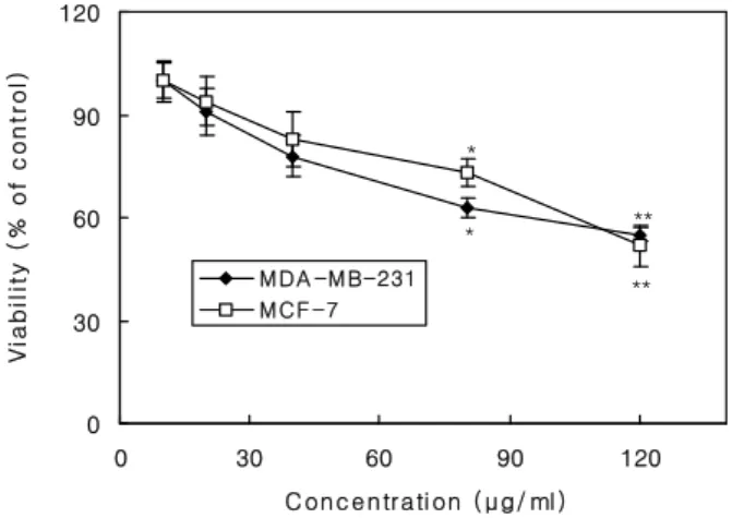
별불가사리 단백추출물이 estrogen-의존성의 유방
암세포(MCF-7)와 estrogen-비의존성 유방암세포 (MDA-MB-231)의 증식에 미치는 영향을 살펴본 결 과, 별불가사리 추출물 농도 10 ~120 ㎍/㎖에 의하 여 estrogen-의존성인 MCF-7 세포의 증식이 억제되 었다 (Fig. 1). 또한 별불가사리 추출물 처리에 의하 여 estrogen-비의존성의 유방암세포 MDA-MB-231의 증식도 억제되었다 (Fig. 1). 별불가사리 단백추출물 이 estrogen-의존성과 estrogen-비의존성의 유방암세 포 모두에 증식 억제효과가 있으므로 별불가사리 단 백추출물은 estrogen receptor를 이용한 유방암세포 증식억제가 아닌 것으로 보인다.
Cytochrome P450 1A1의 활성 저해효과 Cytochrome P450 효소활성 억제효과는 암예방 효 과를 의미하므로 별불가사리 단백추출물이 DMBA 에 의해 유도된 cytochrome P450 1A1 효소활성 억제 율을 측정한 결과, 처리 농도 10~160 ㎍/㎖에서 약 13~34%의 저해율이 나타났으므로 (Fig. 2), 별불가 사리 단백추출물이 CYP 1A1 활성을 저해시키는데 효과가 있음을 알 수 있었다. 암예방물질은 발암과 정의 전단계에서 여러기전으로 작용하며 그 중 중요 한 기전의 하나가 전발암물질(procarcinogen)을 발암 대사물(carcinogenic metabolites)로 전환시키는 cyto- chrome P450 효소와의 상호작용에 의해 암예방효과 를 나타낸다. 특히 cytochrome P450 1A1은 polycyclic
* **
**
*
0 30 60 90 120
0 30 60 90 120
C onc e ntrati on ( μg/ ml )
Viability (% of control)
M DA -M B-231 M CF -7
Fig. 1. Effect of protein extract from Asterina pectinifera on
proliferation of estrogen-dependent MCF-7 and estro- gen-independent MDA-MB-231 human breast cancer cells.Experimental details are described in Material and Methods.
The values are mean±SD (n=3). The value of each group statistically significant as compared with control (* p<0.05,
** p<0.01).
arylamines과 polyaromatic hydrocarbons의 활성에 관 여하는 것으로 알려져 있다[18].
ODC 활성
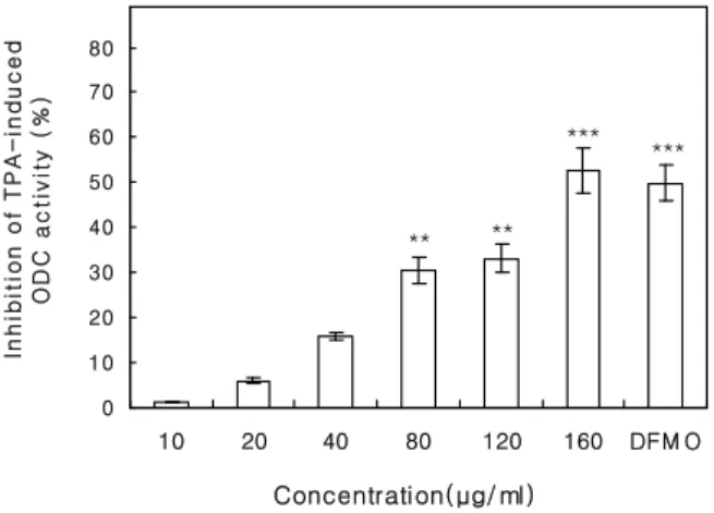
별불가사리 단백추출물에 의한 ODC 활성 억제효 과를 살펴본 결과, 80, 120과 160 ㎍/㎖ 농도에서는 각각 무처리한 세포의 ODC 활성의 30.6, 33.2와 52.6%의 억제효과를 측정할 수 있었다 (Fig. 3). 증가 한 ODC 단백질 표현과 polyamine 함량이 유방암조
***
**
**
*
0 10 20 30 40 50
10 20 40 80 120 160 RV
C onc e ntrati on( μg/ ml)
Inhibition(%)
Fig. 2. Effect of protein extract from Asterina pectinifera on
DMBA-induced cytochrome P450 1A1 activity. Experimental details are described in Material and Methods. Values repre- sent mean±SD (n=3). The value of each group statistically significant as compared with control (* p<0.05, ** p<0.01,*** p<0.005).
** **
*** ***
0 10 20 30 40 50 60 70 80
10 20 40 80 120 160 DFM O
Concentration(μg/ ml) Inhibition of TPA-induced ODC activity (%)
Fig. 3. Effect of protein extract from Asterina pectinifera on
12-O-tetradecanoylphorbol-13-acetate (TPA)-induced orni- thine decarboxylase (ODC) activity. The ODC activity of control is 275±32 pmol 14CO2/h/㎎ protein. DFMO, 0.01 mM difluoromethylornithine. The values are mean±SD (n=3). The value of each group statistically significant as compared with control (** p<0.01, *** p<0.005).직에서 관찰되었고[8], 매우 빨리 증식하는 유방종 양에서도 높은 ODC 표현과 polyamine 함량이 측정 되었으므로[3], ODC 활성이 유방암 발생과 관계가 있음이 증명되었다. DFMO (ODC 저해제)는 estro- gen-비의존적과 estrogen-의존적 종양형성을 위한 생 물학적 진행과정을 방해하고[16], 동물모델에서의 발암물질이나 임의적으로 유도된 유선암 발생을 감 소시키는 효과가 있었다[4,9]. 따라서 별불가사리 단 백추출물 160 ㎍/㎖ 농도에서는 DFMO보다 높은 억 제효과가 측정되었으므로 별불가사리는 발암과정 의 promotion 단계 억제에 의한 암예방 효과가 있으 며 발암물질이나 임의적으로 유도된 유방암 발생을 억제하는 효과가 있을 것으로 기대된다.
요 약
별불가사리 단백추출물을 조제하여 유방암 유발 억제효과를 측정한 결과, 별불가사리 단백추출물은 estrogen-의존성의 유방암세포(MCF-7)와 estrogen-비 의존성 유방암세포(MDA-MB-231)의 증식을 농도 의존적으로 억제하는 효과가 있었으며, 유방암발생 발암물질 활성에 관여하는 cytochrome P450 1A1 활 성도 저해하였다. 또한 유방암발생의 촉진단계에 주 요한 기능을 가진 ODC 활성도 억제하였으므로 별 불가사리 단백추출물은 유방암 발생을 저해할 것으 로 기대된다.
감사의 글
본 연구는 해양수산부 마린바이오21사업의 해양 바이오프로세스연구단 연구비 지원(과제관리번호 B-2004-19)에 의해 수행되었습니다.
참 고 문 헌
1. Fisher, B., Costantino, J. P., Wickerham, D. L., Redmond, C. K., Kavanah, M., Cronin, W. M., Vogel, V., Robidoux, A., Dimitrov, N., Atkins, J., Daly, M., Wieand, S., Tan-Chiu, E., Ford, L., Wolmark, N. and other National Surgical Adjuvant Breast and Bowel Project Investigators. 1998. Tamoxifen for prevention of breast cancer: report of the National Surgical Adjuvant Breast and Bowel Project P-1 Study. J. Natl. Cancer Inst.
18, 1371-1388.
2. Forrester, L. M., Hates, J. D., Millis, R., Barnes, D., Harris, A. L., Schlager, J. J., Powis, G. and Wolf, C.
R. 1990. Expression of glutathione S-transferases and cy- tochrome P450 in normal and tumor breast tissue.
Carcinogenesis 11, 2163-2170.
3. Glikman, P., Vegh, I., Pollina, M. A., Mosto, A. H. and Levy, C. M. 1987. Ornithine decarboxylase activity, pro- lactin blood levels, and estradiol and progesterone re- ceptors in human breast cancer. Cancer (Phila) 60, 2237-2243.
4. Green, J. E., Shibata, M. A., Shibata, E., Moon, R. C., Anver, M. R., Kelloff, G. and Lubet, R. 2001.
2-Difluoromethylornithine and dehydroepiandrosterone inhibit mammary tumor progression but not mammary or prostate tumor initiation in C3(1)/SV40 T/t-antigen transgenic mice. Cancer Res. 61, 7449-7455.
5. Huber, M. and Poulin, R. 1996. Permissive role of poly- amines in the cooperative action of estrogens and insulin or insulin-like growth factor I on human breast cancer cell growth. J. Clin. Endocrinol. Metab. 81, 113-123.
6. Kishimura, H. and Hayashi, K. 2002. Isolation and char- acteristics of trypsin from pyloric ceca of the starfish
Asterina pectinifera. Comp. Biochem. Physiol. B. 132,
485-490.7. Li, L., Carol, T. and Susan, K. G. 2000. Inhibition of ornithine decarboxylase (ODC) decreases tumor vascula- rization and reverse spontaneous tumors in ODC/Ras transgenic mice. Cancer Res. 60, 5696-5730.
8. Manni, A., Astrow, S. H., Gammon, S., Thompson, J., Mauger, D. and Washington, S. 2001. Immunohistochem- ical detection of ornithine decarboxylase in primary and metastatic human breast cancer specimens. Breast
Cancer Res. Treat. 67, 147-156.
9. Marton, L. J. and Pegg, A. E. 1995. Polyamines as targets for therapeutic intervention. Annu. Rev. Pharmacol.
Toxicol. 35, 55-91.
10. Osborn, M. P., Bradlow, H. L., Wong, G. Y. and Telang, N. T. 1993. Upregulation of estradiol C16 alpha-hydrox-
ylation in human breast tissue: a potential biomarker of breast cancer risk. J. Natl. Cancer Inst. (Bethesda) 85, 1917-1920.
11. Pohl, R. J. and Fouts, J. R. 1980. A rapid method for assaying the metabolism of 7-ethoxyresorufin by micro- somal subcellular fractions. Anal. Biochem. 107, 150-155.
12. Rodrigues, A. D. and Prough, R. A. 1991. Induction of cytochromes P450 1A1 and P450 1A2 and measurement of catalytic activities. Methods Enzymol. 206, 423-431.
13. Sharma, S., Stutzman, J. D., Kelloff, G. J. and Steele, V. E. 1994. Screening of potential chemopreventive agents using biochemical markers of carcinogenesis.
Cancer Res. 54, 5848-5855.
14. Shon, Y. H., Jeune, K. H., Choi, S. J. and Chung, S.
R. 1998. Antitumor effect of Asterina pectinifera lectin on ascitic tumor. Yakhak Hoeji 42, 388-394.
15. Shon, Y. H. and Nam, K. S. 2006. Chemopreventive ef- fect of protein extract of Asterina pectinifera in HT-29 human colon adenocarcinoma cells. Arch. Pharm. Res.
29, 209-212.
16. Suh, N., Glasebrook, A. L., Palkowitz, A. D., Bryant, H. U., Burris, L. L., Starling, J. J., Pearce, H. L., Williams, C., Peer, C., Wang, Y. and Sporn, M. B. 2001.
Arzoxifene, a new selective estrogen receptor modulator for chemoprevention of experimental breast cancer.
Cancer Res. 61, 8412-8415.
17. Yamazaki, M., Ikenami, M., Komano, H., Tsunawaki, S., Kamiya, H., Natori, S. and Mizuno, D. 1983.
Polymorphonuclear leukocyte-mediated cytolysis in- duced by animal lectin. Gann 74, 576-583.
18. Yang, C. S., Smith, T. J. and Hong, J. Y. 1994.
Cytochrome P450 enzymes as targets for chemo- prevention against chemical carcinogenesis and toxicity:
opportunities and limitations. Cancer Res. 54 (Suppl.), 1982s-1986s.