Received: November 11, 2015 / Revised: December 18, 2015 / Accepted: December 19, 2015 Corresponding Autor: Joo Hyun Kang
Molecular Imaging Research Center, Korea Institute of Radiological and Medical Sciences (KIRAMS), 75 Nowon-ro, Nowon-gu, Seoul 01812, Korea Tel: +82-2-970-1339, Fax: +82-2-970-1341, E-mail: [email protected]
J R MP
J Radiopharm Mol Probes 2015;1(2):118-122ISSN 2384-1583
ORIGINAL ARTICLE
Suicidal gene therapy with rabbit cytochrome P450 4B1/2-aminoanthracene or 4-ipomeanol system in human colon cancer cell
Su Jin Jang
1,2, Joo Hyun Kang
1,*, Byung Seok Moon
3, Yong Jin Lee
1, Kwang Il Kim
1, Tae Sup Lee
1, Jae Gol Choe
2, and Sang Moo Lim
11Molecular Imaging Research Center, Korea Institute of Radiological and Medical Sciences (KIRAMS), Seoul, Korea;
2Department of Nuclear Medicine, Korea University Anam Hospital, Korea University College of Medicine, Seoul, Korea;
3Department of Nuclear Medicine, Seoul National University Bundang Hospital, Seoul National University College of Medicine, Seoul, Korea
ABSTRACT Suicidal gene therapy is based on the transduction of tumor cells with "suicide" genes encoding for pro- drug-activating enzymes that render target cells susceptible to prodrug treatment. Suicidal gene therapy re- sults in the death of tumor with the expression of gene encoding enzyme that converts non-toxic prodrug into cytotoxic product. Cytochrome P450 4B1 (CYP4B1) activates 4-ipomeanol (4-IPO) or 2-aminoanthracene (2-AA) to cytotoxic furane epoxide and unsaturated dialdehyde intermediate. In this study, therapeutic effects of suicidal gene therapy with rabbit CYP4B1/2-AA or 4-IPO system were evaluated in HT-29 (human colon cancer cell). pcDNA-CYP4B1 vector was transfected into HT-29 by lipofection and stable transfectant was selected by treatment of hygromycin (500 µg/mL) for 3 weeks. Reverse transcription polymerase chain re- action (RT-PCR) analysis was performed for confirmation of CYP4B1 expression in CYP4B1 gene trans- duced cell. The cytotoxic effects of CYP4B1 transduced cell were determined using dye-exclusion assay af- ter treatment of 2-AA or 4-IPO for 96 hrs. Dye-exclusion assay showed that IC50 of HT-29 and CYP4B1 trans- duced HT-29 was 0.01 mM and 0.003 mM after 4-IPO or 2-AA treatment at 96 hrs exposure, respectively. In conclusion, CYP4B1 based prodrug gene therapy probably have the potential for treatment of colorectal adenocarcinoma. J Radiopharm Mol Probes 1(2):118-122, 2015
Key Words: Suicidal gene therapy, Rabbit cytochrome P450 4B1, Prodrug, Human colon cancer cell
Introduction
종양 치료에 있어서 기존의 외과수술에 의한 치료나 방사선 치료에 비해 효과적이며 부작용이 적은 치료 방 법에 대한 다양한 연구가 진행되고 있으며, 자살유전자 발현을 이용한 종양의 치료법은 새로운 치료기법으로서 연구가 활발히 진행되고 있다. Suicidal gene therapy 즉 자살유전자 발현을 이용한 종양 치료는 종양세포에 치료 용 유전자를 발현하고, 발현된 효소단백질에 의해 독성 이 없는 전구체(prodrug)를 독성물질로 전환하여 종양세 포를 선택적으로 치료하는 방법이다(1,2).
대표적인 자살 유전자로는 Herpes Simplex Virus에서 유래된 Thymidine kinase (tk)와 박테리아 또는 이스트로 부터 유래된 Cytosine Deaminase (CD)가 있다. Thymidine kinase (tk)는 독성 없는 ganciclovir (GCV)를 독성이 있는 triphosphorylated GCV (GCV-TP)로 전환시켜 DNA합성 을 저해해 tk를 발현하는 종양세포를 죽이게 된다(3-5).
본 연구에서는 토끼 cytochrome P450 4B1 (CYP4B1)을 자살유전자로 사용하였다. CYP4B의 알려진 전구체인 2-aminoanthracene (2-AA) 또는 4-ipomeanol (4-IPO)를 사 용하였다(Figure 1). CYP4B1은 이들 전구체를 반응성 있 는 furane epoxide와 unsaturated dialdehyde intermediates,
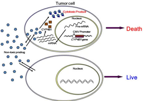
Figure 1. Schematic diagram of CYP4B1 ex- pression mediated prodrug-activating system.
Tumor cells expressing CYP4B1 could convert non-toxic prodrug to cytotoxic drug and result in death. Expression of rabbit CYP4B1 in hu- man colon cancer cell line rendered these cells highly sensitive to the prodrugs 4-IPO or 2-AA.
aromatic amines으로 전환하여 DNA 또는 단백질의 alky- lation을 유발함으로써 세포독성을 나타내는 것으로 알려 져 있다. 또한 사람의 CYP4B1 isozyme은 간과 폐에서 발 현하지만 그 활성이 토끼의 CYP4B1에 비해 1% 미만인 것으로 알려져 있다(6-10). CYP4B1/2-AA와 CYP4B1/4-IPO 시스템을 이용하여 자살유전자의 종양치료효과를 쥐 신 경교종세포인 C6에서 확인한 결과가 보고되었다(11).
종양치료에 있어 HSV-tk/GCV나 CYP4B1/2-AA 또는 CYP4B1/4-IPO 시스템을 이용하면 이들 자살유전자를 발 현하는 종양세포만을 살상하는 치료효과뿐만 아니라 자 살유전자가 이입되지 않은 주변 종양세포까지 죽게하는 bystander effect가 있다고 보고되고 있다(12,13). Bystander effect는 인접한 세포사이에 형성된 gap junction을 통해 자살유전자가 발현되는 종양세포로부터 만들어진 세포독 성 대사물이 전달됨으로써 나타나는 것으로 알려져 있으 며 낮은 유전자 이입효율을 보완하여 자살유전자의 치료 효과를 증진시키는 것으로 알려져 있다.
자살유전자치료 시스템의 종양 세포 살상효과를 향상 시키기 위해서는 자살유전자를 종양세포에 높은 효율로 발현시키기 위한 벡터개발이 중요하다. 자살유전자 전달 용 벡터는 크게 비바이러스성 벡터와 바이러스성 벡터로 나뉘어진다. 비바이러스성 벡터는 제조방법이 비교적 쉽고 면역반응을 유발하지 않으며 유전자의 크기 제한이 없다는 장점이 있으나 이입 효율이 낮고 유전자 발현이 일시적이 라는 단점이 있다. 바이러스성 벡터는 높은 유전자 전달 효 율을 가지나 안전성에 대한 문제가 있다는 단점이 있다.
CYP4B1유전자 발현을 위한 비바이러스성 벡터로 양이온 지질성 리포좀을 이용하여 쥐의 뇌종양 세포주를 치료한 연구와 간암세포주를 치료한 연구 등이 발표되었다(12,14).
현재 HSV-tk를 자살유전자로 적용한 연구는 임상시험 단계까지 갔으나, CYP4B1 유전자는 상대적으로 연구가 덜 진행되었으며 바이러스성 유래가 아닌 비바이러스성 벡터를 사용한다는 점에서 앞으로 연구가 주목받고 있다. 따라서 본 연구에서는 토끼 폐유래 CYP4B1 유전자를 인 간 대장암세포(HT-29)에 이입시켜 안정적인 발현 세포주 를 만들고, 전구체인 2-AA와 4-IPO에 대한 세포독성을 알아보았다.
Materials and Methods
1. 세포주 구축
American Type Cell Collection (ATCC)로부터 분양받은 인간 대장암세포(HT-29)를 10% fetal bovine serum (Invitrogen, Carlsbad, CA) 이 포함된 Dulbecco's Modified Eagle Medium (WelGENE, Korea)을 이용하여 175 mm2 culture flask에서 배양하였다.
종양세포를 6-well tissue culture plate에 well당 3×10⁵개 (100 μL media/well)의 HT-29세포를 분주하여 5% CO2 조건 의 37oC 배양기에서 24시간 배양하고 lipofectamine 2000 (Invitrogen) 10 μL를 serum free Opti-mem media (Invitrogen) 250 μL으로 희석한 후 pcDNA-CYP4B1(11) 4 μL를 혼합
Figure 2. Identification of CYP4B1 expression in CYP4B1 gene trans- duced HT-29. Confirmation of CYP4B1expression in HT-29 cells (CYP4B1
#1, #2, #3, #4) by RT-PCR analysis. RT-PCR products of CYP4B1 were 650 bp. The β-actin product was 540 bp amplified PCR product as an in- ternal control.
Figure 3. 4-IPO or 2-AA induced cytotoxicity in CYP4B1 gene trans- duced HT-29. Dose-dependent cytotoxicity of 2-AA or 4-IPO in CYP4B1 gene transduced HT-29 was determined by trypan-blue dye exclusion assay in HT-29 cells. Cell survival rate was measured at 96 h after 4-IPO treatment (upper panel) or 2-AA treatment (lower panel).
후 상온에서 5분간 반응하고 serum free media 1 mL와 섞어 각 well에 첨가하여 4~6시간 5% CO2 조건의 37oC 배양기에서 배양한다. 배양이 끝나면 10% fetal bovine se- rum이 포함된 media로 갈아준 후 18시간 배양한 후, hy- gromycin 500 μg/mL이 첨가된 media로 3주간 처리하여 발 현 세포주 2종류(CYP4B1#1, CYP4B1#3)를 선별하였다.
2. CYP4B1 발현 확인
무작위로 4개의 세포주를 선발하여 total RNA 분리하 였고, One Step RT-PCR Kit (Qiagen, USA)을 사용하여 CYP4B1 특이 primer인 sense (5'-CTT CCA TTA CGA CGT GCT GA-3')와 antisense (5'-TCA TGC ACA TGG TCA GGT AG-3')을 사용하여 PCR을 시행하였다.
Human β-actin에 특이적인 primer인 sense (5'-GTG GGG CGC CCC AGG CAC CAG GGC-3')와 antisense (5'-CTC CTT AAT GTC ACG CAC GAT TTC-3')을 internal con- trol로 사용하였다.
3. 세포독성 시험
CYP4B1 발현세포와 대조군 HT-29를 6-well tissue cul- ture plate에 1x10⁴ cells/well 분주하여 5% CO2 조건의 37oC 배양기에서 24시간 배양 후 2-aminoanthrancene (0~0.01 mM) 또는 4-ipomeanol (0~1 mM)을 media에 희석하여 농도별로 처리하여 96시간 배양 후 살아있는 세포 개수 를 측정하였다. Trypan-blue solution (Sigma, St Louis, MO, USA) 20 μL와 동량의 세포를 섞어 C-Chip (Digital Bio., Korea)에서 현미경으로 살아있는 세포수를 측정하 였다.
Results
1. 인간 대장암세포에 토끼 CYP4B1 유전자 이입 후 안정적 발현 세포주 선별
pcDNA-CYP4B1을 lipofectamine 2000을 이용해 이입시 킨 후 hygromycin 500 μg/mL을 3주간 처리하여 CYP4B1 의 안정적 발현 세포를 선별하였다. 4종의 세포(CYP4B1
#1, #2, #3, #4)가 무작위로 선별되었으며 각각의 세포로 부터 RNA를 추출하여 RT-PCR을 실시하였다. 양성대조 군으로 β-actin 프라이머를 이용하여 540bp의 밴드를 관
찰하였으며, CYP4B1 프라이머를 이용하여 CYP4B1유전자가 발현된 4종의 세포 중 2종의 세포(CYP4B1#1, CYP4B1#3)에 서 650bp 밴드가 나타났고 음성대조군인 물 또는 유전자 가 이입되지 않은 각 세포에서는 CYP4B1이 발현되지 않 음을 확인하였다(Figure 2).
2. HT-29에서 CYP4B1 발현에 의한 세포독성평가
CYP4B1이 안정적으로 발현하는 세포 CYP4B1 #1 또는 CYP4B1 #3에 독성없는 전구체 (2-AA: 0~0.01 mM 또는 4-IPO: 0~1 mM)를 다양한 농도로 처리하고 96시간 후의 세포생존율을 trypan blue dye-exclusion방법으로 분석하 였다. 대조군(HT-29)에 비해 CYP4B1 유전자 발현세포의 2-AA또는 4-IPO에 대한 50% 세포성장저해 농도가 훨씬
낮은 것으로 확인되었으며, 2-AA를 96시간 처리했을 때 0.003 mM인 경우 대조군의 생존율은 86%이지만 CYP4B1 이 발현하는 세포에서는 67% (CYP4B1 #1) 또는 60%(CYP4B1
#3)였다. 또한 4-IPO 경우 0.1 mM에서 대조군의 생존율은 82%였고, CYP4B1이 발현하는 세포에서는 56%(CYP4B1 #3) 인 것을 확인했다. 반면 CYP4B1 #1의 경우 4-IPO농도 0.8 mM부터 대조군의 생존율(64%)에 비해 생존율(44%) 이 떨어짐을 확인했다(Figure 3).
Discussion
본 연구는 자살유전자인 CYP4B1을 이용한 종양치료 효과를 알아보기 위해 인간 대장암세포인 HT-29를 사용 하였고 자살유전자를 발현시키기 위한 유전자 전달방법 으로 plasmid transfection법을 이용하였다. 본 연구에서 사용한 plasmid transfection법은 유전자 발현이 불안정하 다는 단점이 있다. 이를 보안하고자 자살유전자를 종양 세포에 높은 효율로 발현 또는 감염시키기 위한 방법으 로 바이러스성 벡터(아데노바이러스, 레트로바이러스, 렌 티바이러스등)를 이용할 수 있다. 또한 CYP4B1 유전자 발현에 의한 2-AA 또는 4-IPO에 대한 약제 감수성을 알 아보기 위해 사용한 trypan blue dye exclusion assay는 약 물의 세포독성 혹은 세포 생존율 검사에 쓰이는 방법으 로 죽은 세포를 제외한 살아있는 세포에 의해 측정되는 것으로 살아 있는 세포는 세포막을 통하여 trypan blue 색소가 들어올 수 없기 때문에 사멸세포의 수를 측정함 과 동시에 사멸세포의 세포막에 푸른색으로 염색이 됨으 로 해서 세포막의 손상 정도를 알 수 있는 방법이다. 세 포독성 결과에 따르면 CYP4B1 유전자를 플라스미드 이 입을 통해 발현시켰을 때 4-IPO보다 2-AA에 대한 감수 성이 높은 것을 알 수 있었다. 이는 bystander effect가 4-IPO보다 2-AA에 대해 높아 본 연구에서도 자살유전자 치료효과를 증진시킨 것임을 예측해볼 수 있고, 기존에 CYP4B1 유전자가 발현된 신경교종 세포주중의 하나인 9L을 2-AA로 처리했을 경우 약물의 IC50 값이 1 μM로 보 고된 것에 비교할 만한 수준 (3 μM)을 나타내었다(12).
그리고 자살유전자를 이용한 종양의 치료효과 판정을 위 한 영상을 얻기 위하여 형광 (fluorescence)이나 생물발광 (bioluminescence)같은 광학영상을 사용하거나, 감마카메라, Single Photon Emission Computed Tomography (SPECT) 또 는 Positron Emission Tomography (PET)같은 핵의학영상 등이 이용되고 있는데 본 연구팀의 이전 연구에서 CYP4B1
발현으로 [18F] 또는 [3H]으로 표지된 4-IPO의 섭취율 증 가 결과를 얻었으므로 CYP4B1 유전자 치료에 의한 치료 효과를 다양한 영상으로 모니터링할 수 있는 가능성을 보고한 바 있다(15).
요약하면 본 연구를 통하여 인간 대장암세포에 CYP4B1 유전자의 안정적 발현을 이루었으며, 이를 RT-PCR을 통해 확인하였다. CYP4B1/2-AA 또는 CYP4B1/4-IPO를 이용한 종양의 안정적 치료효과 유도가 가능하였으며, [18F] 또는 [3H]으로 표지 된 4-IPO의 섭취율 증가 결과를 이전 연 구에서 얻었으므로, 이를 종합하여 CYP4B1 유전자 치료에 의한 치료효과를 모니터링할 수 있을 것으로 기대한다.
Acknowledgments
This work was supported by the Korea Science and Engineering Foundation (KOSEF) grant funded by the Korea government (MEST) (NRF-2012M2A2A7013480).
References
1. Springer CJ, Niculescu-Duvaz I. Prodrug-activating systems in suicide gene therapy. J Clin Invest 2000;105:1161-1167.
2. Sasaki M, Plate KH. Gene therapy of malignant glioma: recent advances in experimental and clinical studies. Ann Oncol 1998;9:1155-1166.
3. Moolten FL. Tumor chemosensitivity conferred by inserted her- pes thymidine kinase genes: paradigm for a prospective cancer control strategy. Cancer Res 1986;46:5276-5281.
4. Li Z, Shanmugam N, Katayose D, Huber B, Srivastava S, Cowan K, Seth P. Enzyme/prodrug gene therapy approach for breast cancer using a recombinant adenovirus expressing Escherichia coli cytosine deaminase. Cancer Gene Ther 1997;4:113-117.
5. Jang SJ, Kang JH, Kim KI, Lee TS, Lee YJ, Lee KC, Woo KS, Chung WS, Kwon HC, Ryu CJ, Choi TH, Choi CW, Lim SM, Cheon GJ. Application of bioluminescence imaging to ther- apeutic intervention of herpes simplex virus type I - Thymidine kinase/ganciclovir in glioma. Cancer Lett 2010;297:84-90.
6. Smith PB, Tiano HF, Nesnow S, Boyd MR, Philpot RM, Langenbach R. 4-Ipomeanol and 2-aminoanthracene cytotox- icity in C3H/10 T1/2 cells expressing rabbit cytochrome P450 4B1. Biochem Pharmacol 1995;50:1567-1575.
7. Baer BR, Rettie AE. CYP4B1: an enigmatic P450 at the interface between xenobiotic and endobiotic metabolism. Drug Metab Rev 2006;38:451-476.
8. Rainov NG, Dobberstein KU, Sena-Esteves M, Herrlinger U, Kramm CM, Philpot RM, Hilton J, Chiocca EA, Breakefield XO.
New prodrug activation gene therapy for cancer using cyto-
chrome P450 4B1 and 2-aminoanthracene/4-ipomeanol. Hum Gene Ther 1998;9:1261-1273.
9. Czerwinski M, McLemore TL, Philpot RM, Nhamburo PT, Korzekwa K, Gelboin HV, Gonzalez FJ. Metabolic activation of 4-ipomeanol by complementary DNA-expressed human cyto- chromes P-450: Evidence for species-specific metabolism.
Cancer Res 1991;51:4636-4638.
10. Mohr L, Rainov NG, Mohr UG, Wands JR. Rabbit cytochrome P450 4B1: A novel prodrug activating gene for pharmacogene therapy of hepatocellular carcinoma. Cancer Gene Ther 2000;7:
1008-1014.
11. Jang SJ, Kang JH, Lee TS, Kim SJ, Kim KI, Lee YJ, Cheon GJ, Choi CW, Lim SM. Prodrug-activating gene therapy with rabbit cytochrome P450 4B1/4-ipomeanol or 2-aminoanthracene sys- tem in glioma cells. Nucl Med Mol Imaging 2010;44:193-198.
12. Frank S, Steffens S, Fischer U, Tlolko A, Rainov NG, Kramm
CM. Differential cytotoxicity and bystander effect of the rabbit cytochrome P450 4B1 enzyme gene by two different prodrugs:
implications for pharmacogene therapy. Cancer Gene Ther 2002;
9:178-188.
13. Kievit E, Nyati MK, Ng E, Stegman LD, Parsels J, Ross BD, Rehemtulla A, Lawrence TS. Yeast cytosine deaminase improves radiosensitization and bystander effect by 5-fluorocytosine of human colorectal cancer xenografts. Cancer Res 2000;60:6649-6655.
14. Mohr L, Rainov NG, Mohr UG, Wands JR. Rabbit cytochrome P450 4B1: A novel prodrug activating gene for pharmacogene therapy of hepatocellular carcinoma. Cancer Gene Ther 2000;7:
1008-1014.
15. Moon BS, Jang SJ, Kim SJ, Lee TS, Chi DY, Lee BC, Kang JH, Kim SE. Synthesis and evaluation of a 18F-labeled 4-ipomeanol as an imaging agent for CYP4B1 gene prodrug activation therapy. Cancer Biother Radiopharm 2013;28:588-597.