https://doi.org/10.5468/ogs.2018.61.2.267 pISSN 2287-8572 · eISSN 2287-8580
Introduction
Minimally invasive laparoscopic surgery continues to be the leading surgical method, offering a shorter recovery time, less postoperative pain, better cosmetic outcomes, and lower complication rates compared to open abdominal surgery [1- 3]. Unfortunately, some difficulty remains in removing large- sized specimens via single-port assisted (SPA) laparoscopy. A considerable amount of time and effort must be invested for morcellation, which is the fragmentation of a tissue specimen to reduce it to a compact size, enabling extraction through smaller incisions [4].
In-bag power morcellation technique in single-port laparoscopic myomectomy
Young Bin Won, Hyun Jin Lee, Kyung Jin Eoh, Young Shin Chung, Yong Jae Lee, Seon Hee Park, Jee Whan Kim, Jung Yoon Lee, Eun Ji Nam, Sunghoon Kim, Young Tae Kim, Sang Wun Kim
Division of Gynecologic Oncology, Department of Obstetrics and Gynecology, Women's Cancer Center, Yonsei Cancer Center, Institute of Women's Life Medical Science, Yonsei University College of Medicine, Seoul, Korea
Objective
This study introduces and evaluates the feasibility, safety, and surgical outcomes of the in-bag power morcellation technique during single-port assisted (SPA) laparoscopic myomectomy in comparison with manual scalpel morcellation.
Methods
This is a retrospective review of a total of 58 patients who underwent SPA laparoscopic myomectomy employing in-bag power morcellation (n=27) or manual scalpel morcellation (n=31), performed between December 2014 and December 2016. Surgical outcomes, including total operation time, estimated blood loss, postoperative hemoglobin changes, postoperative hospital stay, postoperative pain (visual analog scale), perioperative and postoperative complications were evaluated.
Results
The demographics and patient characteristics were similar between both groups. The median patient age was 34 years and median body mass index was 20.84 kg/m
2. The median specimen weight was 110 g. The median operating time was 138 minutes. The median estimated blood loss was 50 mL and the median postoperative hemoglobin change was 2.2 g/dL. The median postoperative hospital stay was 2 days and the median postoperative pain scores were 5 after 6 hours, 3 after 24 hours, and 2 after 48 hours. Occult malignancy was not identified in any patients. There were no intraoperative complications such as LapBag ruptures or gross spillage.
Conclusion
In-bag power morcellation for SPA laparoscopic myomectomy is feasible and safe, minimizing the risks of open power morcellation. There were also no statistically significant differences in surgical outcomes.
Keywords: Laparoscopy; Minimally invasive surgical procedures; Myomectomy; Morcellation
Received: 2017.05.31. Revised: 2017.08.04. Accepted: 2017.08.18.
Corresponding author: Sang Wun Kim
Division of Gynecologic Oncology, Department of Obstetrics and Gynecology, Women's Cancer Center, Yonsei Cancer Center, Institute of Women's Life Medical Science, Yonsei University College of Medicine, 50-1 Yonsei-ro, Seodaemun-gu, Seoul 03722, Korea E-mail: [email protected]
https://orcid.org/0000-0002-8342-8701
Articles published in Obstet Gynecol Sci are open-access, distributed under the terms of the Creative Commons Attribution Non-Commercial License (http://creativecommons.
org/licenses/by-nc/3.0/) which permits unrestricted non-commercial use, distribution, and reproduction in any medium, provided the original work is properly cited.
Copyright © 2018 Korean Society of Obstetrics and Gynecology
The prototype for power morcellation was first introduced in 1993 by Rolf Steiner of Switzerland [5]. Electromechanical morcellation is an easy and efficient method for removing tissue from the abdominal cavity compared to hand-manip- ulated morcellation [6]. However, power morcellation related complications such as injury from morcellator blades to vessels or adjacent visceral organs is a concern [7].
This method also introduces the risk of unintentional dis- semination of removed tissue, which could lead to benign myoma seeding or spillage of malignant material into the peritoneal cavity if that tissue is pathologically confirmed to be endometrial cancer or sarcoma [8,9]. A safety communication warning published by the U.S. Food and Drug Administration in April 2014 advised against the use of power morcellation due to the risk of intra-abdominal malignancy dissemination during laparoscopy [10].
The purpose of this study is to introduce and evaluate the feasibility, safety, and surgical outcomes of the in-bag power morcellation technique during SPA laparoscopic myomectomy in comparison with manual scalpel morcellation.
Materials and methods
This study is a retrospective review of a total of 58 patients who underwent SPA laparoscopic myomectomy employing in-bag power morcellation (n=27) or manual scalpel morcel- lation (n=31), performed between December 2014 and De- cember 2016 at Severance Hospital, Yonsei University College of Medicine, in Seoul, Korea. All surgeries were performed by a single gynecologic surgeon specializing in minimally in- vasive gynecologic surgical techniques. We received written consent from all patients prior to surgery. Inclusion criteria for this study limited subjects to women undergoing laparoscopic surgical treatment for a benign disease of the uterus such as myoma, adenomyosis, and/or adenomyoma. Exclusion crite- ria included patients with known or suspected malignancies.
Pathologic confirmation was done using tissue extracted dur- ing surgery. Patients were categorized into 2 groups according to whether they received specimen extraction using in-bag power morcellation or manual morcellation using a scalpel.
Demographic and physical characteristics such as age, coital history, parity, body mass index (BMI), previous surgical his- tory, and surgical indication were investigated. Parity included both vaginal and cesarean section delivery. Preoperative evalu-
ation included cervical cytology and imaging evaluation such as ultrasonography, magnetic resonance imaging, or abdomi- nal and pelvic computed tomography scans. Myoma size and location were ascertained by imaging. Surgical outcomes such as estimated blood loss during surgery (mL), serum hemo- globin (Hb) differences (the change between pre-surgical Hb and postoperative day 1 Hb), total operation time (minutes), postoperative hospital stay (days), and the dry weight of the extracted specimen (g) were also investigated. Total operation time was calculated as the time from initial incision to final closure. Estimated blood loss during surgery was approxi- mated as the total suctioned volume. Hospital discharge was determined according to whether patients were afebrile for 24 hours, could tolerate a soft diet, could ambulate without assistance, and were able to urinate independently after uri- nary catheter removal. Postoperative pain was appraised ac- cording to the visual analog scale. Patients were asked to rate postoperative pain 6, 24, and 48 hours following surgery.
1. Surgical techniques
All SPA laparoscopic surgeries were performed according to a technique previously described by our group [11]. The SPA system entails a single multichannel port at the umbilicus. In- bag power morcellation was performed by morcellating the removed uterine tissue inside an insufflated endoscopic bag within the abdominal cavity (Supplementary Video 1).
With this single-port surgery technique, an Alexis wound re- tractor (Applied Medical, Rancho Santa Margarita, CA, USA) was inserted into the abdomen through a 2 cm trans-umbil- ical incision site. A 7½ left-handed surgical glove was affixed to the wound retractor. Incisions were made in the glove fin- gertips. A 12-mm trocar was inserted into the thumb, a 5-mm trocar into the pinky, and a 5-mm mini trocar into the middle finger. A 45-cm rigid, 30 degrees 5-mm endoscope was used.
Instruments used during surgery included a monopolar L- hook, atraumatic graspers, scissors, biopsy forceps with a slightly-bent shaft, a suction-irrigation system, laparoscopic needle holders, and energy devices such as Enseal (Ethicon, Somerville, NJ, USA) and Thunderbeat (Olympus Medical Sys- tems, Tokyo, Japan) [11].
Standard procedure was used for myomectomy. A monopo- lar hook was used to make a vertical incision in the prominent part of the myoma. Dissection of the cleavage plane between the myoma and surrounding tissue was then completed.
Enucleation and removal were achieved by fixing and imple-
menting traction on the myoma. Thunderbeat was used for dissection in several cases. The myomectomy site was repaired
in 2 layers with an intracorporeal continuous suture and tie.
For in-bag power morcellation, a 30 cm
2laparoscopic speci- men retrieval bag, XXL or 3XL LapBag (Sejong Medical Co., Seoul, Korea), was used to remove the myoma without spill- age. After inserting the LapBag into the abdominal cavity, it was opened intra-abdominally and the myoma specimen was placed into the LapBag and pulled out midway through the wound retractor by drawing on the thread of the bag.
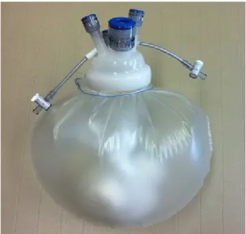
The surgical glove was then detached from the Alexis wound retractor. The wound retractor and surgical glove were reat- tached together by folding 3 or 4 times with the LapBag inserted in between (Fig. 1, Supplementary Video 2). After completely attaching the LapBag to the wound retractor and surgical glove, CO
2gas was pumped into the bag. The 12- mm trocar was separated from the surgical glove, and the power morcellator (Chungang Medical Co., Seoul, Korea) was inserted into the thumb of the surgical glove. The entire my- oma was morcellated while contained within the insufflated bag (Fig. 2, Supplementary Video 3). After morcellation was complete, the surgeon examined the bag for tears and in- spected the pelvis for visible spillage of fluid or uterine tissue.
Fig. 1. Simulation of 3XL LapBag insufflated with CO2 gas at- tached to single-port wound retractor.
Fig. 2. Intraoperative laparoscopic view of myoma power morcellation while contained within insufflated LapBag. (A) 3XL LapBag is opened intra-abdominally. (B) Myoma is inserted into the LapBag. (C) The myoma is grabbed with morcellator instrument. (D) The myoma is power morcellated within the insufflated LapBag.
A B
C D
In patients with small myomas that can be easily removed through the umbilical incision, manual morcellation technique was usually selected. But in patients with relatively large myo- mas (>5 cm), power morcellation was predominantly selected except when the power morcellator was not available.
2. Statistical analysis
The demographic and physical characteristics of all patients were obtained retrospectively. Surgical outcome results were obtained pre- and post-operatively. All statistical analyses were performed using SPSS ver. 23 (SPSS Inc., Chicago, IL, USA).
Descriptive statistics were used to summarize the results of the patients’ characteristics and operation outcomes. All data was analyzed using the Student’s t-test or Pearson’s χ
2test and reported as a value of number (%) or a median (range).
Results
The in-bag morcellation technique for specimen extraction was successfully performed in all 27 cases. The demographic and physical characteristics of the patients are shown in Table 1.
A known huge myoma (n=12, 44.4%) was the leading in- dicator for surgery. Other indications included hypermenor- rhea (n=5, 18.5%), an increase in myoma size (n=5, 18.5%), and pelvic pain (n=5, 18.5%). Four (14.8%) and 9 (29.0%) women had received previous abdominal surgeries in the in- bag morcellation group and the manual scalpel morcellation group, respectively. The median patient age was 34 years (range, 26–54), and the median BMI was 20.84 kg/m
2(range,
18.26–28.89) for the in-bag morcellation group. For the man- ual morcellation group, the median patient age was 37 years (range, 26–59) and the median BMI was 21.93 kg/m
2(range, 19.07–29.41). There were no significant differences in age, parity, coital history, or BMI (Table 1) between the 2 groups.
The surgical outcomes of the patients are shown in Table 2.
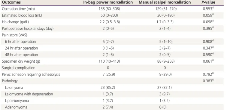
The sizes of the largest uterine tissue removed differed from 4 to 10 cm vs. 3 to 9 cm, and the median specimen weight was 110 g (range, 40–413) vs. 88 g (range, 9–258). The me- dian operating time was 138 minutes (range, 60–308) vs. 129 minutes (range, 51–270). The average time taken to prepare a device for in-bag power morcellation was approximately 5 to 10 minutes. No surgical procedures were converted from laparoscopy to laparotomy. Estimated blood loss was 50 mL (range, minimal–200) vs. 30 mL (range, minimal–180), and the median postoperative Hb change was 2.2 g/dL (range, 0.5–3.8) vs. 1.7 g/dL (range, 0–3.3). Severe adhesions requir- ing adhesiolysis during surgery were seen in 7 women (25.9%) in the in-bag morcellation group and 9 women (29.0%) in the manual morcellation group. The mean postoperative hospital stay was 2 days (range, 0–5) vs. 2 days (range, 1–4), and the median postoperative pain scores were 5 (range, 2–7) after 6 hours, 3 (range, 1–5) after 24 hours, and 2 (range, 1–5) after 48 hours vs. 5 (range, 1–10) after 6 hours, 3 (range, 2–7) after 24 hours, and 2 (range, 0–5) after 48 hours. There were no significant differences in surgical outcomes such as total dry specimen weight, total operating time, estimated blood loss, Hb difference, postoperative hospital stays, or postopera- tive pain scores (Table 2).
Pathologic confirmation following the surgical removal of
Table 1. Demographics and physical characteristicsCharacteristics In-bag power morcellation Manual scalpel morcellation P-value
Age (yr) 34 (26–54) 37 (26–59) 0.372a)
BMI (kg/m2) 20.84 (18.26–28.89) 21.93 (19.07–29.41) 0.067a)
Parity 9.0 (33.3) 10.7 (35.5) 0.864b)
Previous abdominal surgery history 4 (14.8) 9 (29.0) 0.195b)
Indication for surgery 0.635b)
Known huge myoma 12 (44.4) 12 (38.7)
Hypermenorrhea 5 (18.5) 7 (22.6)
Myoma size increase 5 (18.5) 9 (29.0)
Pelvic pain 5 (18.5) 3 (9.7)
Values are presented as median (range) or number (%).
BMI, body mass index.
a)Student's t-test; b)Pearson's χ2 test.
Table 2. Surgical outcomes
Outcomes In-bag power morcellation Manual scalpel morcellation P-value
Operation time (min) 138 (60–308) 129 (51–270) 0.553a)
Estimated blood loss (mL) 50 (0–200) 30 (0–180) 0.059a)
Hb change (g/dL) 2.2 (0.5–3.8) 1.7 (0–3.3) 0.098a)
Postoperative hospital stays (day) 2 (0–5) 2 (1–4) 0.395a)
Pain score (VAS)
6 hr after operation 5 (2–7) 5 (1–10) 0.908a)
24 hr after operation 3 (1–5) 3 (2–7) 0.347a)
48 hr after operation 2 (1–5) 2 (0–5) 0.590a)
Specimen dry weight (g) 110 (40–413) 88 (9–258) 0.061a)
Surgical complication 0 0
Pelvic adhesion requiring adhesiolysis 7 (25.9) 9 (29.0) 0.792b)
Pathology 0.383b)
Leiomyoma 23 (85.2) 27 (87.1)
Leiomyoma with degeneration 1 (3.7) 3 (9.7)
Lipoleiomyoma 1 (3.7) 1 (3.2)
Adenomyoma 2 (7.4) 0 (0)
Values are presented as median (range) or number (%).
Hb, hemoglobin; VAS, visual analog scale.
a)Student's t-test; b)Pearson's χ2 test.