Articles published in Obstet Gynecol Sci are open-access, distributed under the terms of the Creative Commons Attribution Non-Commercial License (http://creativecommons.
org/licenses/by-nc/3.0/) which permits unrestricted non-commercial use, distribution, and reproduction in any medium, provided the original work is properly cited.
Copyright © 2015 Korean Society of Obstetrics and Gynecology http://dx.doi.org/10.5468/ogs.2015.58.4.314
pISSN 2287-8572 · eISSN 2287-8580
Introduction
Benign metastatic leiomyomatosis (BML) is defined as a muscle tumor in association with one or more smooth muscle tumors of the uterus and without evidence of any extrauterine primary site. The group of smooth muscle tumors resembling uterine leiomyoma with unusual growth patterns and distant metas- tasis includes diffuse peritoneal leiomyomatosis (DPL) and, lymphatic spreading and intravenous leiomyomatosis (IVL) [1].
IVL usually invades the lumen of veins and extends into the uterine vein, ovarian vein, common iliac vein, inferior vena cava and right atrium, whereas DPL has characteristics consistent with direct peritoneal seeding [2]. Leiomyomatosis is treated by observation, hormonal suppression, and/or surgical debulk- ing via laparotomy or laparoscopy [3]. Laparoscopic surgery decreases postoperative pain and is associated with a shorter recovery period than laparotomic surgery. Although single-port laparoscopy (SPL) with a small single transumbilical incision is technically more difficult than multi-port laparoscopy, SPL has the advantages of better cosmesis and easier tissue extraction via transumbilical morcellation than multi-port laparoscopy [4].
We report a 39-year-old woman with leiomyomatosis that had features of both multiple peritoneal seeding and lymph node metastasis who underwent SPL laparoscopic debulking surgery.
Case report
A 39-year-old woman (gravida-1, para-1) was admitted to the emergency department due to severe abdominal pain. She had a history of Cesarean section 15 years prior and total hysterectomy due to a 15-cm-diameter uterine leiomyoma 4 years prior. Physi- cal examination revealed a distended abdomen and palpable hard mass. Abdominopelvic 3-dimensional computed tomography (CT) revealed conglomerated and enlarged para-aortic lymph nodes with multiple enlarged common iliac and external iliac lymph nodes and multiple nodules scattered in the pelvis and abdomen (Fig. 1A). Chest CT showed several small nodules in both lower
Received: 2014.11.20. Revised: 2015.1.13. Accepted: 2015.3.6.
Corresponding author: Joo Hee Yoon
Department of Obstetrics and Gynecology, St. Vincent Hospital, the Catholic University of Korea School of Medicine, 93 Jungbu- daero, Paldal-gu, Suwon 442-723, Korea
Tel: +82-31-249-8302 Fax: +82-31-254-7481 E-mail: [email protected]
Single-port laparoscopic debulking surgery of variant benign metastatic leiomyomatosis with simultaneous lymphatic spreading and intraperitoneal seeding
Yoo Hyun Chung
1, Suk Woo Lee
2, So Young Shin
1, Chae Chun Rhim
2, Soyoung Im
3, Sie Hyeon Yoo
4, Joo Hee Yoon
1Department of Obstetrics and Gynecology, 1The Catholic University of Korea School of Medicine, Seoul; 2Hallym University College of Medicine, Chuncheon; 3Department of Pathology, The Catholic University of Korea School of Medicine, Seoul; 4Department of Anesthesiology and Pain Medicine, Soonchunhyang University Cheonan Hospital, Soonchunhyang University College of Medicine, Cheonan, Korea
Benign metastatic leiomyomatosis (BML) is a rare disease characterized by smooth muscle cell proliferation in extrauterine sites including the lung, abdomen, pelvis, and retroperitoneum. Depending on location, BML is classified as intravenous leiomyomatosis and diffuse peritoneal leiomyomatosis. Pathogenesis of BML can be iatrogenic after previous myomectomy or hysterectomy, hormonal, or coelomic metaplasia. Treatment options are observation, hormonal suppression, and/or surgical debulking via laparotomy or laparoscopy. Laparoscopic surgery is gaining in popularity in the gynecologic field compared to laparotomic surgery and single-port laparoscopy has the benefits of cosmesis and early tissue extraction by transumbilical morcellation. We report a 39-year-old woman with BML who underwent single-port laparoscopy debulking surgery.
Keywords: Hysterectomy; Laparoscopy; Leiomyomatosis
lobes and lingual without lymph node enlargement in the thorax (Fig. 1). We consulted a thoracic surgeon who recommended a lung biopsy was not performed, for the patient had not repiratory symptoms and she refused it. Ultrasonography-guided biopsy of the omental mass lesion showed benign spindle cell proliferation with smooth muscle differentiation. The patient was therefore transferred to the obstetrics and gynecology department. We de- cided to perform minimal invasive surgery, mainly for diagnostic purpose. SPL was started under general anesthesia with the pa- tients in the Trendelburg position. A 2.5-cm vertical intraumbilical incision was made and OCTO Port system (Dalim SurgNET Corpo- ration, Seoul, Korea) was placed in the incision. OCTO Port has a capability to do multi-tasking by using multiple tentacles that al- low three laparoscopic instruments. OCTO Port was inserted and fixed to the outer ring of the wound retractor. After insufflations of carbon dioxide, a rigid 30-degree 10-mm telescope was in- serted into the abdomen. We confirmed the absence of a uterus due to the previous hysterectomy. Numerous firm, smooth nod- ules with diameters ranging in size from 2 to 8 cm were present on the surface of the omentum, Douglas pouch, peritoneal wall, both adnexae, small bowel and lymph nodes: paraaortic, com- mon illiac, and external illiac lymph nodes. We performed bilateral salpingo-oophorectomy, partial omentectomy, para-aortic, com- mon iliac, and external iliac lymph node dissection and multiple mass excisions (Fig. 2). After excision of leiomyomatosis adherent to the small bowel, primary closure of the serosal layer of the small bowel was performed by an extracorporeal approach. His- tological evaluation demonstrated nodular structures with spindle cells. Signs of malignancy, including nuclear atypia or mitotic ac- tivity, were not found.
The patient was given a gonadotropin-releasing hormone ago- nist (GnRHa) for 6 months after the surgery. There have been no subsequent signs of recurrence. There was no interval change of several small nodules in both lower lobes and lingual on the follow-up low-dose chest CT after treatment of GnRHa. There were also no subsequent signs of recurrence on the follow-up abdominopelvic CT.
Discussion
The term BML was initially coined by Steiner in 1937 and re- fers to multiple well-differentiated leiomyomas at sites distant from the uterus. Although this tumor type is associated with multiple distant metastases, it appears benign histologically as it is characterized by a low mitotic index, lack of nuclear pleo-
morphisms, and no evidence of invasion of adjacent organs [5]. Depending on tumor location, leiomyoma found outside the anatomic confines of the uterus is referred to as IVL (when
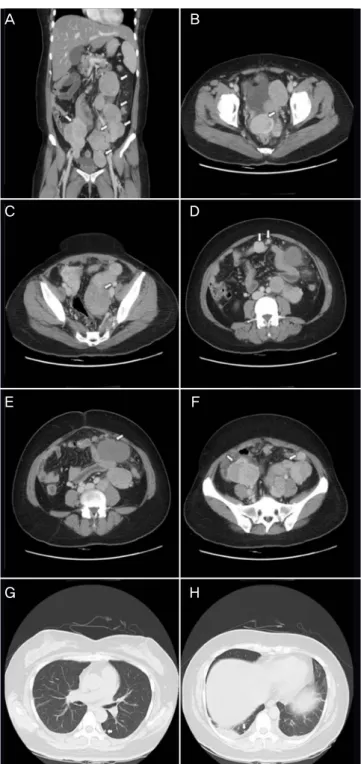
Fig. 1. Multiple metastatic leiomyomatosis in a 39-year-old woman. Con- trast enhanced 3-dimensional multi-detector computed tomography images show several well-defined masses (arrows) in abdomino-pelvic cavity (A).
Multiple masses are sacttered in the Douglas pouch (B), near the left adnex- ae (C), peritoneum (D), omentum (E), and near both external iliac vessel (F).
A chest computed tomography scan revealed multiple peripheral nodules in both lower lobe and lingula of lung (G,H).
A B
C D
E F
G H
found in vascular channels) and DPL (when found in the peritoneal cavity) [1]. BML metastasize to the lung most com- monly, and can metastasize to other distant sites such as the heart, inferior vena cava, bone, lymph nodes, muscular tissue, and retroperitoneum infrequently [6].
There are some experimental evidences to suggest that BML and uterine leiomyoma are actually same disease. Patton et al. [7] reported that uterus and pulmonary nodule tumors had identical histology patterns (benign), immunohistochemical profiles (estrogen receptor positive, progesterone receptor positive, and very low proliferative index), and androgen recep- tor allelic inactivation based on molecular cytogenetic analysis.
Tietze et al. [8] reported that uterine leiomyoma and pulmo- nary tumors showed an identical pattern of X-chromosome inactivation in comparative genomic hybridization, consistent
with a monoclonal origin. These results indicate that BML is a benign disease and originates from a uterine leiomyoma.
IVL is a rare disease which is characterized by smooth muscle tumor cells invading the lumen of veins. The most common metastatic site of IVL is the lung, however it may extend to right atrium and ventricle, inferior vena cava, and tje ovarian vein. Although IVL is a benign smooth muscle tumor, exten- sion into the cardiac cavity can cause sudden death due to me- chanical obstruction [9]. Lymphatic spread of leiomyomatosis may be associated with previous surgical trauma during uter- ine dilatation and curettage, myomectomy, or hysterectomy.
A previous study reported BML with lymphatic spread in a 27-year-old woman [10]. The authors of that study postulated that fragments of leiomyoma, detached at the time of endo- metrial curettage, entered the dilated lymphatic channel, and
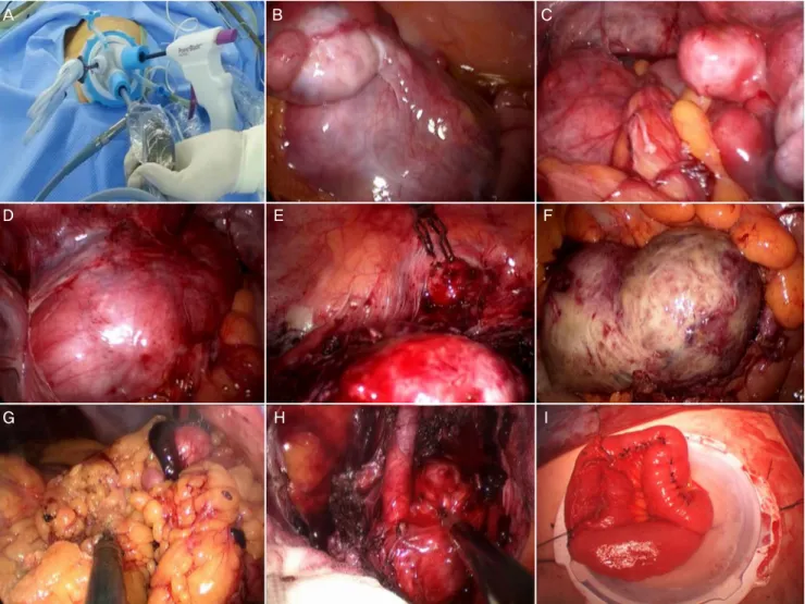
Fig. 2. Operative findings. Single port system with OCTO Port (A). Multiple leiomyoma of near the left adnexae (B), Douglas pouch (C), near the left external iliac vessel (D), and peritoneum (E). Leiomyoma with red degeneration attached to bowel serosa and mesentery (F). Partial omentectomy (G). Para-aortic lymph node dissection (H). Extracorporeal primary repair of serosal area of small bowel after removal mass attached the small bowel (I).
A B C
D E F
G H I